Plancia ëd Dasymutilla Ashmead 1899
Descrission:
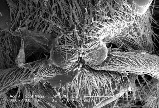
At a low magnification of only 46X, this scanning electron micrograph (SEM) showed the head region from an anterior view of a female velvet ant, Dasymutilla sp.. Note the two laterally positioned eyes which are partially visible at the topmost area of the photograph, but its the two anteriorly-placed antennae with their rounded "scapes" that are the most apparent head appendages. Like the antennae, the numerous hairs or setae adorning almost all of the insects exterior surfaces, act as sensory structures, supplying the organism with information about its environmental parameters. The jointed legs, from which the insects Phylum Arthropoda is derived, i.e., Arthro = jointed, and poda leg, are also partially visible, emanating from the thoracic region.
Created: 2007
Ancludù an coste pàgine-sì:
- Life
- Cellular
- Eukaryota
- Opisthokonta
- Metazoa
- Bilateria
- Protostomia
- Ecdysozoa
- Arthropoda
- Pancrustacea
- Hexapoda
- Insecta
- Pterygota
- Neoptera
- Endopterygota
- Hymenoptera
- Apocrita
- Aculeata
- Vespoidea
- Mutillidae
- Dasymutilla
- Panarthropoda
Costa plancia a compariss an gnun-e colession.
Anformassion an sla sorgiss
- licensa
- cc-publicdomain
- fotògraf
- Janice Carr
- fornidor
- Public Health Image Library
- original
- archivi ëd mojen original
- visité la sorgiss
- sit compagn
- Public Health Image Library
- ID