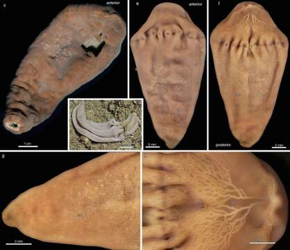
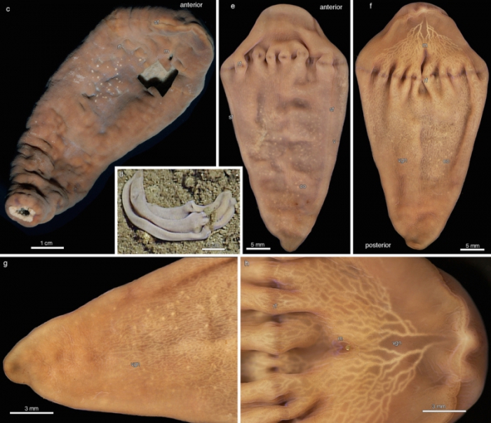
Xenoturbella monstrosa
Descrission:
c, Ventral view of the holotype SIO-BIC BI1037. Although highly contracted and incomplete (the posterior end was removed and frozen and the ventral area removed for histology), the specimen is still over 10?cm long. The mouth (m), ring furrow (rf) and side furrow (sf) are visible. d, Frame grab of paratype SIO-BIC BI1039, a female in situ. e, Dorsal view of paratype SIO-BIC BI1039 (relaxed) showing ring furrow (rf), side furrow (sf), oocytes in body wall (oo) and part of the ventral surface (v). f, Ventral view of paratype SIO-BIC BI1039 showing the mouth (m), ring furrow (rf), oocytes in body wall (oo) and part of the epidermal ventral glandular network (vgn). g, Close-up of the ventral posterior of paratype SIO-BIC BI1039 showing the trailing off of the ventral glandular network (vgn) and oocytes clearly visible in the body wall. h, Close-up of the ventral anterior of paratype SIO-BIC BI1039 showing the mouth (m), ring furrow (rf) and the beginning of the ventral glandular network (vgn) near the anterior tip of the animal.
Ancludù an coste pàgine-sì:
- Life
- Cellular
- Eukaryota
- Opisthokonta
- Metazoa
- Bilateria
- Xenacoelomorpha
- Xenoturbellidae
- Xenoturbella
- Xenoturbella monstrosa
Costa plancia a compariss an gnun-e colession.
Anformassion an sla sorgiss
- licensa
- cc-by-nc-sa-4.0
- drit d'autor
- WoRMS Editorial Board
- original
- archivi ëd mojen original
- visité la sorgiss
- sit compagn
- World Register of Marine Species
- ID