Electron micrograph of poxvirus particles in synovium of a big brown bat and negative staining of poxvirus particles in cell culture supernatant
Descrission:
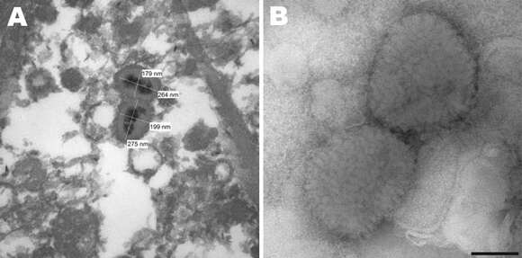
Description: English: Figure 1.. A) Electron micrograph of poxvirus particles in synovium of a big brown bat, northwestern United States. B) Negative staining of poxvirus particles in cell culture supernatant. Scale bar = 100 nm. Date: 26 May 2013, 17:45:07. Source: Emerging Infectious Diseases Journal, Volume 19, Number 6—June 2013,. Author: Ginny L. Emerson, Robert Nordhausen, Michael M. Garner, John R. Huckabee, Steven Johnson, Ron D. Wohrle, Whitni B. Davidson, Kimberly Wilkins, Yu Li, Jeffrey B. Doty, Nadia F. Gallardo-Romero, Maureen G. Metcalfe, Kevin L. Karem, Inger K. Damon, and Darin S. Carroll.
Ancludù an coste pàgine-sì:
Costa plancia a compariss an gnun-e colession.
Anformassion an sla sorgiss
- licensa
- cc-publicdomain
- creator
- Ginny L. Emerson, Robert Nordhausen, Michael M. Garner, John R. Huckabee, Steven Johnson, Ron D. Wohrle, Whitni B. Davidson, Kimberly Wilkins, Yu Li, Jeffrey B. Doty, Nadia F. Gallardo-Romero, Maureen G. Metcalfe, Kevin L. Karem, Inger K. Damon, and Darin S. Carroll
- sorgiss
- Emerging Infectious Diseases Journal, Volume 19, Number 6—June 2013,
- original
- archivi ëd mojen original
- visité la sorgiss
- sit compagn
- Wikimedia Commons
- ID