Rotavirus infected gut
Descrission:
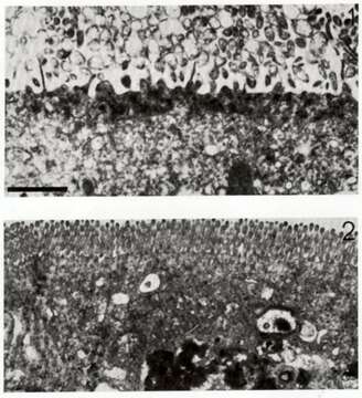
Description: Transmission electron micrograph of an enterocyte 3 days after infection by rotavirus (top). The microvilli have been destroyed. An unifected enterocyte is shown for comparison at the bottom. The bar = approx 500 nm. Date: 5 November 2007. Source: English Wikipedia. Author: Graham Beards. English Wikipedia user Graham Beards, the copyright holder of this work, hereby publishes it under the following license: : Permission is granted to copy, distribute and/or modify this document under the terms of the GNU Free Documentation License, Version 1.2 or any later version published by the Free Software Foundation; with no Invariant Sections, no Front-Cover Texts, and no Back-Cover Texts. A copy of the license is included in the section entitled GNU Free Documentation License.http://www.gnu.org/copyleft/fdl.htmlGFDLGNU Free Documentation Licensetruetrue. : This file is licensed under the Creative Commons Attribution 3.0 Unported license.:. You are free: to share – to copy, distribute and transmit the work to remix – to adapt the work Under the following conditions: attribution – You must attribute the work in the manner specified by the author or licensor (but not in any way that suggests that they endorse you or your use of the work). http://creativecommons.org/licenses/by/3.0 CC BY 3.0 Creative Commons Attribution 3.0 truetrue.
Ancludù an coste pàgine-sì:
Costa plancia a compariss an gnun-e colession.
Anformassion an sla sorgiss
- licensa
- cc-by-3.0
- drit d'autor
- Graham Beards
- creator
- Graham Beards
- sorgiss
- English Wikipedia
- original
- archivi ëd mojen original
- visité la sorgiss
- sit compagn
- Wikimedia Commons
- ID