7829 lores
Descrission:
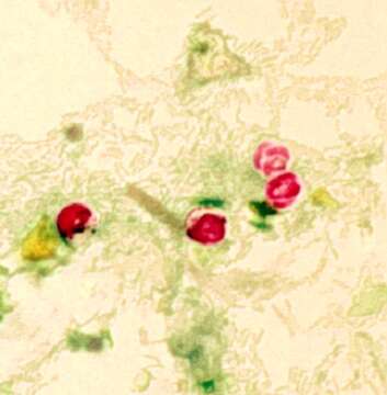
Summary.mw-parser-output table.commons-file-information-table,.mw-parser-output.fileinfotpl-type-information{border:1px solid #a2a9b1;background-color:#f8f9fa;padding:5px;font-size:95%;border-spacing:2px;box-sizing:border-box;margin:0;width:100%}.mw-parser-output table.commons-file-information-table>tbody>tr,.mw-parser-output.fileinfotpl-type-information>tbody>tr{vertical-align:top}.mw-parser-output table.commons-file-information-table>tbody>tr>td,.mw-parser-output table.commons-file-information-table>tbody>tr>th,.mw-parser-output.fileinfotpl-type-information>tbody>tr>td,.mw-parser-output.fileinfotpl-type-information>tbody>tr>th{padding:4px}.mw-parser-output.fileinfo-paramfield{background:#ccf;text-align:right;padding-right:0.4em;width:15%;font-weight:bold}.mw-parser-output.commons-file-information-table+table.commons-file-information-table,.mw-parser-output.commons-file-information-table+div.commons-file-information-table>table{border-top:0;padding-top:0;margin-top:-8px}@media only screen and (max-width:719px){.mw-parser-output table.commons-file-information-table,.mw-parser-output.commons-file-information-table.fileinfotpl-type-information{border-spacing:0;padding:0;word-break:break-word;width:100%!important}.mw-parser-output.commons-file-information-table>tbody,.mw-parser-output.fileinfotpl-type-information>tbody{display:block}.mw-parser-output.commons-file-information-table>tbody>tr>td,.mw-parser-output.commons-file-information-table>tbody>tr>th,.mw-parser-output.fileinfotpl-type-information>tbody>tr>td,.mw-parser-output.fileinfotpl-type-information>tbody>tr>th{padding:0.2em 0.4em;text-align:left;text-align:start}.mw-parser-output.commons-file-information-table>tbody>tr,.mw-parser-output.fileinfotpl-type-information>tbody>tr{display:flex;flex-direction:column}.mw-parser-output.commons-file-information-table+table.commons-file-information-table,.mw-parser-output.commons-file-information-table+div.commons-file-information-table>table{margin-top:-1px}.mw-parser-output.fileinfo-paramfield{box-sizing:border-box;flex:1 0 100%;width:100%}} Description: English: This photomicrograph revealed the morphologic details of Cryptosporidium parvum oocysts, which had been stained using the modified acid-fast method. These oocysts exhibit a bright red coloration when using this staining technique, and in this case, you’ll note the sporozoites that were made visible inside the two oocysts on the right. Sporozoites are the nucleated, motile stage of development through which many protozoans pass such as C. parvum, on their way to becoming adults, and represent a very infectious form of these organisms. When mature, the sporozoites will be liberated from the oocysts. Date: Unknown dateUnknown date. Source: https://phil.cdc.gov/Details.aspx?pid=7829. Author: CDC.
Ancludù an coste pàgine-sì:
- Life
- Cellular
- Eukaryota
- SAR (Stramenopiles, Alveolates, Rhizaria)
- Alveolata
- Apicomplexa
- Conoidasida
- Coccidia
- Eucoccidiorida
- Eimeriorina
- Cryptosporidiidae
- Cryptosporidium
- Cryptosporidium parvum
Costa plancia a compariss an gnun-e colession.
Anformassion an sla sorgiss
- licensa
- cc-publicdomain
- creator
- CDC
- sorgiss
- https://phil.cdc.gov/Details.aspx?pid=7829
- original
- archivi ëd mojen original
- visité la sorgiss
- sit compagn
- Wikimedia Commons
- ID