Abstract:
Morganella morganii (M. morganii) is one of the important nosocomial pathogen associated with the urinary tract infections and bacteremia. The aim of this study was to evaluate the effect of Mr. Trivedi’s biofield energy treatment on M. morganii in the lyophilized as well as revived state for antimicrobial susceptibility pattern, biochemical characteristics, biotype number and genotype. M. morganii cells were procured from MicroBioLogics Inc., USA in sealed packs bearing the American Type Culture Collection (ATCC 25829) number and stored according to the recommended storage protocols until needed for experiments. M. morganii strain was divided into two groups, Group (Gr.) I: control and Gr. II: treated. Gr. II was further subdivided into two groups, Gr. IIA and Gr. IIB. Gr. IIA was analyzed on day 10, while Gr. IIB was stored and analyzed on day 142 (Study I). After retreatment on day 142, the sample (Study II) was divided into three separate tubes. First, second and third tube was further analyzed on day 5, 10 and 15 respectively. All experimental parameters were studied using the automated MicroScan Walk-Away® system. The 16S rDNA sequencing of lyophilized treated sample was carried out to correlate the phylogenetic relationship of M. morganii with other bacterial species. Antimicrobial susceptibility results showed 32.14% alterations, while minimum inhibitory concentration results showed 18.75% alterations of the tested antimicrobials. Biochemical study also showed altered positive reactions in nitrofurantoin and indole with respect to control. Biotype study showed alteration in Gr. IIB, study II, on day 15 (4005 1446) as compared to the control (4004 1446). 16S rDNA sequencing analysis showed similar results with the identified microbe as M. morganii (GenBank accession number: AB210972) having 80% identity of the gene sequencing data. Total 1507 base nucleotide of 16S rDNA gene sequences were analyzed by multiple alignments, while nearest homolog genus-species of M. morganii was found as Providencia rettgeri (accession number: AM040492). These results suggested that biofield treatment has a significant impact on M. morganii in lyophilized as well as revived state.
Morganella morganii (M. morganii) is one of the important nosocomial pathogen associated with the urinary tract infections and bacteremia. The aim of this study was to evaluate the effect of Mr. Trivedi’s biofield energy treatment on M. morganii in the lyophilized as well as revived state for antimicrobial susceptibility pattern, biochemical characteristics, biotype number and genotype. M. morganii cells were procured from MicroBioLogics Inc., USA in sealed packs bearing the American Type Culture Collection (ATCC 25829) number and stored according to the recommended storage protocols until needed for experiments. M. morganii strain was divided into two groups, Group (Gr.) I: control and Gr. II: treated. Gr. II was further subdivided into two groups, Gr. IIA and Gr. IIB. Gr. IIA was analyzed on day 10, while Gr. IIB was stored and analyzed on day 142 (Study I). After retreatment on day 142, the sample (Study II) was divided into three separate tubes. First, second and third tube was further analyzed on day 5, 10 and 15 respectively. All experimental parameters were studied using the automated MicroScan Walk-Away® system. The 16S rDNA sequencing of lyophilized treated sample was carried out to correlate the phylogenetic relationship of M. morganii with other bacterial species. Antimicrobial susceptibility results showed 32.14% alterations, while minimum inhibitory concentration results showed 18.75% alterations of the tested antimicrobials. Biochemical study also showed altered positive reactions in nitrofurantoin and indole with respect to control. Biotype study showed alteration in Gr. IIB, study II, on day 15 (4005 1446) as compared to the control (4004 1446). 16S rDNA sequencing analysis showed similar results with the identified microbe as M. morganii (GenBank accession number: AB210972) having 80% identity of the gene sequencing data. Total 1507 base nucleotide of 16S rDNA gene sequences were analyzed by multiple alignments, while nearest homolog genus-species of M. morganii was found as Providencia rettgeri (accession number: AM040492). These results suggested that biofield treatment has a significant impact on M. morganii in lyophilized as well as revived state
Morganella morganii (M. morganii) is one of the important nosocomial pathogen associated with the urinary tract infections and bacteremia. The aim of this study was to evaluate the effect of Mr. Trivedi’s biofield energy treatment on M. morganii in the lyophilized as well as revived state for antimicrobial susceptibility pattern, biochemical characteristics, biotype number and genotype. M. morganii cells were procured from MicroBioLogics Inc., USA in sealed packs bearing the American Type Culture Collection (ATCC 25829) number and stored according to the recommended storage protocols until needed for experiments. M. morganii strain was divided into two groups, Group (Gr.) I: control and Gr. II: treated. Gr. II was further subdivided into two groups, Gr. IIA and Gr. IIB. Gr. IIA was analyzed on day 10, while Gr. IIB was stored and analyzed on day 142 (Study I). After retreatment on day 142, the sample (Study II) was divided into three separate tubes. First, second and third tube was further analyzed on day 5, 10 and 15 respectively. All experimental parameters were studied using the automated MicroScan Walk-Away® system. The 16S rDNA sequencing of lyophilized treated sample was carried out to correlate the phylogenetic relationship of M. morganii with other bacterial species. Antimicrobial susceptibility results showed 32.14% alterations, while minimum inhibitory concentration results showed 18.75% alterations of the tested antimicrobials. Biochemical study also showed altered positive reactions in nitrofurantoin and indole with respect to control. Biotype study showed alteration in Gr. IIB, study II, on day 15 (4005 1446) as compared to the control (4004 1446). 16S rDNA sequencing analysis showed similar results with the identified microbe as M. morganii (GenBank accession number: AB210972) having 80% identity of the gene sequencing data. Total 1507 base nucleotide of 16S rDNA gene sequences were analyzed by multiple alignments, while nearest homolog genus-species of M. morganii was found as Providencia rettgeri (accession number: AM040492). These results suggested that biofield treatment has a significant impact on M. morganii in lyophilized as well as revived state
Morganella morganii (M. morganii) is one of the important nosocomial pathogen associated with the urinary tract infections and bacteremia. The aim of this study was to evaluate the effect of Mr. Trivedi’s biofield energy treatment on M. morganii in the lyophilized as well as revived state for antimicrobial susceptibility pattern, biochemical characteristics, biotype number and genotype. M. morganii cells were procured from MicroBioLogics Inc., USA in sealed packs bearing the American Type Culture Collection (ATCC 25829) number and stored according to the recommended storage protocols until needed for experiments. M. morganii strain was divided into two groups, Group (Gr.) I: control and Gr. II: treated. Gr. II was further subdivided into two groups, Gr. IIA and Gr. IIB. Gr. IIA was analyzed on day 10, while Gr. IIB was stored and analyzed on day 142 (Study I). After retreatment on day 142, the sample (Study II) was divided into three separate tubes. First, second and third tube was further analyzed on day 5, 10 and 15 respectively. All experimental parameters were studied using the automated MicroScan Walk-Away® system. The 16S rDNA sequencing of lyophilized treated sample was carried out to correlate the phylogenetic relationship of M. morganii with other bacterial species. Antimicrobial susceptibility results showed 32.14% alterations, while minimum inhibitory concentration results showed 18.75% alterations of the tested antimicrobials. Biochemical study also showed altered positive reactions in nitrofurantoin and indole with respect to control. Biotype study showed alteration in Gr. IIB, study II, on day 15 (4005 1446) as compared to the control (4004 1446). 16S rDNA sequencing analysis showed similar results with the identified microbe as M. morganii (GenBank accession number: AB210972) having 80% identity of the gene sequencing data. Total 1507 base nucleotide of 16S rDNA gene sequences were analyzed by multiple alignments, while nearest homolog genus-species of M. morganii was found as Providencia rettgeri (accession number: AM040492). These results suggested that biofield treatment has a significant impact on M. morganii in lyophilized as well as revived state.
Morganella morganii is a species of Gram-negative bacteria.[2] It has a commensal relationship within the intestinal tracts of humans, mammals, and reptiles as normal flora.[2] Although M. morganii has a wide distribution, it is considered an uncommon cause of community-acquired infection, and it is most often encountered in postoperative and other nosocomial infections, such as urinary tract infections.[3]
Morganella morganii was first described by a British bacteriologist H. de R. Morgan in 1906 as Morgan's bacillus. Morgan isolated the bacterium from stools of infants who were noted to have had "summer diarrhea".[4] Later in 1919, Winslow et al. named Morgan's bacillus, Bacillus morganii. In 1936, though, Rauss renamed B. morganii as Proteus morganii. Fulton, in 1943, showed that B. columbensis and P. morganii were the same and defined the genus Morganella, due to the DNA-DNA hybridization.[5] In 1943, Fulton attempted to define a subspecies, M. m. columbensis.[6] However, in 1962, a review article by Ewing reported that M. columbensis had been re-identified as Escherichia coli, thereby removing that organism from the genus Morganella.[6]

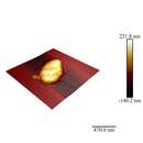
Morganella morganii is facultatively anaerobic and oxidase-negative. Its colonies appear off-white and opaque in color, when grown on agar plates.[7] M. morganii cells are straight rods, about 0.6–0.7 μm in diameter and 1.0–1.7 μm in length. This organism moves by way of peritrichous flagella, but some strains do not form flagella at 30 °C (86 °F).[8]
M. morganii is split into two subspecies: M. morganii subsp. morganii and M. morganii subsp. sibonii.[6] M. morganii subsp. sibonii is able to ferment trehalose, whereas subsp. morganii cannot, and this is the primary phenotype used to differentiate them.[6]
M. morganii can produce the enzyme catalase, so it is able to convert hydrogen peroxide to water and oxygen. This is a common enzyme found in most living organisms. In addition, it is indole test-positive, meaning that this organism can split tryptophan to indole, pyruvate, and ammonia. M. morganii also produces urease, allowing it to break down urea.[9] Methyl red tests positive in M. morganii, an indicator dye that turns red due to the bacterium's acid production during fermentation.[7]
Although a rare human pathogen, M. morganii has been reported as a cause of urinary tract infections, nosocomial surgical wound infections, peritonitis, central nervous system infection, endophthalmitis, pneumonia, chorioamnionitis, neonatal sepsis, pyomyositis, necrotizing fasciitis, and arthritis. Numerous cases of nosocomial infection have been described, usually as postsurgical wound infections or urinary tract infections. Patients in whom bacteremia develops are typically immunocompromised, diabetic, or elderly, or have at least one serious underlying disease. M. morganii has been regarded as a normally harmless opportunistic pathogen, but some strains carry "antibiotic-resistant plasmids" and have been associated with nosocomial outbreaks of infections.[10] Several reports indicate M. morganii causes sepsis, ecthyma, endophthalmitis, and chorioamnionitis, and more commonly urinary tract infections, soft tissue infections, septic arthritis, meningitis, and bacteremia, in the latter 2 cases with frequent fatal consequences.[11]
In a rare case published in 2003, a patient presented with bilateral necrosis of both upper and lower eyelids. Upon microbial analysis, the areas were shown to have heavy growth of M. morganii.[12]
Treatment of M. morganii infections may include:
A study conducted at the University Hospital at Heraklion, Crete, Greece, showed a 92% success rate in the use of these antibiotics.[13]
However, some M. morganii strains are resistant to penicillin, ampicillin/sulbactam, oxacillin, first-generation and second-generation cephalosporins, macrolides, lincosamides, fosfomycin, colistin, and polymyxin B.[3] The emergence of highly resistant strains of M. morganii have been associated with use of third-generation cephalosporins.[3]
Polymicrobial infections are most abundantly caused by this microbe which additionally damages the skin, soft tissues, and urogenital tract; these can be cured through use of the aforementioned antibiotics.[13]
Morganella morganii is a species of Gram-negative bacteria. It has a commensal relationship within the intestinal tracts of humans, mammals, and reptiles as normal flora. Although M. morganii has a wide distribution, it is considered an uncommon cause of community-acquired infection, and it is most often encountered in postoperative and other nosocomial infections, such as urinary tract infections.