The pupae are sensitive to fluctuations of carbon dioxide in the environment and also to vibrations. They use these environmental cues to time their emergence from their cocoons. Fleas possess a sensory organ called a pygidium on the posterior portion of their bodies, which allows them to detect vibrations and air currents. No information is available on how these fleas communicate with one another.
Perception Channels: tactile ; vibrations ; chemical
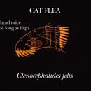
Like all fleas, Ctenocephalides felis is laterally compressed and wingless. Cat fleas are 2 mm long and reddish-brown to black, with the females being a bit larger than males and a slightly different color. Aside from the slight difference in size and color, the other main distinguishing feature between males and females is the presence of complex, snail-shaped genitalia in males. Ctenocephalides felis is distinguished from other fleas by its characteristic ctenidia, or combs; it has a pronotal ctenidium and a genal ctenidium with more than 5 teeth. The morphology of cat fleas is similar to that of dog fleas, Ctenocephalides canis, but cat fleas have a characteristic sloping forehead. The hind tibia is also different from other flea species in that it lacks an outer apical tooth. All members of the order Siphonaptera have powerful muscles containing resilin, a highly elastic protein, in their legs, which allows these fleas to leap as high as 33 cm.
Flea larvae resemble tiny maggots with short bristles and mandibles for chewing. Pupae live encased in silky debris-studded cocoons.
Average length: 2 mm.
Other Physical Features: ectothermic ; bilateral symmetry
Sexual Dimorphism: female larger; sexes colored or patterned differently; sexes shaped differently
Optimal conditions for survival of Ctenocephalides felis are described by a temperature range of 26.7 to 31.5 degrees Celsius and a relative humidity between 50 and 92 percent. Given these favorable conditions and a steady food supply, fleas can survive for two to three years.
Range lifespan
Status: wild: 3 (high) years.
Cat fleas live in the nests and resting places of their hosts when they are not feeding, and on their hosts when they are feeding. They live in just about any type of habitat, as long as it is warm and humid enough to promote development.
Habitat Regions: temperate ; tropical ; terrestrial
Terrestrial Biomes: taiga ; desert or dune ; savanna or grassland ; chaparral ; forest ; rainforest ; scrub forest ; mountains
Other Habitat Features: urban ; suburban ; agricultural
Ctenocephalides felis is one of the few flea species that is truly cosmopolitan. In the United States, these fleas are ubiquitous in all areas except the mid- to north- Rocky Mountain area. Throughout the rest of the world, cat fleas are found wherever suitable hosts reside.
Other Geographic Terms: cosmopolitan
After piercing the skin of the host, adult cat fleas use their mouthparts to suck up blood. The bloodmeal then passes through epithelial cells in the gut that are elongated into spines, collectively called the proventriculus, where it is broken up. As their name implies, cat fleas prefer to feed on domestic cats, Felis silvestris. Cat fleas also feed on dogs, rabbits, horses, skunks, foxes, mongooses, koalas, and poultry. They are known to bite humans in the absence of other hosts. In contrast to adult Ctenocephalides felis, larvae feed on the feces of the adult cat fleas and detritus in the environment.
Animal Foods: blood
Other Foods: detritus
Primary Diet: carnivore (Sanguivore )
Ctenocephalides felis is an obligate ectoparasite whose main hosts are cats. Cat fleas also parasitize dogs, rabbits, horses, skunks, foxes, mongooses, koalas, and poultry. Cat fleas are vectors for murine typhus and intermediate hosts of the most common tapeworm that infects cats and dogs, Dipylidium caninum. They have been known to carry Burrelia burgdorferi, the spirochaete that causes Lyme disease.
Ecosystem Impact: parasite
Species Used as Host:
Ctenocephalides felis is of some medical and economic importance. Most prevalent, but least serious, is the allergic reactions that fleas and their feces induce in some humans and animals. Itching and redness may occur, but with no serious results. Ctencephalides felis is a vector of murine typhus in humans, caused by Rickettsia mooseri. It is also the intermediate host of the most common tapeworm that infects domestic cats and dogs, Dipylidium caninum. It has been known to carry Burrelia burgdorferi, the spirochaete that causes Lyme disease, but it does not transfer the disease. All of these conditions require medical attention. The amount of damage in dollars per year is not available, but with the numerous variety of diseases that Ctenocephalides felis carries, the additive amount is not likely to be low.
Negative Impacts: injures humans (bites or stings, carries human disease); causes or carries domestic animal disease ; household pest
The life cycle of Ctenocephalides felis is a holometabolous one; that is, it involves complete metamorphosis. The entire life cycle lasts from 30 to 75 days depending on environmental conditions. At 13 degrees Celsius, the larvae emerge from the eggs in 6 days. Lower temperatures and low humidity slow development. After going through three larval instars, or molts, larval fleas spin loose cocoons of silk and enter their pupal stage. The pupae are sensitive to fluctuations of carbon dioxide in the environment and also to vibration. When an appropriate change in either of these factors occurs, the adult emerges and finds a host on which to live.
Development - Life Cycle: metamorphosis
No information is available on the mating system of these fleas.
Flea eggs are fertilized internally. The adult females lay their eggs on their host, but the eggs soon fall into the host's nest, where they develop. The eggs are white, translucent, and approximately 0.5 mm in length.
Breeding interval: Females lay eggs at frequent intervals while feeding, as long as the temperature and humidity are favorable
Breeding season: These fleas can breed year-round; optimal temperatures and humidity occur in the months of June, July, and August in most of the U.S.
Key Reproductive Features: iteroparous ; year-round breeding ; gonochoric/gonochoristic/dioecious (sexes separate); sexual ; fertilization (Internal ); oviparous
Female fleas carry their eggs inside of them, providing them with nourishment until they are laid. After they are laid, there is no further investment on the part of the parents.
Parental Investment: pre-fertilization (Provisioning, Protecting: Female)
Fleas have a pattern for choosing certain places to hide or lay their eggs in.
Inside a household:
Outside:
On live hosts such as cats and dogs:
La puça del gat (Ctenocephalides felis) és una espècie d'insecte sifonàpter de la família Pulicidae, una de les més abundants i de major distribució mundial.[1]
Unes poques puces en els gossos o gats adults no causen dany seriós tret que l'hoste sigui al·lèrgic a les substàncies en la saliva.[2] Aquesta malaltia es diu dermatitis al·lèrgica de puces. Els animals petits atacats per gran nombre de puces poden sofrir seriosos danys com a deshidratació.[3][4]
Les puces dels gats poden transmetre altres paràsits i infeccions als gats, els gossos i fins i tot als humans. Les més destacades són el bacteri Bartonella,[5] el cuc intestinal Dipylidium caninum, el tifus.
La puça del gat (Ctenocephalides felis) és una espècie d'insecte sifonàpter de la família Pulicidae, una de les més abundants i de major distribució mundial.
Blecha kočičí (Ctenocephalides felis) je druh parazitického hmyzu patřící do řádu blechy. Je parazitem zejména koček a psů, ale byla nalezena i na ovcích, telatech, kozách, hlodavcích, králících a také na člověku. Je rozšířena po celém světě čili kosmopolitně.
Dospělá blecha je zbarvena červenohnědě, někdy černě, larva je bílá se zřetelnou hnědou hlavou. Dospělec je velký 1–3 mm a má silné zadní nohy, které slouží ke skákání a běhu; beznohá larva měří okolo 0,5 mm. Velké skoky umožňuje bleše mj. látka resilin[1], avšak před dalším skokem musí blecha určitou dobu počkat, než se látka doplní. Dospělec žije čtyři až pětadvacet dní. Dříve, než se larva promění v dospělce, musí projít stadiem kukly. Vlákno kokonu (zámotku) je lepkavé a lehce se na něj nalepují různé nečistoty, které chrání kuklu před vysycháním a také slouží jako maskování.
Od podobné blechy psí (Ctenocephalides canis) lze blechu kočičí odlišit mikroskopicky – (1) v krajině čelního štítku má plošší hlavu, (2) má delší první zub v lícním hřebenu.[2]
Životní cyklus blechy kočičí patří k proměně dokonalé (vajíčko, larva, kukla, dospělý jedinec). Bleší samička denně snese 20–50 vajíček. Vajíčka jsou hladká, neudrží se na těle hostitele a spadnou někam, kde se z nich za jeden až deset dní vylíhnou larvy. Larvy se živí biologickým materiálem v prostředí včetně výkalů dospělých blech, zakuklí se a z kukly se líhne dospělý jedinec. To, že většina vývojových stadií blechy (vajíčka, larvy, kukly) žije mimo hostitele, je jedním z problémů při boji proti blechám. Vajíčka, larvy i kukly jsou většinou odolné vůči nepříznivým podmínkám, přičemž larvy jsou méně odolné proti vyschnutí než vajíčka. Kokon (zámotek) slouží larvám jako ochrana před predátory (např. mravenci). Dospělá blecha již parazituje na svém hostiteli, živí se krví, ale využije jen malou část – většinu krve vyloučí s výkaly, které jsou po rozmáčknutí typicky červené. Pouze dospělá blecha je schopná reprodukce, jen v tomto stadiu můžeme rozlišit samičku a samečka. Délka vývoje závisí na vnějších podmínkách, v létě vývoj trvá 4–5 týdnů, v zimě déle.
Blecha kočičí je parazit, který může hostiteli způsobit více různých nepříjemností – může být přenašečem chorob, může hostitele celkově oslabit aj. Bleší sliny obsahují látky, které zabraňují srážení krve, a díky tomu se může blecha dostatečně nasytit. Látky vyvolávají alergickou reakci hostitele, hostitel má tendenci se škrábat a může si způsobit hnisání či ekzémy. Dále mohou blechy přenést na svého hostitele tasemnici psí. Vzhledem k tomu, že se blechy živí krví, mohou být nebezpečné pro malá nebo křehká zvířata – mohou způsobit chudokrevnost štěňat, koťat či dospělých zvířat malých plemen, která mohou následkem anémie i uhynout. Vzácněji blechy přenášejí tyfus a mor myší.
Pokud má kočka nebo pes delší srst, je těžké najít dospělou blechu. Ovšem lze najít černé kuličky – výkaly blech se zbytky nestrávené krve, které po rozmáčknutí zčervenají. Další indicií je hubnutí nebo ekzém. Pokud se zvíře škrábe, pečlivě prohlédneme kořen ocasu, příp. je postavíme na bílý podklad a kartáčem pročešeme srst. Přítomnost blech prokazují bleší výkaly.
Nejlepší způsob jak zjistit jestli zvíře má blechy, je vyčesat zvíře kartáčem nebo hřebenem a vyčesané chlupy položit na savý papír (kuchyňská papírová utěrka) a postříkat vlažnou vodou. Pokud se na paíře objeví červené skvrny (zbytky výkalů blechy, krev zvířete) je zvíře napadeno blechami.
Mezi nejběžnější prostředky proti blechám patří spreje, šampóny, tablety a speciální obojky. Souběžně je třeba uklidit celou domácnost – vyklepat a vyluxovat pelíšky, deky, čalounění (sáček neponechávat ve vysavači!). Prostředky proti blechám můžeme rozdělit do dvou skupin: adulticidy hubí dospělé jedince, zatímco růstové regulátory zabraňují vývoji vajíček. Oba typy prostředků mají své výhody i nevýhody. V prvním případě musí dojít k přímému kontaktu hubicí látky s blechou, takže je nutné kromě zvířete ošetřit i domácnost. Nevýhodou růstových regulátorů je, že nezabrání usazení blechy na kočce či psu; ovšem jakmile blecha začne sát krev, nasaje látky zabraňující vývoji vajíček, která naklade. Optimálním řešením je proto kombinovat oba typy prostředků.
Dnes existují čtyři poddruhy Ctenocephalides felis a všechny tyto poddruhy jsou ektoparaziti převážně masožravých savců. Ctenocephalides felis strongylus a Ctenocephalides felis damarensis žije pouze v Africe, Ctenocephalides felis orientis se nachází v jihovýchodní Asii a východní Indii a poddruh Ctenocephalides felis felis je rozšířen po celém světě.
Blecha kočičí (Ctenocephalides felis) je druh parazitického hmyzu patřící do řádu blechy. Je parazitem zejména koček a psů, ale byla nalezena i na ovcích, telatech, kozách, hlodavcích, králících a také na člověku. Je rozšířena po celém světě čili kosmopolitně.
Der Katzenfloh (Ctenocephalides felis) ist ein parasitierendes flügelloses Insekt der Ordnung der Flöhe (Siphonaptera). Weibchen sind 2 bis 3 mm, Männchen etwa 1 mm lang und braun bis rot-braun gefärbt. Der Katzenfloh hält sich vorwiegend auf befallenen Tieren auf und ernährt sich vom Blut des Wirtes, seine Entwicklungsstadien sind dagegen vorwiegend in Verstecken anzutreffen. Durch sein weites Wirtspektrum ist der Katzenfloh eine sehr häufige Flohart. Er befällt auch den Menschen und spielt unter anderem als Krankheitsüberträger eine Rolle.
Wie alle Flöhe ist der Katzenfloh ein flügelloses Insekt mit drei Beinpaaren, von denen das letzte zu kräftigen Sprungbeinen umgebildet ist, sowie mit einem kombinierten Stech- und Saugrüssel als Mundwerkzeug. Er ist braun bis rot-braun gefärbt. Weibchen sind 2 bis 3 mm, Männchen etwa 1 mm lang.
Der Katzenfloh besitzt einen länglichen Kopf, der etwa doppelt so lang wie hoch ist. Er lässt sich anhand der aus Chitindornen bestehenden Stachelkämme (Ctenidien) von anderen Floharten unterscheiden. Der Kopfstachelkamm (Genalctenidium) trägt sieben bis acht Dornen, wobei der erste und zweite Dorn etwa gleich lang sind. Der Stachelkamm des Prothorax (Pronotalctenidium) trägt etwa 16 Dornen.
Der fußrückenseitige Rand des Sprungbeins hat sechs Einkerbungen, aus denen Borsten entspringen.
Befruchtete Weibchen legen 24 Stunden nach der Begattung etwa 30 Eier in das Fell befallener Tiere. Die etwa 0,5 mm großen Eier fallen jedoch bald vom Tier ab und verteilen sich, meist in der direkten Umgebung der Schlafstätten.[1] Nach 4–5 Tagen schlüpft die Larve 1, die sich vor allem von verdautem Blut adulter Flöhe und von organischem Material aus der Umgebung ernährt. Da die Larven sehr empfindlich gegen Austrocknung sind, leben sie meist versteckt im Gras, Sand, Teppichböden und Ritzen. Nach Häutung zur Larve 2 und 3 folgt die Verpuppung in einem 5 mm großen Kokon.
Im Puppenstadium sind die Parasiten weitgehend resistent gegenüber Austrocknung, Licht und auch den meisten Insektiziden. Der Schlupf des adulten Flohs (Imago) erfolgt durch verschiedene Reize wie Wärme, Kohlenstoffdioxid-Gehalt oder Vibrationen. Der gesamte Entwicklungszyklus dauert unter günstigen Bedingungen etwa einen Monat, kann aber bei niedriger Umgebungstemperatur mehrere Monate in Anspruch nehmen.
Unmittelbar nach dem Schlupf sucht er springend einen neuen Wirt. Ab dieser Phase leben Flöhe gewöhnlich permanent auf ihren Wirten, bei starkem Befall wechseln sie auch auf andere vorbeikommende Wirte. Die Lebensdauer der Imago beträgt etwa einen, in Ausnahmefällen bis zu drei Monate.
Den Winter können nur die adulten Flöhe auf ihren Wirten und die Puppen überleben, in beheizten Räumen kann der Entwicklungszyklus allerdings ganzjährig erfolgen.
Durch sein weites Wirtspektrum ist der Katzenfloh eine der am weitesten verbreiteten Floharten. Trotz des Einsatzes vorbeugender Flohmittel gehen Schätzungen davon aus, dass bis zu 20 % der Hunde und Katzen einen Flohbefall aufweisen, wobei das Befallsmaximum im Spätsommer auftritt. In urbanen Gebieten ist der Katzenfloh auch bei Haushunden der häufigste Parasit, der stärker spezialisierte Hundefloh ist eher in ländlichen Gebieten anzutreffen. Auch andere Tiere und Menschen werden vom Katzenfloh nicht verschont.
Ausgewachsene (adulte) Flöhe ernähren sich durch ihre saugend-stechenden Mundwerkzeuge vom Blut ihres Wirtes. Sie nehmen pro Tag etwa 13 µl Blut auf. Der Flohkot besteht vorwiegend aus unverdauten Blutbestandteilen und ist schwarz und krümelig. Bringt man ihn auf ein angefeuchtetes Stück Papier, so färbt er sich rötlich-bräunlich, was ein wichtiger Nachweis für einen geringgradigen Befall ist, da insbesondere Katzen sehr effektiv in der Flohbeseitigung sind, so dass man unter Umständen keine adulten Flöhe auf dem Wirt findet.
Durch den Flohbiss entsteht eine punktförmige Rötung mit hellem Hof, unter Umständen eine Papel. Bei stärkerem Befall kann sich eine ausgedehntere Hautentzündung (Dermatitis) entwickeln. Häufiger und klinisch auffälliger sind jedoch allergische Reaktionen (Flohallergie), die auch bei nur geringem Befall zu schweren Hautveränderungen führen können.
Auch bei der indirekten Krankheitsübertragung spielt dieser Floh eine Rolle, denn er ist beispielsweise ein wichtiger Zwischenwirt für den Gurkenkernbandwurm. Auch andere Krankheitserreger wie Mykoplasmen (Erreger der felinen infektiösen Anämie), Bartonella henselae (Erreger der Katzenkratzkrankheit) oder Rickettsia felis werden durch den Katzenfloh verbreitet.
Gegen adulte Flöhe können Insektizide wie Afoxolaner, Fipronil, Imidacloprid, Metaflumizon, Nitenpyram, Selamectin, Fluralaner oder Spinosad eingesetzt werden. Zur Verhinderung der Larvenentwicklung eignen sich Chitininhibitoren wie Lufenuron. Zur chemischen Umgebungsbehandlung werden Chlorpyrifos, Permethrin, Propoxur, Fenoxycarb, Pyrethrine, Methopren bzw. Kombinationen dieser Wirkstoffe eingesetzt. Permethrin ist für Katzen giftig.[2]
Piperonylbutoxid ist ein häufiger Synergist, der die Wirkung von Insektiziden wie z. B. Pyrethrine verstärkt. Allerdings behindert es auch „den Entgiftungsmechanismus im menschlichen Körper, vergrößert daher die Gefahr anderer Insektizide“.[3]
Haben Haushalte mit Haustieren Flöhe, sollten gleichzeitig die Umgebung und die Tiere selbst behandelt werden. Zur Behandlung von Katzen gibt es Spot-ons und andere Medikamente gegen Flöhe, zur Vorbeugung von Befall/Neubefall eignen sich Halsbänder. Pulver oder Sprays werden dagegen eingesetzt, um die Umgebung – insbesondere den Schlafplatz – schädlingsfrei zu bekommen.[4]
Die Wirkung von Hausmitteln ist oft nicht wissenschaftlich belegt.[5] Sie helfen am besten in Kombination und teilweise mit individueller Anpassung nach der Trial-and-error-Methode.
Um die Vermehrung zu unterbrechen, sollten sowohl die Hausmittel, die tierärztliche Behandlung von allen Tieren im Haushalt als auch chemische Bekämpfungsmaßnahmen je nach Befallsstärke über mehrere Wochen aufrechterhalten werden, da noch vorhandene Floheier in dieser Zeit neue Populationen hervorbringen können.[6]
Der Katzenfloh (Ctenocephalides felis) ist ein parasitierendes flügelloses Insekt der Ordnung der Flöhe (Siphonaptera). Weibchen sind 2 bis 3 mm, Männchen etwa 1 mm lang und braun bis rot-braun gefärbt. Der Katzenfloh hält sich vorwiegend auf befallenen Tieren auf und ernährt sich vom Blut des Wirtes, seine Entwicklungsstadien sind dagegen vorwiegend in Verstecken anzutreffen. Durch sein weites Wirtspektrum ist der Katzenfloh eine sehr häufige Flohart. Er befällt auch den Menschen und spielt unter anderem als Krankheitsüberträger eine Rolle.
Mačija buha (latinski: Ctenocephalides felis) je najrasprostranjenija vrsta buha u svijetu.
Mačija buha je eksterni parazit domaće mačke, ali se također može naći i na psima. Životni ciklus može obaviti i na drugim mesojedima, a može se naći i na zečevima, stoci, glodarima i ljudima ali na njima ne može dugo opstati.
Ženka mačije buhe leže jaja na domaćina, ali jaja nakon što se osuše ispadaju iz krzna u vanjsku sredinu, najčešće na mjesto gdje životinja spava. Iz jaja izlaze larve koje posjeduju osobinu negativne fototaksije, što znači da se kreću suprotno od izvora svjetlosti. Larve se hrane različitim organskim supstancama, ali najvažnije u ishrani je osušena krv koja opada sa krzna domaćina kao fekalna materija odraslih buha. Nakon tri larvalne faze, prelazi u fazu lutke. U fazi lutke, odnosno u kukuljici ostaje sve do pojave prisustva topline, vibracija ili povećane koncentracije ugljik dioksida od strane potencijalnog domaćina. Prisustvo novog domaćina stimuliše novoizlegle buhe da skoče na domaćina. Mlade buhe nakon tek nekoliko minuta počinju da se hrane krvlju.[1]
Mačije buhe ne nanose neku veliku štetu odraslim domaćinima na kojim parazitiraju, osim u slučaju da domaćin postane alergičan na neke supstance u pljuvački buhe. Ovo dovodi do alergijskog dermatitisa. Kod mlađih jedinki sa velikim infestacijama, može doći do povećanog gubitka krvi što dovodi do anemije.
Mačije buhe također mogu biti vektori u prenosu nekih bolesti, kao i u prenosu nekih parazita.
Kao prevencija od pojave mačijih buha može se koristiti ogrlica protiv buha, kao i redovno češljanje. U slučaju pojave buha koriste se različiti sprejevi ili šamponi namjenjeni za tu svrhu.[2]
Iščešljana mačija dlaka sa fekalnim otpacima mačijih buha
Jaja mačije buhe
Larva mačije buhe
Mačija buha u stadiju kukuljice
Mačija buha (latinski: Ctenocephalides felis) je najrasprostranjenija vrsta buha u svijetu.
Mačija buha je eksterni parazit domaće mačke, ali se također može naći i na psima. Životni ciklus može obaviti i na drugim mesojedima, a može se naći i na zečevima, stoci, glodarima i ljudima ali na njima ne može dugo opstati.
The cat flea (scientific name Ctenocephalides felis) is an extremely common parasitic insect whose principal host is the domestic cat, although a high proportion of the fleas found on dogs also belong to this species.[3] This is despite the widespread existence of a separate and well-established "dog" flea, Ctenocephalides canis. Cat fleas originated in Africa[4] but can now be found globally.[5] As humans began domesticating cats, the prevalence of the cat flea increased and it spread throughout the world.
Of the cat fleas, Ctenocephalides felis felis is the most common, although other subspecies do exist, including C. felis strongylus, C. orientis, and C. damarensis. [5] Over 90% of fleas found on both dogs and cats are Ctenocephalides felis felis. [3]
The cat flea belongs to the insect order Siphonaptera which in its adult stage is an obligatory hematophage. Adults of both sexes range from 1–2 mm long and are usually a reddish-brown colour, although the abdomens of gravid females often swell with eggs causing them to appear banded in cream and dark brown. Like all fleas, the cat flea is compressed laterally allowing it to slip between the sometimes dense hairs of its host just above the top layer of the skin, resulting in an extremely thin insect that may be difficult to observe even if the host's coat is pure white.[6] Cat fleas are wingless.[3]
The cat flea affects both the cat and the dog worldwide.[7] The cat flea can also maintain its life cycle on other carnivores and on omnivores, but these are only chosen when more acceptable hosts become unavailable.[6] Adult cat fleas do not willingly leave their hosts, and inter-animal transfer of adult fleas is rare except in animals that share sleeping quarters. A flea which becomes separated from its host will often die within hours from starvation.[8] It has been found that mortality differs between male and female cat fleas when separated from the host. It was found that within two days all male cat fleas were dead, while females became inactive after three days.[5]
In addition to their role as pests in dogs and cats, cat fleas are responsible for a number of diseases. They can cause flea bite dermatitis and the transmission of dog tapeworm[9] to name a few.
Cat fleas are holometabolous (undergo complete metamorphosis) insects and therefore go through four life cycle stages of egg, larva, pupa, and imago (adult). Adult fleas must feed on blood before they can become capable of reproduction.[10]
Flea populations are distributed with about 50% eggs, 35% larvae, 10% pupae, and 5% adults.[11] Cat fleas may live up to two years.
An adult gravid female flea that has consumed a full blood meal will begin to produce between 20 and 30 microscopic (0.5 mm) non-adhesive white ovoid eggs per day, laying them individually and continually at a rate of about one per hour until she dies (under ideal conditions it might be possible for her to produce between 2,000 and 8,000 eggs in her lifetime, though most only manage to produce around 100 before being consumed by their host during grooming activity[8]). The eggs are dispersed freely into the environment. Within two to seven weeks a certain proportion will then hatch into larvae.[3] Hatching is at its highest when temperature is 27 °C and humidity is greater than 50%.[9]
Given that eggs are non-adhesive, they do not stick to the host (70% are lost from the host in the first 8 hours).[9]

The larva of the cat flea has a grub-like appearance and is ~2 mm in length. The larvae are negatively phototaxic/phototropic, avoiding light and hiding in the substrate around them. The larvae require adequate ambient moisture and warmth, and will die at temperatures near freezing.[8] Cat fleas prefer soil moisture content between 1-10%.[12] While in this developmental stage the larvae will feed on a variety of organic substances, but the most important dietary item for them is the crumbs of dried blood that continually fall like snow out of the haircoat of the host after it has been excreted by the adult fleas as fecal material. Thus, the adult flea population continually feeds the larval population in the animal's environment. Adult feces is an important part of the larval diet. When reared in the lab, flea larvae provided with adult feces have a higher survival rate (67%) than those provided diets of dried bovine blood (39%) or meat flour (55%).[13]
Flea larvae metamorphose through four stages before spinning a cocoon and entering the pupal stage. The cocoon is adhesive, and quickly acquires a coat of camouflage from surrounding dirt and dust. Pupation depends heavily on temperature and moisture, and takes a week or more to complete, though a fully pupated adult can remain inside of its cocoon in a state of semi-dormancy (called the "pupal window") awaiting signs of the presence of a host.[8][14][15][16]
Newly emerged fleas use variations in light and shadow along with increases in warmth and CO2 to detect the presence of a potential host, and will jump to a new host within seconds of emerging from the cocoon. The new flea begins feeding on host blood within minutes.[14][15][16]
A few fleas on adult dogs or cats cause little harm unless the host becomes allergic to substances in the flea's saliva. There are 15 substances that can cause allergy in flea saliva.[3] The disease that results from allergy is called flea allergy dermatitis. Small animals with large infestations can lose enough bodily fluid to fleas feeding that dehydration may result. Cat fleas also may be responsible for disease transmission through humans, and have been suspected as transmission agents of plague. Severe flea infestations can result in anemia due to blood loss.
Cat fleas can transmit other parasites and infections to dogs and cats and also to humans. The most prominent of these are Bartonella, murine typhus, and atopic dermatitis. The tapeworm Dipylidium caninum can be transmitted when an immature flea is swallowed by pets or humans. In addition, cat fleas have been found to carry Borrelia burgdorferi, the etiologic agent of Lyme disease, but their ability to transmit the disease is unclear.[17] Finally, cat fleas are vectors for Rickettsia felis.[18]
Since more than three-quarters of a flea's life is spent somewhere other than on the host animal, it is not adequate to treat only the host; it is important also to treat the host's environment. Thorough vacuuming, washing linens in hot water, and treating all hosts in the immediate environment (the entire household, for example) is essential for successful eradication. These steps should be performed on a regular basis[19] as the flea life cycle is complex. Treatment should be implemented every five to ten days.[20] Pet safe insecticides may also be an option in treating a pet with fleas, and soap is sufficient as an insecticide for adult fleas.[20]
Cat fleas have developed insecticide resistance to many of the common insecticides used to control them environmentally, including carbamates, organophosphates, and pyrethroids.[12][21] Additionally, it has been found that larvae are more resistant to certain insecticides than adults. Targets of juvenile hormone may be successful to limit growth in the larval stages. When administering insecticides to pets for flea treatment, it is critically important to finish the full dose to limit the spread of resistance.[12][21]
Cat fleas are generally tolerant to a wide range of environmental conditions.[22] As the climate warms, however, it is predicted that the tropical haplotype will displace the temperate haplotype. Climate change often drives changes in species range. In Australia, it is predicted that warming temperatures will drive the cat flea distribution south.[22]
The cat flea (scientific name Ctenocephalides felis) is an extremely common parasitic insect whose principal host is the domestic cat, although a high proportion of the fleas found on dogs also belong to this species. This is despite the widespread existence of a separate and well-established "dog" flea, Ctenocephalides canis. Cat fleas originated in Africa but can now be found globally. As humans began domesticating cats, the prevalence of the cat flea increased and it spread throughout the world.
Of the cat fleas, Ctenocephalides felis felis is the most common, although other subspecies do exist, including C. felis strongylus, C. orientis, and C. damarensis. Over 90% of fleas found on both dogs and cats are Ctenocephalides felis felis.
La pulga del gato (Ctenocephalides felis) es una especie de insecto sifonáptero de la familia Pulicidae, una de las más abundantes y de mayor distribución mundial.
Esta pulga primariamente se hospeda en el gato doméstico, pero también infesta perros en todo el mundo; y puede mantener su ciclo de vida en otros carnívoros y en Didelphis virginiana. Conejos, roedores, rumiantes y humanos pueden infestarse o ser picados, aunque no es común que esta especie complete su ciclo en ellos.[1]
La hembra oviposiciona sus huevos en el huésped, pero los huevos, una vez secos pueden caer de la piel de sus huéspedes al terreno. Los huevos eclosionan en larvas, que tienen fototactismo negativo, es decir que se ocultan de la luz en el sustrato. La larva se alimenta de una variedad de materiales orgánicos, pero especialmente de sangre seca que queda en la piel del huésped al ser depositada por las pulgas adultas junto con la materia fecal. Así la población adulta alimenta a la población larvaria en el ambiente del hospedante.
La larva se metamorfosea pasando por cuatro estadios antes de tejer un capullo y de entrar en el estadio de pupa. La duración del estadio de pupa es variable. El adulto joven sólo emerge de la pupa cuando detecta la presencia de un hospedante potencial, al percibir las vibraciones o el calor. Las jóvenes pulgas tienen el estímulo de saltar hacia el huésped apenas segundos después de su emergencia del capullo. Comienzan a alimentarse en unos pocos minutos. De no encontrar huésped adecuado pueden vivir mucho tiempo en el hogar, sea en tierra, o plantas como en muebles de algarrobo o ropa de cama[2] [3] .[4]
Unas pocas pulgas en los perros o gatos adultos no causan daño serio a menos que el huésped sea alérgico a las substancias en la saliva. Esta enfermedad se llama dermatitis alérgica de pulgas. Los animales pequeños atacados por gran número de pulgas pueden sufrir serios daños como deshidratación.
Las pulgas de los gatos pueden transmitir otros parásitos e infecciones a los gatos, los perros e incluso a los humanos. Las más destacadas son la bacteria Bartonella, el gusano intestinal Dipylidium caninum, el tifus.
La pulga del gato (Ctenocephalides felis) es una especie de insecto sifonáptero de la familia Pulicidae, una de las más abundantes y de mayor distribución mundial.
Kassikirp (Ctenocephalides felis) on kirbuliste seltsi kuuluv putukaliik.
Putukat on ka Eestis.[1]
Kassikirp (Ctenocephalides felis) on kirbuliste seltsi kuuluv putukaliik.
Putukat on ka Eestis.
Puce du chat
Ctenocephalides felis, la puce du chat[2],[3] est une espèce d'insectes piqueurs holométaboles siphonaptères de la famille des Pulicidae. Cette puce est connue comme l'ectoparasite le plus fréquent du chat et du chien en zone tempérée et subtropicale.
On peut aussi la rencontrer sur au moins 50 espèces de mammifères à travers le monde[4]. Elle peut parfois pulluler sur des animaux de ferme (veaux par exemple[5]).
Une autre puce, Ctenocephalides canis (Curtis) semble spécifique du chien mais elle est peu fréquente en France.
D'après BioLib (15 janvier 2022)[6] et GBIF (15 janvier 2022)[1] cette espèce comprend trois sous-espèces :
Comme chez tous les insectes, le corps comprend trois parties[7] : la tête, le thorax et l’abdomen.
Cet insecte hématophage porte une paire d’yeux, une paire d’antennes (situées en arrière des yeux, assez difficiles à distinguer sans préparation spéciale) et, entourant la bouche, les pièces buccales comprenant un labre, impair, deux mandibules, deux maxilles, munies d’un long palpe, et un labium, impair muni de deux palpes. L’ensemble de ces pièces constitue un organe piqueur qui permet de percer la peau de l’hôte et d’absorber son sang.
De chaque côté de la tête on remarque un rang de fortes épines noires, légèrement tournées vers l’arrière, c’est le peigne de la joue nommé « cténidie génale ».
Il est composé de trois segments (métamères) dont chacun possède une paire de pattes terminées par des griffes. La paire postérieure, la plus développée sert au saut, qui est effectué avec un angle de 50° avec l'horizontale[8].
Le premier segment thoracique porte, à l’arrière de sa partie dorsale une rangée de dents puissantes : le peigne du pronotum, nommé « cténidie pronotale ».
Composé de 9 segments dont seuls les 8 premiers sont aisément identifiables. Son contour est arrondi. Sur les côtés des segments se trouvent les orifices respiratoires ou stigmates. À l’arrière, l’abdomen porte l’anus, surmonté du pygidium, et les orifices génitaux mâles ou femelles équipés de pièces génitales résultant de la transformation des derniers segments. Les sexes sont séparés.
La forme aplatie du corps, les peignes, les nombreuses soies, les griffes des pattes, permettent aux puces de progresser et de s’ancrer efficacement dans le pelage de leurs hôtes.
L’adulte produit des excréments (fèces) qui ont la forme de bâtonnets courbes, plus ou moins nettement cannelés, de couleur rouge foncé (« crottes de puces ») constitués de sang partiellement digéré. Ces excréments tombent aux mêmes endroits que les œufs.
Les œufs, presque cylindriques, arrondis aux extrémités, sont de couleur blanche, ils mesurent environ 0,5 mm de long. Ils ne sont pas adhésifs et tombent donc au sol dans les endroits fréquentés par le chat ou le chien (principalement sur leur couchage). Une puce femelle pond environ 25 œufs par jour et de 1 000 à 2 000 au cours de son existence. Le rythme maximum de ponte se situe entre 0 et 3 heures du matin[4].
L’incubation dure 6 jours à 13 °C mais seulement 32 heures à 32 °C. Cette durée est également fonction de l’hygrométrie (voir Degré jour de croissance).
À l’issue de l’incubation l’œuf libère une première larve (L1) qui a l’aspect d’une chenille dépourvue de pattes, longue d’environ 1 à 2 mm, constituée d’une tête, d’un thorax et d’un abdomen. La tête porte deux courtes antennes et la bouche équipée des mêmes pièces buccales que l’adulte mais les appendices sont cette fois du type broyeur. Le corps est muni de soies raides, plus longues et plus nombreuses à la partie postérieure. Les déplacements s’effectuent en prenant alternativement appui sur la tête et sur la partie arrière du corps. Du fait de l’absence des pattes ou d’organes adhésifs la larve est incapable de grimper le long de surfaces verticales lisses.
Les larves grandissent grâce à des mues. Les dépouilles qu’elles abandonnent à cette occasion sont les exuvies. La larve L1 mue en donnant une larve L2 qui est suivie elle-même d’une larve L3. L1, L2 et L3 ont la même forme générale. L3 atteint une taille d’environ 5 mm. Grâce à leurs pièces buccales broyeuses, les larves se nourrissent des excréments solides des adultes. Le tube digestif est de ce fait coloré en rouge plus ou moins foncé. À la fin du stade L3, la larve cesse de s’alimenter, thorax et abdomen prennent alors une couleur blanche. À 15−20 °C, l’ensemble des 3 stades larvaires dure environ 15 jours.
La larve L3 blanche tisse un cocon de soie, ovoïde, long d’environ 5 mm, auquel se collent des éléments étrangers (poils, grains de sable, débris divers). Lorsque le cocon est terminé, L3 mue et se transforme en nymphe, pratiquement immobile, les appendices collés au corps et dont la forme évoque déjà celle de l’adulte. Elle ne s’alimente pas. La nymphe est encore une larve, mais sa forme particulière, bien différente de celle des précédentes, lui vaut d’être classée à part.
Au terme de son évolution la nymphe mue (le cocon contient donc deux exuvies : celle de L3 et celle de la nymphe) pour libérer l’adulte ou imago qui se trouve encore à l’intérieur du cocon. Dans cette situation l’adulte, qui ne dispose pas de nourriture, peut attendre plusieurs jours, voire plusieurs semaines (50 à 11 °C), qu’un animal, chien, chat… ou homme, passe à proximité. Il déchire alors le cocon et se lance vers son hôte (c’est l’émergence). La chaleur, la pression mécanique, l’augmentation du taux de dioxyde de carbone, peut-être les vibrations, sont les principaux stimuli qui déclenchent cette réaction.
À 15−20 °C, il s’écoule environ 20 jours entre le début du tissage du cocon et les premières émergences.
La durée totale du cycle de développement varie entre 12 et 174 jours selon la température et l’hygrométrie du lieu[4].
En plus du mode le plus commun d'infestation décrit ci-dessus, les chats peuvent s'infester par transfert direct de puces de chat à chat. Certaines puces femelles peuvent changer d’hôtes en un temps aussi bref qu’une heure, ce qui peut jouer un rôle dans l’introduction rapide de nouvelles populations de puces dans l’environnement des chats [9].
Les traitements chimiques, si efficaces qu’ils soient, ne le sont jamais à 100 % sur toutes les étapes du développement des puces (œufs, larves, nymphes ou adultes). Ils doivent donc être répétés pour éviter une recontamination par les puces issues des formes survivantes.
Puce du chat
Ctenocephalides felis, la puce du chat, est une espèce d'insectes piqueurs holométaboles siphonaptères de la famille des Pulicidae. Cette puce est connue comme l'ectoparasite le plus fréquent du chat et du chien en zone tempérée et subtropicale.
On peut aussi la rencontrer sur au moins 50 espèces de mammifères à travers le monde. Elle peut parfois pulluler sur des animaux de ferme (veaux par exemple).
Une autre puce, Ctenocephalides canis (Curtis) semble spécifique du chien mais elle est peu fréquente en France.
Pinjal kucing (Ctenocephalides felis) adalah salah satu jenis pinjal yang paling umum ditemukan di dunia.[2] Sesuai namanya, pinjal kucing merupakan parasit pada kucing yang hidup dari menghisap darah.[3] Meskipun demikian, pinjal kucing relatif tidak berbahaya jika dibandingkan dengan pinjal tikus karena jarang membawa agen penyakit.[3]
Seperti jenis pinjal lainnya, pinjal kucing memiliki bentuk tubuh pipih vertikal dan berwarna cokelat kemerahan atau cokelat kehitaman.[4][2] Pinjal kucing juga tidak memiliki sayap, tetapi memiliki kaki belakang yang kuat sehingga mampu melompat dan berlari melewati rambut pada permukaan tubuh kucing.[2]
Pinjal kucing sering hidup pada bagian punggung kucing, yaitu daerah pangkal ekor sampai leher.[4] Selain bagian tersebut, pinjal kucing juga terkadang ditemukan pada paha bagian dalam.[4]
Gigitan pinjal kucing dapat menyebabkan alergi pada kulit kucing yang ditandai dengan rasa gatal, perubahan warna kulit menjadi kemerahan, dan penipisan rambut kucing pada daerah gigitan.[2] Selain itu, pinjal kucing sering menjadi perantara cacing pita (Dipylidium canium), sehingga kucing yang menjadi inangnya akan ikut terinfeksi oleh cacing pita.[2][4][5]
|coauthors= yang tidak diketahui mengabaikan (|author= yang disarankan) (bantuan)
Pinjal kucing (Ctenocephalides felis) adalah salah satu jenis pinjal yang paling umum ditemukan di dunia. Sesuai namanya, pinjal kucing merupakan parasit pada kucing yang hidup dari menghisap darah. Meskipun demikian, pinjal kucing relatif tidak berbahaya jika dibandingkan dengan pinjal tikus karena jarang membawa agen penyakit.
Ctenocephalides felis, o pulce del gatto è un insetto cosmopolita, nella fase adulta parassita esterno ematofago dei felini domestici, ed occasionalmente di molti altri mammiferi, principalmente cane e uomo. Come tutti i congeneri, è relativamente poco specie specifica, e non esita a parassitare ospiti differenti in assenza di quello elettivo. Può essere vettore di diverse parassitosi ed infezioni.
La fase larvale si svolge nel ricovero dell'ospite, con dieta a base di detriti organici, prevalentemente sangue disseccato e deiezioni degli adulti. Tra le patologie causate, anche solo occasionalmente, si ricordano le bartonellosi, la parassitosi da tenia Dipylidium caninum, il tifo murino e alcune dermatiti allergiche.
De kattenvlo (Ctenocephalides felis) is in Nederland de meest voorkomende soort vlo, ook op mensen en honden.[1]
De vlo laat haar eieren (ca. 20 per dag, tot 600 gedurende haar leven) gewoon vallen, die dan meestal in de slaapplaats van de kat zullen terechtkomen. De larven, die eruitzien als kleine borstelige maden, maken drie vervellingen door en voeden zich met dierlijk afval en met de ontlasting van hun ouders. Na de verpopping komen ze uit en gaan liggen wachten tot er een gastheer langskomt, die ze aan trillingen en diens lichaamswarmte kunnen waarnemen. Met een sprong (tot enige decimeters) kunnen ze deze vervolgens bereiken. De ontwikkelingscyclus hangt af van temperatuur en voedselaanbod maar kan ergens tussen 3 weken en 8 maanden liggen. Vooral mensen met katten die na een periode van warm weer van vakantie terugkomen worden weleens massaal besprongen door kattenvlooien bij het weer betreden van het huis. Deze plaag is overigens niet te voorkomen door stofzuigen. Door de kat voor vertrek te behandelen met een goed op de huid werkend vlooienmiddel, zal de kat gedurende de afwezigheid van de eigenaren de pas ontpopte vlooien opvangen. De vlooien zullen dan voordat ze de kat opnieuw bijten en eieren leggen, sterven. Hiermee wordt meteen de besmetting van het huis verminderd.
De kattenvlo heeft een kosmopolitische verspreiding en is nauw verwant aan de hondenvlo. Kattenvlooien zijn een tussengastheer van de lintwormen Hymenolepis nana en Dipylidium caninum.
Bronnen, noten en/of referentiesDe kattenvlo (Ctenocephalides felis) is in Nederland de meest voorkomende soort vlo, ook op mensen en honden.
Pchła kocia (Ctenocephalides felis) – owad z rodziny Pulicidae. Pasożyt zewnętrzny kota. Poza kotem pchła ta może pasożytować na człowieku[2], psie, szczurze wędrownym oraz innych zwierzętach domowych i dzikich[3]. Jest pasożytem kosmopolitycznym.
Występują następujące podgatunki[4]:
Samiec C. felis osiaga wielkość 1,5 mm długości[2], samica jest większa i mierzy 2,5 – 3,2 mm długości[2]. Budowa bardzo podobna do pchły psiej z tym że głowa jest bardziej wydłużona a stosunek długości do szerokości głowy wynosi 2:1[2]. Różnią się dodatkowo długością grzebyków (ctenidium) na policzkach, które u pchły kociej są dłuższe[2] Pchły te są w stanie skakać na odległość nawet 150 razy dłuższą od długości ich ciała[5].
Jest podobny do pchły ludzkiej oraz pchły psiej. Zapłodnione samice po napiciu się krwi składają jaja. Dziennie około 15 sztuk[6]. Jaja mają kształt elipsoidalny, są barwy białawej[6]. Po 2 – 8 dniach[2] wykluwają się larwy, które są beznogie, posiadają aparat gębowy typu gryzącego i poruszają się ruchem robakowatym. Odżywiają się ekskrementami i resztkami organicznymi[6]. Stadium larwalne trwa 7 – 18 dni[2]. Stadium poczwarki zaś 2 – 14 dni[2]. Po dwukrotnym linieniu następuje przepoczwarczenie w imago w luźnym oprzędzie zmieszanym z ziarnami kurzu. [6].
Pchła kocia (Ctenocephalides felis) – owad z rodziny Pulicidae. Pasożyt zewnętrzny kota. Poza kotem pchła ta może pasożytować na człowieku, psie, szczurze wędrownym oraz innych zwierzętach domowych i dzikich. Jest pasożytem kosmopolitycznym.
Występują następujące podgatunki:
Ctenocephalides felis darmarensis Ctenocephalides felis felis Ctenocephalides felis orientis Ctenocephalides felis strongylusSamiec C. felis osiaga wielkość 1,5 mm długości, samica jest większa i mierzy 2,5 – 3,2 mm długości. Budowa bardzo podobna do pchły psiej z tym że głowa jest bardziej wydłużona a stosunek długości do szerokości głowy wynosi 2:1. Różnią się dodatkowo długością grzebyków (ctenidium) na policzkach, które u pchły kociej są dłuższe Pchły te są w stanie skakać na odległość nawet 150 razy dłuższą od długości ich ciała.
Bọ chét mèo (tên khoa học Ctenocephalides felis) là một trong những loài bọ chét phân bố rộng rãi và có số lượng quần thể đông đảo nhất trên địa cầu.
Ký chủ chính của bọ chét mèo là mèo nhà, nhưng loài này cũng lây nhiễm cho phần lớn các loài chó trên thế giới. Bọ chét mèo cũng có thể có chu trình sinh học trên các loài động vật ăn thịt khác và trên loài thú có túi Virginia. Loài người cũng có thể bị bọ chét mèo cắn nhưng không thể bị lây nhiễm vì lý do loài bọ chét này không thích ứng được với môi trường trên ký chủ lạc chỗ là người.[2]
Bọ chét cái đẻ trứng trên ký chủ, tuy nhiên thường thì sau đó trứng lại rơi và phát triển trên đất hoặc nơi ký chủ sinh sống và đi qua.
Trứng sau đó nở ra ấu trùng, vốn có tính quang ứng động âm, có nghĩa là các ấu trùng này tránh ánh sáng trong môi trường chất nền. Ấu trùng bọ chét sử dụng nhiều loại mảnh vụn của các sinh vật khác làm thức ăn, nhưng nguồn thức ăn quan trọng nhất là máu khô của ký chủ đã được tiêu hóa và thải ra phân của bọ chét trưởng thành. Do đó quần thể bọ chét trưởng thành ký sinh trên ký chủ có vai trò nuôi sống quần thể ấu trùng sống xung quanh môi trường sống của ký chủ.
Ấu trùng bọ chét biến thái qua 4 bước trước khi cuộn mình vào trong kén và bước vào giai đoạn nhộng. Thời gian của giai đoạn nhộng biến đổi rất lớn; con bọ chét hoàn chỉnh trong nhộng bình thường sẽ không thoát ra ngoài trở thành bọ chét trưởng thành cho đến khi nào xuất hiện dấu hiệu của ký chủ tiềm năng như nhiệt độ cao hay sự rung động. Bọ chét trưởng thành thường được kích thích để xâm nhập ký chủ mới chỉ trong vòng vài giây sau khi rời khỏi kén. Con bọ chét mới này bắt đầu hút máu ký chủ chỉ vài phút sau đó.[3][4][5]
Bọ chét mèo có thể lây truyền các loài ký sinh khác và lây nhiễm cho chó, mèo cũng như con người. Các vi khuẩn chính và bệnh lây nhiễm qua bọ chét mèo gồm Bartonella, sốt phát ban chuột, và apedermatitis. Sán dải Dipylidium caninum có thể lây nhiễm cho người khi bọ chét bị nuốt bởi người hay vật nuôi trong nhà. Ngoài ra, bọ chét mèo đã được tìm thấy mang Borrelia burgdorferi, tác nhân gây bệnh Lyme, tuy nhiên khả năng lan truyền bệnh này của bọ chét mèo hiện nay vẫn chưa rõ.[6]
Bọ chét mèo (tên khoa học Ctenocephalides felis) là một trong những loài bọ chét phân bố rộng rãi và có số lượng quần thể đông đảo nhất trên địa cầu.
Блоха кошачья (лат. Ctenocephalides felis) — вид блох из семейства Pulicidae (Archaeopsyllinae). Встречается повсеместно[2]. Опасный паразит кошек, собак, грызунов, человека и других животных. В местах укуса вызывают сильный зуд и дерматофилиазы, а при расчесах развиваются гнойничковые заболевания. При массовом нападении приводят к анемии и истощению животных. Переносчики возбудителей различных инфекционных заболеваний (чумы, риккетсиоза, бруцеллеза), которые способны при кровососании передаваться человеку[3][4]. Переходя на крыс вытесняет оттуда крысиную блоху (Nosopsyllus fasciatus)[5].
Длина тела — от 0,75 до 5 мм, у самок с яйцами длина может достигать 16 мм. Взрослые блохи питаются кровью (ротовой аппарат колюще-сосущего типа), развиваются в шерсти животных или в подстилке, личинки питаются растительными остатками, испражнениями, сухой кровью. Развитие происходит круглогодично, одно поколение развивается в нормальных условиях за 10-50 дней, суммарная продолжительность жизни до 2 лет[3][6].
Тело с боков сплюснуто и покрыто многочисленными направленными назад волосками, щетинками и гребнями из плоских зубчиков. Лоб пологий и у самок и у самцов. Задние ноги прыгательные. Между средней и апикальной группой шпор на заднем крае задней голени расположена одна толстая щетинка. Хоботок длинный. Не более двух щетинок находится на метэпистерне. Яйца мелкие (до 0,5 мм) белого цвета. Личинки червеобразные (до 5 мм), безглазые, состоят из 13 сегментов (десяти брюшных и трёх грудных), трижды линяют. Куколки коричневые, неподвижные. Близкий вид собачья блоха (Ctenocephalides canis; имаго) отличается следующими признакам: тремя щетинками на метэпистерне, пологим закруглённым лбом, у самца рукоятка половой клешни сильно расширена на конце[3][7]. Сроки развития различных стадий собачьей блохи зависят от внешних условий. При оптимальных условиях (температура от +18 до +24°С и влажность более 60 %) могут жить от 3 месяцев до 1,5 лет: яйцо (развивается от 2 до 14 дней), личинка (от 9 дней до 8 месяцев), куколки могут переживать неблагоприятные условия до года (от 6 дней до года), имаго (от 3 месяцев до 3 лет)[3][6][8]. Вид был впервые описан в 1835 году немецким натуралистом Петером Карлом Буше (1783—1856)[1].
По мнению Центра правовой зоозащиты самым эффективным способом борьбы с кошачьими блохами в квартирах первых этажей, проникающими из подвала, является постоянное присутствие в этом помещении кошки (возможно и собаки) обработанной инсектицидным средством длительного действия. На такое животное очень быстро перемещаются все блохи и погибают. Преимущество этого способа перед другими в том, что при обработке путем опрыскивания поверхностей помещения, всегда остаются куколки из которых потом опять вылупляются блохи, которые один раз укусив человека или животное, опять размножаются. В присутствии кошки обработанной инсектицидным средством, эти новорожденные блохи уже не будут размножаться, а будут погибать на теле кошки[9][10].
Блоха кошачья (лат. Ctenocephalides felis) — вид блох из семейства Pulicidae (Archaeopsyllinae). Встречается повсеместно. Опасный паразит кошек, собак, грызунов, человека и других животных. В местах укуса вызывают сильный зуд и дерматофилиазы, а при расчесах развиваются гнойничковые заболевания. При массовом нападении приводят к анемии и истощению животных. Переносчики возбудителей различных инфекционных заболеваний (чумы, риккетсиоза, бруцеллеза), которые способны при кровососании передаваться человеку. Переходя на крыс вытесняет оттуда крысиную блоху (Nosopsyllus fasciatus).
貓蚤(學名:Ctenocephalides felis),亦稱貓栉头蚤,是分佈最廣泛、數量最多的一種跳蚤。以其主要宿主為家貓而得名。
貓蚤是櫛頭蚤屬的一種小型跳蚤,成年個體長度為1.5~4毫米(0.059~0.157英寸),通常為紅褐色,有口器以吸食血液。其身體扁平,因此不容易發現。它們可能起源於非洲,現在已經遍佈世界各地,也是科學研究最常用的跳蚤之一。[2]
貓蚤的主要宿主是家貓,且小貓一般比老貓身上的跳蚤更多[2],它們也是狗身上的一種主要跳蚤。此外,貓蚤也可以在其他的食肉動物和雜食動物身上存活[2]。人類有時也會被貓蚤叮咬,但不会被大量寄生[2]。
和其他跳蚤一樣,貓蚤有四個生命週期,分別是卵、幼蟲、蛹和成蟲。成蟲在繁殖前必以血液為食[3][4]。其卵外表乾燥,呈奶油白色,長度為0.5毫米(0.020英寸),需要3周左右孵化。幼蟲有13節,無足、眼,使用剛毛移動,以成蟲的糞便為食。身體是透明的白色。頭鞘發達,有口器。成熟的幼蟲體長是成蟲的兩倍,3~5.2毫米(0.12~0.20英寸),1至2周後會變成蛹,并使用各種碎屑偽裝自己。4至14周後變成成蟲。貓蚤偏好生活在27~32 °C(81~90 °F)、相對濕度在75-92%的環境里,正常情況下一隻貓蚤即使沒有寄主,也可以在這樣的環境里生活數周[2]。
除非宿主有跳蚤過敏性皮膚炎,少量的貓蚤並不會對貓或狗產生嚴重危害。不過貓蚤可以傳播其他寄生蟲和疾病,例如巴爾通氏體屬(Bartonella)生物、鼠型斑癥傷寒等給其宿主,甚至人類有時也會被傳染。如果被宿主吞下,它們也會傳染犬复孔绦虫。此外還發現貓蚤攜帶有萊姆病的病原體伯氏疏螺旋体,但尚不清楚是否會傳播。[5][6]
貓蚤偏好生活在溫暖濕潤的環境里,這種氣候中的貓蚤更容易繁殖也更難殺滅。為了防止貓狗不感染貓蚤,平時要限制其出入骯髒狹窄的地方,并定期洗澡,清理其住所。[2]
一旦貓蚤等跳蚤在宿主身上存活超過三分之四個生命週期,單純除去寄主身上的跳蚤的方法便不會完全奏效。這時還應該對周圍環境進行除蟲處理,以防再次被傳染。[7]
氯芬奴隆、吡虫啉、氟虫腈、西维因、苄呋菊脂、甲氧滴滴涕和马拉硫磷等物質可以用來殺滅貓蚤[2]。除噴灑殺蟲藥劑外,除蚤項圈也可用來為貓狗除去貓蚤,但是曾有使用貓蚤項圈導致貓狗中毒的案例。[8]


ネコノミ(学名:Ctenocephalides felis )とは、ノミ目(隠翅目)ヒトノミ科に属する昆虫である。 成虫の体長はオス約2mmメス約3ミリメートルになり[1][2]、ネコなど恒温性の小動物の体毛・外表皮に寄生し、口器を皮膚に刺して吸血する。飼育などで小動物と接触する機会があればヒトでも吸血する場合がある。
幼虫は体長2mm程で芋虫形状であり蛹で完全変態をなす[3]。羽化した成虫は、寄生する動物が通りかかるなどで近くに来るまで待機し、二酸化炭素や体温などで動物の気配を察知すると狙いをつけ髙さ30センチメートル程跳躍する[3]。 成虫のメス1匹が1日に10個程の卵を産み[3]、生涯に凡そ1000個に及ぶ。産卵は体表面にされ、ネコの生活する場所の地表に落ちる。幼虫は塵埃のなかにある虫の糞便やフケのような有機物を餌にして成長する[4]。
また猫ひっかき病の要因となる細菌バルトネラ・ヘンセラ菌の保菌者であり、ネコの体表面に付着した本種糞便をブルーミング行動で舐めてしまうことから媒介することでも知られる。
ネコノミ(学名:Ctenocephalides felis )とは、ノミ目(隠翅目)ヒトノミ科に属する昆虫である。 成虫の体長はオス約2mmメス約3ミリメートルになり、ネコなど恒温性の小動物の体毛・外表皮に寄生し、口器を皮膚に刺して吸血する。飼育などで小動物と接触する機会があればヒトでも吸血する場合がある。
幼虫は体長2mm程で芋虫形状であり蛹で完全変態をなす。羽化した成虫は、寄生する動物が通りかかるなどで近くに来るまで待機し、二酸化炭素や体温などで動物の気配を察知すると狙いをつけ髙さ30センチメートル程跳躍する。 成虫のメス1匹が1日に10個程の卵を産み、生涯に凡そ1000個に及ぶ。産卵は体表面にされ、ネコの生活する場所の地表に落ちる。幼虫は塵埃のなかにある虫の糞便やフケのような有機物を餌にして成長する。
また猫ひっかき病の要因となる細菌バルトネラ・ヘンセラ菌の保菌者であり、ネコの体表面に付着した本種糞便をブルーミング行動で舐めてしまうことから媒介することでも知られる。

.jpg)


