-
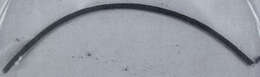
The foram in life position. The upper third protrudes from the seafloor, while the rest remains buried. Image courtesy of Andrew J. Gooday, Southampton Oceanography Centre. This image first appeared in J. Foram. Res. 22:129-146 (1992) and is used with permission.
-
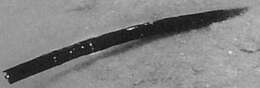
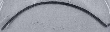
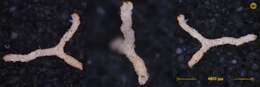
This approx. 10 cm. long individual was taken from the seafloor at 840 m. depth, 144 km. east of Cape Lookout, North Carolina. Image courtesy of Andrew J. Gooday, Southampton Oceanography Centre. This image first appeared in J. Foram. Res 22:129-146 (1992) and is used with permission.
-
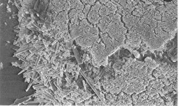
This SEM image shows the two distinct particle types that make up the foram's agglutinated wall. The outer surface is made of fine, black or brown particles, and is underlain by a much thicker layer of sponge spicules and small quartz particles. Image courtesy of Andrew J. Gooday, Southampton Oceanography Centre. This image first appeared in J. Foram. Res 22:129-146 (1992) and is used with permission.
-
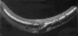
The white surface of this 6-cm-long foraminiferan is partially hidden by sediment trapped in mucus. The mucus was probably secreted by the polychaete worm Nicolea, which has been found on the test surface and is only about 20% as long as the foram is. Image courtesy of Andrew J. Gooday, Southampton Oceanography Centre. This image first appeared in J. Foram. Res 22:129-146 (1992) and is used with permission.
-
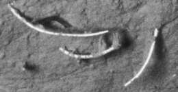
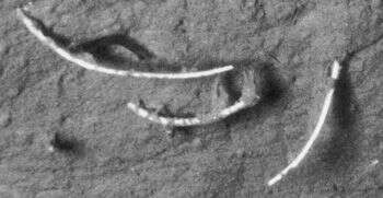
Three individuals photographed on the seafloor, at 850 m. depth, 72 km. NE of Cape Hatteras, North Carolina.Image courtesy of Andrew J. Gooday, Southampton Oceanography Centre. This image first appeared in J. Foram. Res 22:129-146 (1992) and is used with permission.
-
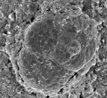
A closeup of a smaller foram (a member of the genus Trochammina) which is living on the surface of the Bathysiphon test. The Trochammina test is 100 microns in diameter. Image courtesy of Andrew J. Gooday, Southampton Oceanography Centre. This image first appeared in J. Foram. Res 22:129-146 (1992) and is used with permission.
-
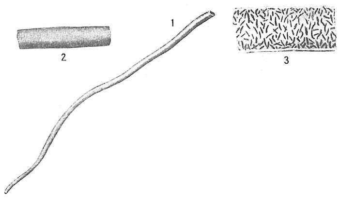
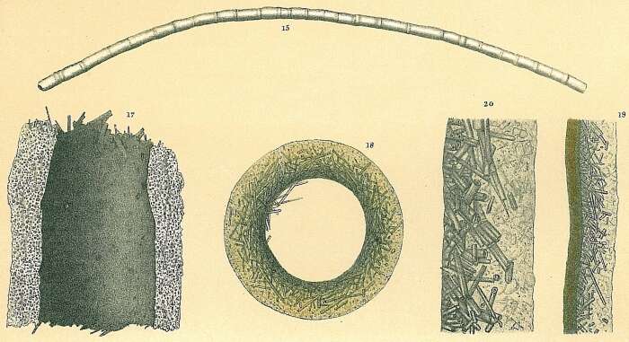
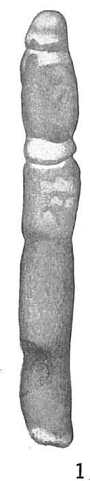
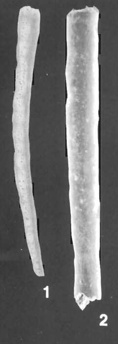
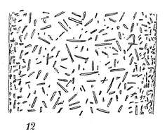
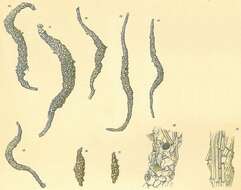
Image source: Cushman, J.A. 1918. The Foraminifera of the Atlantic Ocean. Part I. Astrorhizidae. Bull. U.S. Natl. Mus. 104.
-
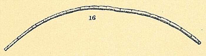
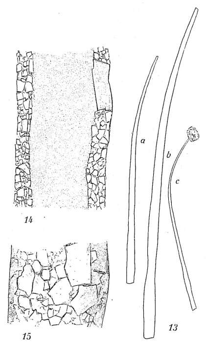
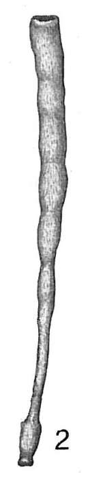
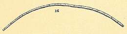
Image source: Höglund, H. 1947. Foraminifera in the Gullmar fjord and the Skagerak. Zoologiska Bidrag från Uppsala 26: 1-328 + 32 pls. Uploaded with written permission from the copyright owners Annalena Höglund and Jan Höglund.
-
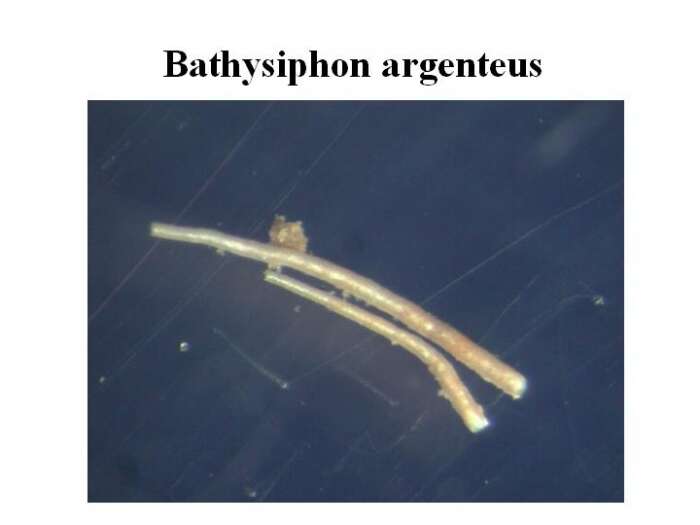
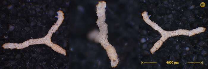
Specimens were isolated from surface sediments samples collected in Kongsfjorden, Isfjorden and Adventfjorden during the cruise of r/v « Oceania » between 22 July and 2 August 2004. The sediment samples were sieved at 500 um and 125 um sized meshes, and living specimens were picked under dissecting microscope on board. The specimens were photographed, measured and fixed for further DNA extraction. Source: http://www.iopan.gda.pl/projects/biodaff/EMBS-06.html
-
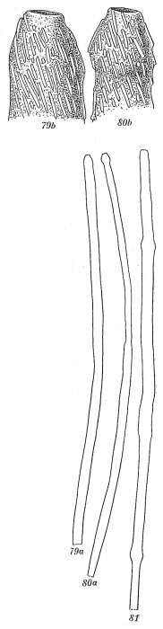
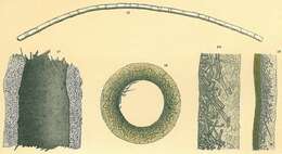
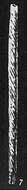
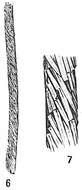
Bathysiphon capillare sensu Jones, R.W. 1994. The Challenger Foraminifera. Image source: Brady, H.B. (1884) Pl. 26
-
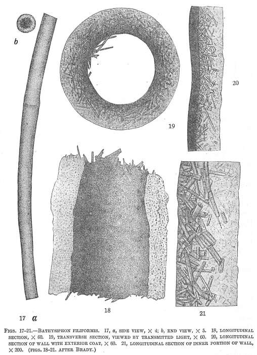
Bathysiphon filiformis sensu Jones, R.W. 1994. The Challenger Foraminifera. Image source: Brady, H.B. (1884) Pl. 26
-
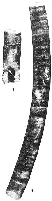
Image source: Cushman, J.A. 1918. The Foraminifera of the Atlantic Ocean. Part I. Astrorhizidae. Bull. U.S. Natl. Mus. 104.
-
Image source: Cushman, J.A. 1921. Foraminifera of the Philippine and adjacent seas. Bull. U.S. Natl. Mus. 100(4).
-
Image source: Cushman, J.A. 1910. A Monograph of the Foraminifera of the North Pacific Ocean. Part I. Astrorhizidae and Lituolidae. Bull. U.S. Nation. Mus 71: xiv+134 pp.
-
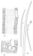
Image source: Höglund, H. 1947. Foraminifera in the Gullmar fjord and the Skagerak. Zoologiska Bidrag från Uppsala 26: 1-328 + 32 pls. Uploaded with written permission from the copyright owners Annalena Höglund and Jan Höglund.
-
Publication: Loeblich, A.R., Tappan, H., 1994. Foraminifera of the Sahul Shelf and Timor Sea. Cushman Foundation for Foraminiferal Research Special Publication 31, 13–630 Plate 1 Figures 1-2 Figure caption: 1, Hypotype (USNM 469470) from V-323, x 25.2, Hypotype (USNM 469471) from V-322. x40, (p, 13)
-
Image source: Cushman, J.A. 1921. Foraminifera of the Philippine and adjacent seas. Bull. U.S. Natl. Mus. 100(4).
-
Image source: Cushman, J.A. 1910. A Monograph of the Foraminifera of the North Pacific Ocean. Part I. Astrorhizidae and Lituolidae. Bull. U.S. Nation. Mus 71: xiv+134 pp.
-
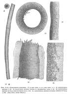
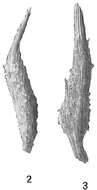
Marsipella elongata sensu Jones, R.W. 1994. The Challenger Foraminifera. Image source: Brady, H.B. (1884) Pl. 24
-
Image source: Cushman, J.A. 1918. The Foraminifera of the Atlantic Ocean. Part I. Astrorhizidae. Bull. U.S. Natl. Mus. 104.
-
Image source: Cushman, J.A. 1918. The Foraminifera of the Atlantic Ocean. Part I. Astrorhizidae. Bull. U.S. Natl. Mus. 104.
-
Image source: Cushman, J.A. 1918. The Foraminifera of the Atlantic Ocean. Part I. Astrorhizidae. Bull. U.S. Natl. Mus. 104.
-
Image source: Höglund, H. 1947. Foraminifera in the Gullmar fjord and the Skagerak. Zoologiska Bidrag från Uppsala 26: 1-328 + 32 pls. Uploaded with written permission from the copyright owners Annalena Höglund and Jan Höglund.
-
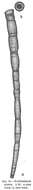
found at 192m depth Greenland Sea, Arctic Source: (http://www.foraminifera.eu/rhabdammina-abyssorum-greenland.html)