-
Hjerritsdal mølle, ved Hobro, Danmark
-
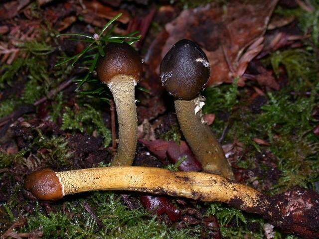
Buchtrops Plantage, Mariager, Danmark
-
Midtsjælland, Danmark
-
Midtsjælland, Danmark
-
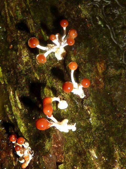
Sebberkloster Hede, Nibe Jylland, Danmark
-
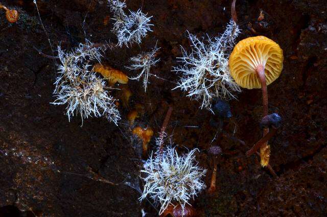
ØLS KRAT VED Hobro
-
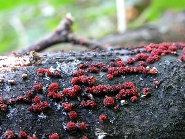
Mushroom Observer Image 691254: Nectriopsis parmeliae (Berk. & M.A. Curtis) M.S. Cole & D. Hawksw.
-
Mushroom Observer Image 29272: Hypocrea nybergiana T. Ulvinen & H. Chamb.
-
Mushroom Observer Image 876539: Cordyceps tuberculata (Lebert) Maire
-
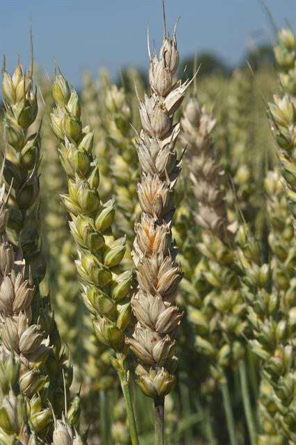
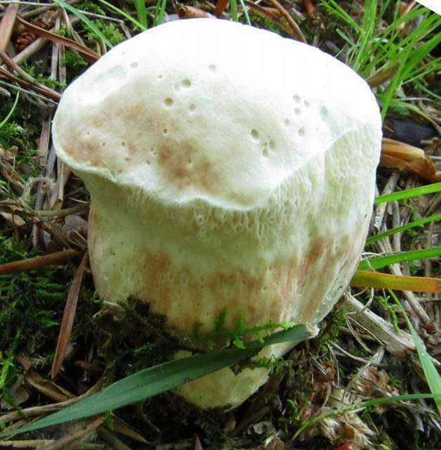
Mushroom Observer Image 210488: Hypocrea pulvinata Fuckel
-
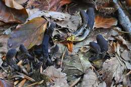
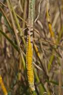
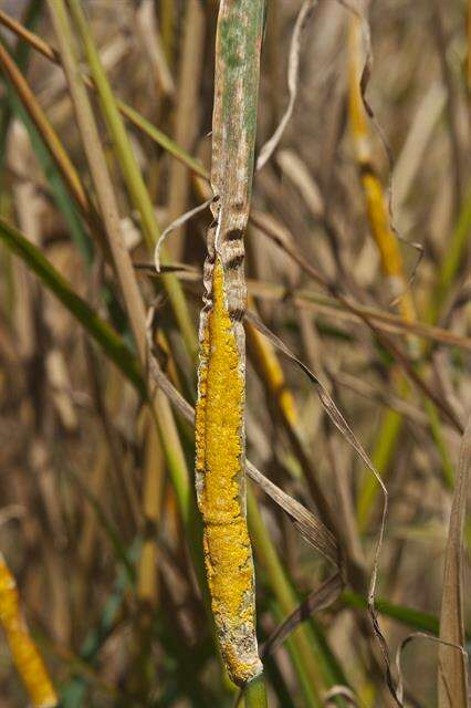
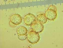
Mushroom Observer Image 42002: Claviceps purpurea (Fr.) Tul.
-
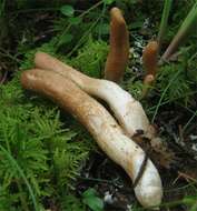
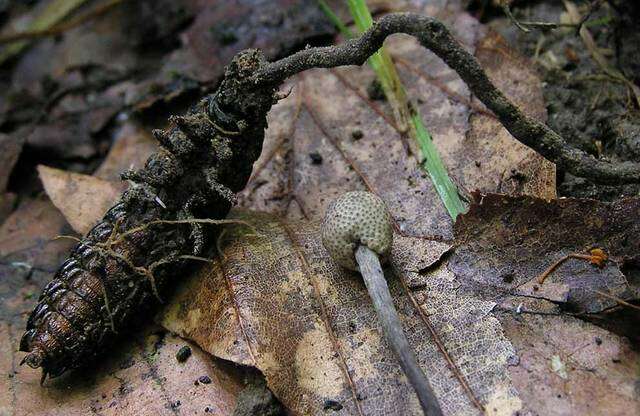
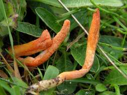
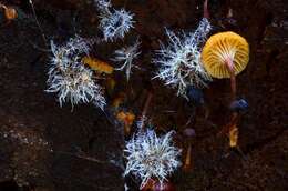
Mushroom Observer Image 64307: Cordyceps entomorrhiza (Dicks.) Fr.
-
Mushroom Observer Image 690781: Trichoderma Pers.
-
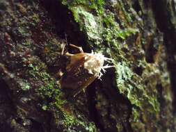
Mushroom Observer Image 301132: Hypomyces cervinigenus Rogerson & Simms
-
Mushroom Observer Image 407579: Hypomyces chlorinigenus Rogerson & Samuels
-
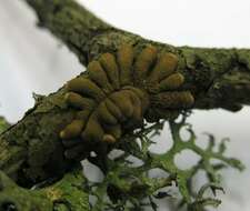
Mushroom Observer Image 124656: Hypocreopsis P. Karst.
-
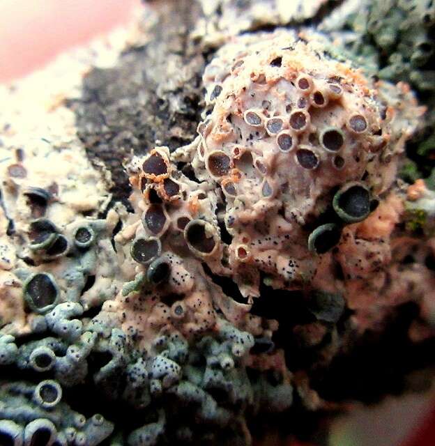
Mushroom Observer Image 158064: Neobarya agaricicola (Berk.) Samuels & Lowen
-
Mushroom Observer Image 165018: Nectriopsis tremellicola (Ellis & Everh.) W. Gams
-
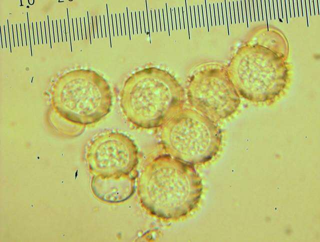
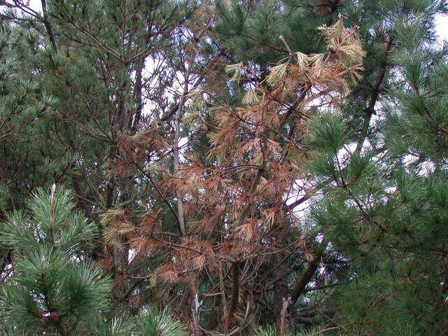
Mushroom Observer Image 196466: Fusarium circinatum Nirenberg & O'Donnell
-
Mushroom Observer Image 264492: Calostilbella calostilbe Hohnel
-
Mushroom Observer Image 498266: Tilachlidium Preuss
-
Mushroom Observer Image 452367: Trichoderma alutaceum Jaklitsch
-
Mushroom Observer Image 431251: Hypocreopsis amplectens T.W. May & P.R. Johnst.
-
Mushroom Observer Image 190163: Tolypocladium capitatum (Holmsk.) Quandt, Kepler & Spatafora