-
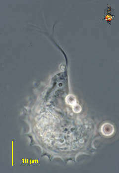
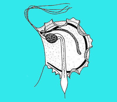
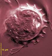
Trichomitopsis (trick-owe-mite-us) is a trichomonad flagellate. this genus has five flagella, four pointing forward and in this micrograph these adhere to each other along most of their lengths. There is also a recurrent flagellum which adheres to the surface of the cell and when it beats causes the margin of the cell to undulate. Flagellates ranging in size from 11-150 microns Costa stout, axostyle stout with a terminal segment often expanded into a pointed bulbous enlargement. The trichomonads often wrap around debris in the gut. They ingest particles of wood which gives some cells a very refractile appearance. From the termite Zootermopsis, supplied by Wards Natural Science Establishment, Rochester, New York, USA. Phase contrast.
-
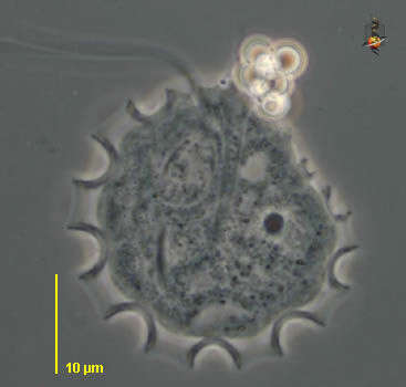
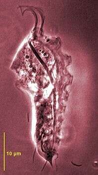
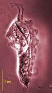
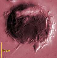
Trichomitopsis (trick-owe-mite-us) is a trichomonad flagellate. this genus has five flagella, four pointing forward. There is also a recurrent flagellum which adheres to the surface of the cell and when it beats causes the margin of the cell to undulate - the feature that is emphasized in this image. The axostyle leading from the front to the rear is also evident as the stiff dark internal structure. From the termite Zootermopsis, supplied by Wards Natural Science Establishment, Rochester, New York, USA. Phase contrast.
-
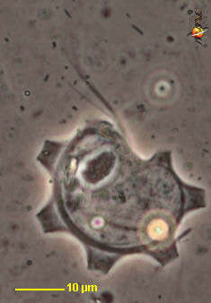
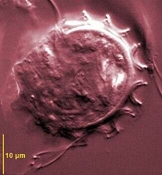
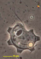
Trichomitopsis (trick-owe-mite-us) is a trichomonad flagellate. this genus has five flagella, four pointing forward. There is also a recurrent flagellum which adheres to the surface of the cell and when it beats causes the margin of the cell to undulate. Flagellates ranging in size from 11-150 microns Costa stout and very obvious here as a curving rod arising anterior to the nucleus. The trichomonads often wrap around debris in the gut. They ingest particles of wood which gives some cells a very refractile appearance. From the termite Zootermopsis, supplied by Wards Natural Science Establishment, Rochester, New York, USA. Phase contrast.
-
-
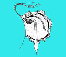
Trichomitpsis termopsidis from Hodotermopsis sjoestedti - all cells have two flagellar apparatuses comprising two undulating membranes and two costa and axostyles. Phase contrast microscopy.
-
Undulating membrane, DIC image
-
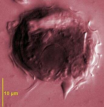
DIC image showing bulbous posterior axostyle
-
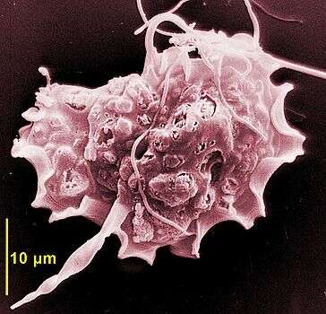
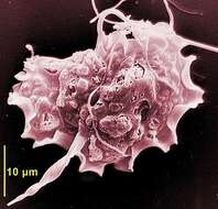
Scanning electron micrograph showing recurrent flagellum associated with the conspicuous undulating membrane, posterior axostyle and cytoplsam filled with wood particles.