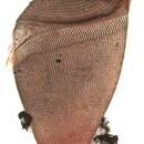
Description of Metopus fuscus
provided by BioPedia
This is one of the largest species in the genus, with body size near 190-220 µm X 60-70 µm M. fuscus may be easily distinguished from M. es in living condition; its cytoplasm is very clear; dorsoventral body flattening more developed; contractile vacuole is of unusual shape, like in Spirostomum. Body is of clear brownish tint and irregular shape; posterior extremity is flattened, so that it seems acute when viewed laterally but rectangular from the ventral and dorsal aspect. This asymmetry is well seen in the free-swimming cells. M. fuscus is devoid of trichocysts; the pellicle is finely striated. Near 40-50 kineties are encountered along the equator; they are distinct in living cells also. Both the perizonal ciliary stripe and adoral zone of membranelles are comparatively longer than those in M. es since the left-side anterior torsion is more developed in M. fuscus. The cytostome is subanterior. The macronucleus is well-seen in the living condition; it is sharply outlined due to accumulation of brownish refractile granules on its surface. This feature is peculiar for this species only. Colourless food vacuoles are scattered throughout the cytoplasm; food remnants are gathered into a large amorphous accumulation that presses against the contractile vacuole in the posterior body half from which it is excreted outwards. From: Jankowski, A.W. Arch Protistenk. 107:185-294, 1964.