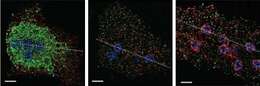
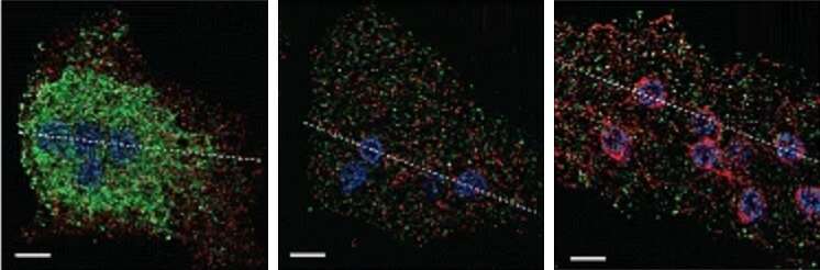
Description: English: Localization for Mastigamoebe balamuthi COPI-β. Structured illumination microscopy of M. balamuthi labelled with antibodies againstCOPI and PDI (left, ER structure),COPI and MDH (middle, hydrogenosomes), andCOPI and α tubulin (right).The COPI signal is observed in numerous vesicles scattered within the M. balamuthi cells. α tubulin antibody labelled the tubular conus around nuclei and network of fibers. Signal for PDI network is concentrated around multiple nuclei. Graphs show line scans for fluorescence intensities corresponding to the dotted lines in merged images. Scale bar, 5 μm. Date: 7 March 2018. Source: Fig. 2 at
https://bmcbiol.biomedcentral.com/articles/10.1186/s12915-018-0492-9 A sophisticated, differentiated Golgi in the ancestor of eukaryotes
doi:10.1186/s12915-018-0492-9 (rearranged extract). Author: Lael D. Barlow, Eva Nývltová, Maria Aguilar, Jan Tachezy, Joel B. Dacks.