Comprehensive Description
provided by Smithsonian Contributions to Zoology
Sphalloplana (Speophila) holsingeri
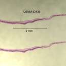
TYPE MATERIAL.—Holotype, set of sagittal sections on 5 slides, USNM 53436. Paratypes, eight sets of sagittal and transverse sections on 35 slides, USNM 53437–53443, 53478.
EXTERNAL FEATURES (Figures 7, 14).—Sphalloplana holsingeri is a blind, white species, measuring, when gliding quietly, up to 15 mm in length and 1.5 mm in width. The anterior end is truncate, with a bulging frontal margin flanked by a pair of short, rounded auricles projecting anterolaterally. In the center of the frontal margin, the location of the adhesive organ is visible as an opaque spot. When protruded, the organ appears as a short conical projection. Behind the auricles, the body narrows slightly, then widens again to attain its greatest width a short distance behind the head. In the greater part of the body, the lateral margins run parallel up to the region of the copulatory complex, where they converge again to meet at the pointed posterior end. An opaque marginal rim, corresponding to the modified marginal epithelium, is conspicuous in the living animal. The pharynx is inserted behind the middle of the body and measures in length about one-seventh the length of the body. The copulatory apparatus occupies the anterior half of the postpharyngeal region. The anterior end of the intestine forms a median projection behind the adhesive organ (Figure 18); there is no V-shaped extension of the lateral branches to the sides of the organ such as is seen in some other species of the subgenus Speophila, e.g., S. pricei (Hyman) (Figure 19).
ANATOMY.—The epidermal epithelium of the lateral margins is thickened and contains rhabdites considerably larger than those of either the dorsal or ventral epidermis, as is seen generally in the genus Sphalloplana.
The adhesive organ (Figure 36) is well developed, consisting of a rounded subterminal depression, the central part of which forms a deep invagination when the organ is retracted. The muscular differentiations of the organ correspond to those described in other species of Sphalloplana with conspicuous adhesive organs, such as S. pricei (see Hyman, 1937:464–465), S. mohri Hyman (see Mitchell, 1968:611–613), and S. weingartneri Kenk (see Kenk, 1970:315). The glandular equipment of the organ, however, differs from the conditions seen in other species of the genus. Two kinds of glands open through the epithelial lining: eosinophilic gland ducts passing through the infranucleate epithelium of the invaginated portion and cyanophilic glands through the remaining field of the depression surrounding the inverted canal. The eosinophilic secretions are of a homogeneous or coarsely granular nature, while the cyanophilic secretions, staining deeply with hematoxylin, appear as threadlike or rod-shaped filaments. The gland cells producing these threads form voluminous masses occupying both the dorsal and ventral portions of the mesenchyme and the spaces between the intestinal branches in the medial part of the anterior two-thirds of the prepharyngeal region. In other species of Sphalloplana, only eosinophilic secretions are known to occur. It is difficult to interpret the functions of the two types of glands. The eosinophilic secretions in the central, eversible part of the organ presumably correspond to those described in other triclads that are equipped with adhesive organs, e. g., Procotyla fluviatilis Leidy, where the secretions dissolve into a sticky mucous mass that is used in the capture of living prey. The function of the cyanophilic secretions, so prominent in S. holsingeri, is not clear. It is interesting to note that a similar differentiation of the glands of the adhesive organ has been observed in a European planarian, Dendrocoelum album (Steinmann), where erythrophilic glands open through the epithelium of the adhesive pit and cyanophilic gland ducts at the peripheral rim of the organ (Steinmann, 1910:190).
The musculature of the pharynx follows the plan of the genus Sphalloplana. The internal pharyngeal epithelium is surrounded by a thick layer of circular muscle fibers, followed by a thinner layer of longitudinal muscles.
In the reproductive system, the two ovaries are situated on the medial sides of the ventral nerve cords, behind the third or fourth lateral branch of the anterior intestinal trunk. The numerous testes (Figure 43) are arranged in two longitudinal rows, one on either side of the midline, occupying the posterior half of the prepharyngeal region. They are located essentially ventral, medial to the nerve cord. Only a few testes may be pushed toward the dorsal side in the “septa” between the intestinal branches, or may extend through the entire dorsoventral diameter of the body. Many follicles are attached directly to the sperm ducts or vasa deferentia without the mediation of efferent ductules. In the region of the testes, the vasa deferentia run parallel to the ventral nerve cords, somewhat removed from the ventral integumental muscle layers. At the level of the pharynx, they widen to form the sinuous spermiductal vesicles (or false seminal vesicles) that keep their expanded shape up to their entrance into the penis bulb.
The copulatory apparatus (Figure 54) was analyzed in 12 series of sagittal sections. The penis consists of a relatively small bulb (bp) with rather feeble musculature, embedded in the mesenchyme, and a larger, generally plug-shaped, papilla, sometimes constricted at its base, protruding into the genital atrium. The shape of the papilla is rather variable in the specimens studied, as it is easily distorted when the animal is being killed. It lacks any rigid supporting structures and its musculature is confined to two rather thin layers of muscle fibers, a circular layer underlying the external epithelium and a weak longitudinal layer below it. Only a few scattered longitudinal fibers run through the parenchyma of the papilla. The bulb contains an elongated cavity, the seminal vesicle (vs), lined with an epithelium pierced by gland ducts with a finely granular, faintly cyanophilic secretion that enter the bulb from the surrounding mesenchyme. Toward the penis papilla, the cavity continues as a narrow, nonglandular canal, the ejaculatory duct (de), which opens into the atrium on the dorsal side of the papilla, about halfway between its base and its tip.
The genital aperture or gonopore (gp) leads into two cavities, anteriorly the male atrium (am) and posteriorly the cavity of the large vagina (v). There is no common atrium developed.
The two vasa deferentia (vd), which approach the penis bulb as enlarged spermiductal vesicles, enter the bulb ventrolaterally, diminishing in diameter, and open separately into short lateral extensions of the anterior part of the seminal vesicle.
The two oviducts, in the region of the copulatory complex, ascend from their course above the ventral nerve cords, pass to the midline, and unite above the male atrium to form the common oviduct (odc), which arches ventrally and opens into the posterior part of the atrium. The copulatory bursa (b) is an elongated sac lying close to the anterior face of the penis bulb. Its outlet is a rather narrow, straight canal (bd) running in the midline dorsally to the penis and atrium and extending rather far posteriorly. Behind the level of the gonopore, the canal curves ventrally and widens into a voluminous cavity, the vagina (v). In the preserved specimens, the outline of the vagina is very variable, usually irregularly folded. In fully mature worms, the lining of the vagina is infranucleate while that of the narrow part of the bursal canal contains nuclei. In young specimens, however, nuclei are present in the vaginal epithelium; it appears, therefore, that the nuclei pass from the cellular lining only at the approach of full sexual maturity. The vagina is surrounded by a thick coat of interlaced circular and longitudinal muscle fibers. The musculature of the anterior, narrow part of the bursal canal is very feeble, probably also made up of intermingled longitudinal and circular muscles.
DISTRIBUTION AND ECOLOGY.—Sphalloplana holsingeri is known from only one locality, the spring on the property of J. W. Biggers in Fairfax County, Virginia, the same spring where S. subtilis occurs (see above and Figures 27, 28). Two specimens, both mature, were collected first by Dr. John R. Holsinger on 23 May 1965 but were fixed in alcohol, which made an analysis of their anatomy rather difficult. Additional (over 80) specimens were obtained by Dr. Holsinger, Bill Biggers, and the writer by baiting with shrimp meat on 18 March, 25 March, 2 April, and 12 August 1973, among them many mature animals.
TAXONOMIC POSITION.—The presence of a deeply invaginated, protrusible adhesive organ places the species in the subgenus Speophila. Among the outstanding characteristics of the species is the differentiation of the glands of the adhesive organ, eosinophilic glands in the inverted part, surrounded by a field of cyanophilic gland openings. In the reproductive system, it shares the ventral position of the testes with several other species of the genus. The most important specific characters are in the anatomy of the copulatory complex: the opening of the ejaculatory duct on the dorsal side of the penis papilla and the development of a voluminous vagina in the terminal part of the bursal duct. The species is named in honor of my distinguished colleague, Dr. John R. Holsinger, who collected the first specimens and collaborated in the procurement of additional material of the species.
When the manuscript of this description was completed, I received notice that the American Museum of Natural History had, among the materials left by the late Libbie H. Hyman, a set of three slides of sagittal sections marked “Sphalloplana holsingeri.” The condition of the sections is rather bad, so that no identification of the species can be made. Apparently, Hyman's S. holsingeri is an unpublished manuscript name and has no nomenclatorial standing.
- bibliographic citation
- Kenk, Roman. 1977. "Freshwater triclads (Turbellaria) of North America, IX, the genus Sphalloplana." Smithsonian Contributions to Zoology. 1-38. https://doi.org/10.5479/si.00810282.246