-
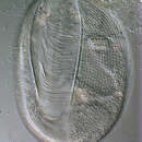
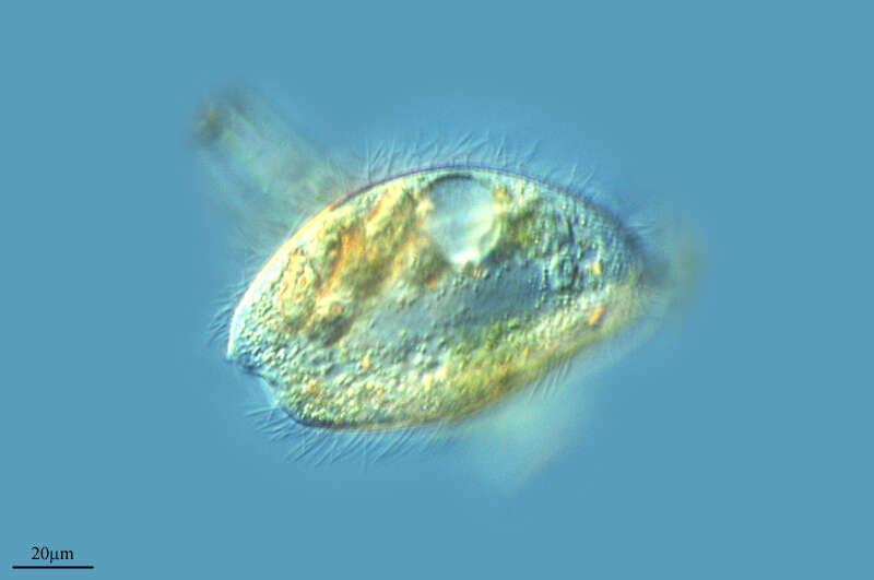
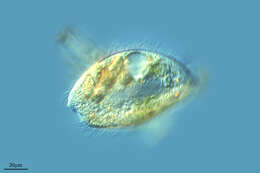
Portrait (ventral view) of the oligohymenophorean ciliate, Lembadion bullinum (Müller,1786;Perty,1849). Cell outline is oval. The ventral surface is concave and the dorsum convex. The very large scoop-like peristome occupies most of the ventral surface (seen well here). The cytostome is at the posterior end of the peristome. There is a small undulating membrane on the right margin of the peristome. A large sheet-like adoral membranelle arises from the left margin of the peristome (seen well here). There are 40-60 evenly spaced longitudinal somatic kineties. The pellicle of L. bullinum, divided into small roughly rectangular depressions, has a distinct cribriform pattern (seen clearly to the left of the peristome). This feature distinguishes L. bullinum from L. magnum which has a striate pellicular pattern. There is usually a tuft of longer caudal cilia. The contractile vacuole connects with its excretory pore by a long curved canal (not seen here). The single ovoid macronucleus and micronucleus are posterior (not seen here). Collected from a freshwater pond near Boise, Idaho May 2004. DIC optics.
-
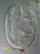
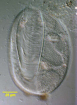
Portrait (optical section) of the oligohymenophorean ciliate, Lembadion bullinum (Müller,1786;Perty,1849). Cell outline is oval. The ventral surface is concave and the dorsum convex. The very large scoop-like peristome occupies most of the ventral surface. The cytostome is at the posterior end of the peristome. There is a small undulating membrane on the right margin of the peristome. A large sheet-like adoral membranelle arises from the left margin of the peristome. There are 40-60 evenly spaced longitudinal somatic kineties. The pellicle of L. bullinum, divided into small roughly rectangular depressions, has a distinct cribriform pattern. This featurte distinguishes L. bullinum from L. magnum which has a striate pellicular pattern. There is usually a tuft of longer caudal cilia. The contractile vacuole (seen just anterior to the macronucleus here) connects with its excretory pore by a long curved canal (not seen here). The single ovoid macronucleus (seen here) and micronucleus (not seen in this image) are posterior. Collected from a freshwater pond near Boise, Idaho May 2004. DIC optics.
-

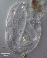
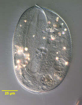
Portrait (dorsal view) of the oligohymenophorean ciliate, Lembadion bullinum (Müller,1786;Perty,1849). Cell outline is oval. The ventral surface is concave and the dorsum convex. The very large scoop-like peristome occupies most of the ventral surface (not seen here). The cytostome is at the posterior end of the peristome. There is a small undulating membrane on the right margin of the peristome. A large sheet-like adoral membranelle arises from the left margin of the peristome (not seen here). There are 40-60 evenly spaced longitudinal somatic kineties. The pellicle of L. bullinum, divided into small roughly rectangular depressions, has a distinct cribriform pattern (seen clearly in this image). This feature distinguishes L. bullinum from L. magnum which has a striate pellicular pattern. There is usually a tuft of longer caudal cilia. The contractile vacuole connects with its excretory pore by a long curved canal (not seen here). The single ovoid macronucleus and micronucleus are posterior (not seen here). Collected from a freshwater pond near Boise, Idaho May 2004. DIC optics.
-
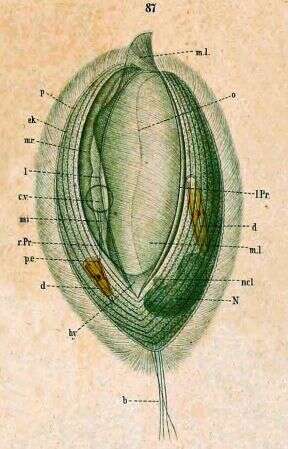
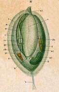
Key to Schewiakoff's abbreviations: b -- Sensory bristle cv -- Contractile vacuole d -- Ingested diatom ek -- Ectoplasm hy -- Hypostome l.Pr -- Left edge of peristome mi -- Inner undulating membrane ml -- Left undulating membrane mr -- Right undulating membrane N -- Macronucleus ncl -- Micronucleus o -- Mouth P -- Peristome pe -- Excretory pore r.pr -- Right edge of peristome
-
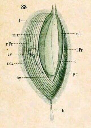
Right side view. Key to Schewiakoff's abbreviations: cv -- Contractile vacuole ccv -- Canal of the contractile vacuole hy -- Hypostome l -- Border, trim l.Pr -- Left edge of Peristome ml -- Left undulating membrane mr -- Right undulating membrane o -- Mouth pe -- Excretory pore r.Pr -- Right edge of Peristome
-
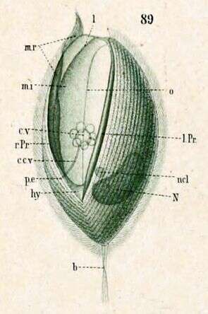
Left side view, with left undulating membrane removed to better show the shape of the right and inner undulating membranes. Key to Schewiakoff's abbreviations: b - Sensory bristle ccv -- Canal of the contractile vacuole cv -- Contractile vacuole hy -- Hypostome l -- Border, rim l.Pr -- Left edge of the Peristome mi -- Inner undulating membrane mr -- Right undulating membrane N -- Macronucleus ncl -- Micronucleus pe -- Excretory pore r.Pr -- Right edge of peristome
-
Matute, La Rioja, Spain