-
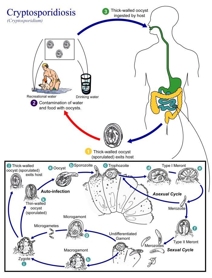
This illustration depicts the life cycle of different species of Cryptosporidium, the causal agents of Cryptosporidiosis.Created: 2002
-
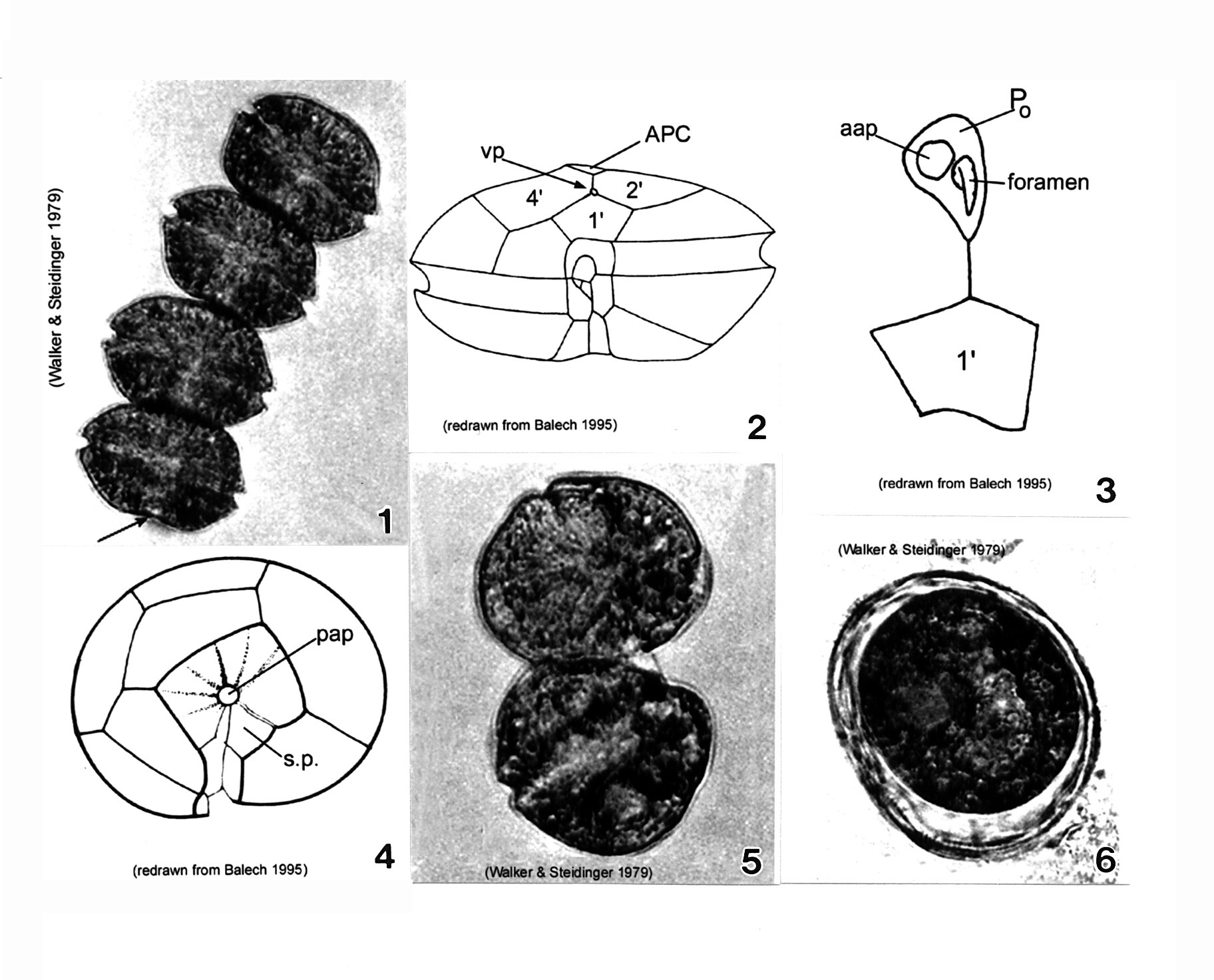
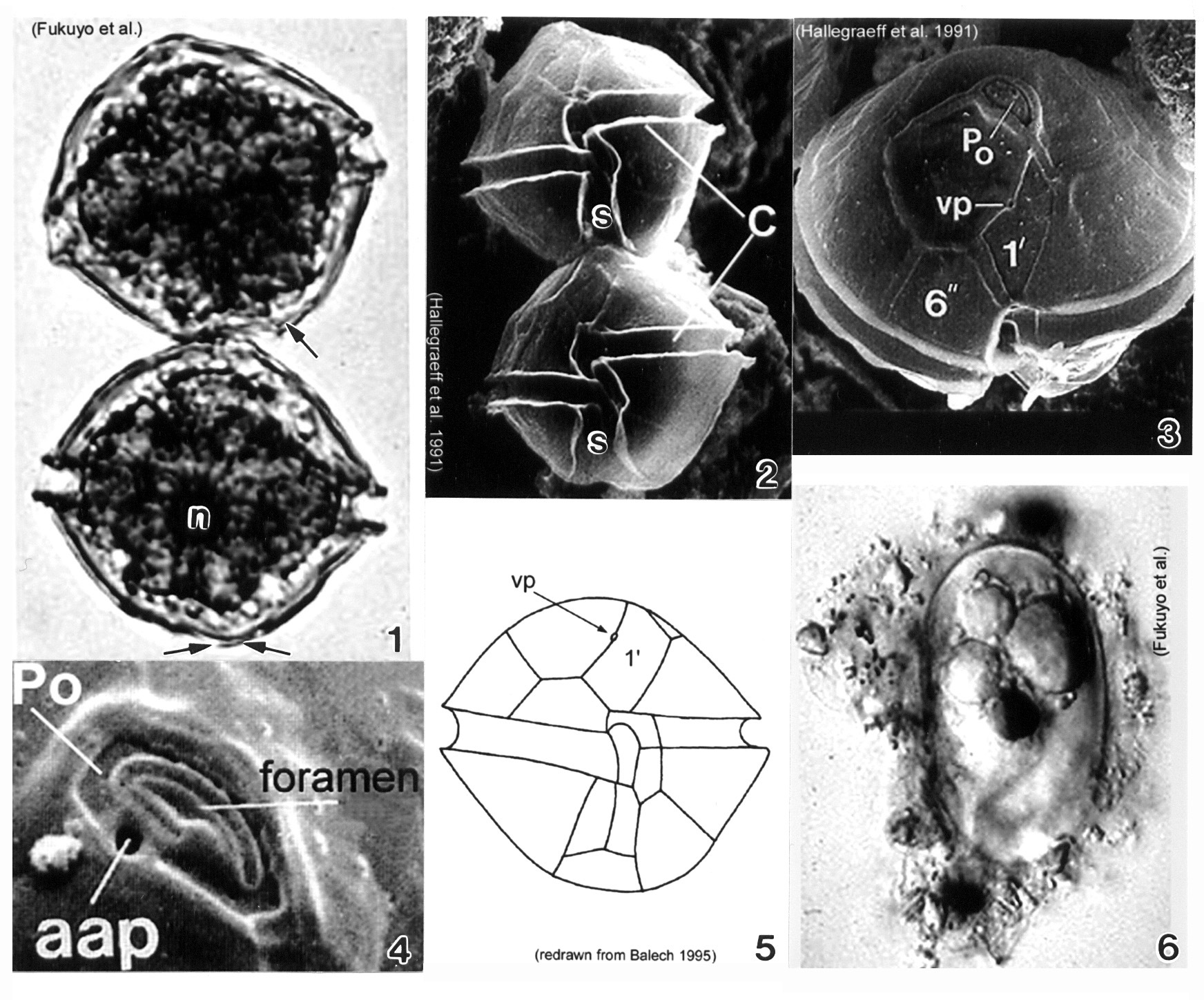
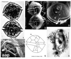
Plate 4. Alexandrium monilatum. Fig. 1. LM: four-cell chain. Cells large, wider than long, flattened anterio-posteriorly. Antapex slightly concave (arrow). Figs. 2-4. Line drawings. Fig. 2. Ventral pore (vp) depicted (Florida specimens) at anterior margin of 1' plate where it comes in contact with plates 2' and 4'. Cingulum (C) deeply excavated, wide, descending; displaced one time its width. Fig. 3. Apical pore plate (Po) does not come in contact with 1' plate. Anterior attachment pore (aap) large, round and dorsally situated in the APC. Foramen comma-shaped. Fig. 4. Antapical view: posterior sulcal plate (sp) large, rhomboid and concave with radial markings. Posterior attachment pore (pap) large and centrally located. Figs. 5-6. LM. Fig. 5. Two isogamous gametes fusing at oblique angles. Fig. 6. Mature resting cysts: dark and round, with a triple layered wall.
-
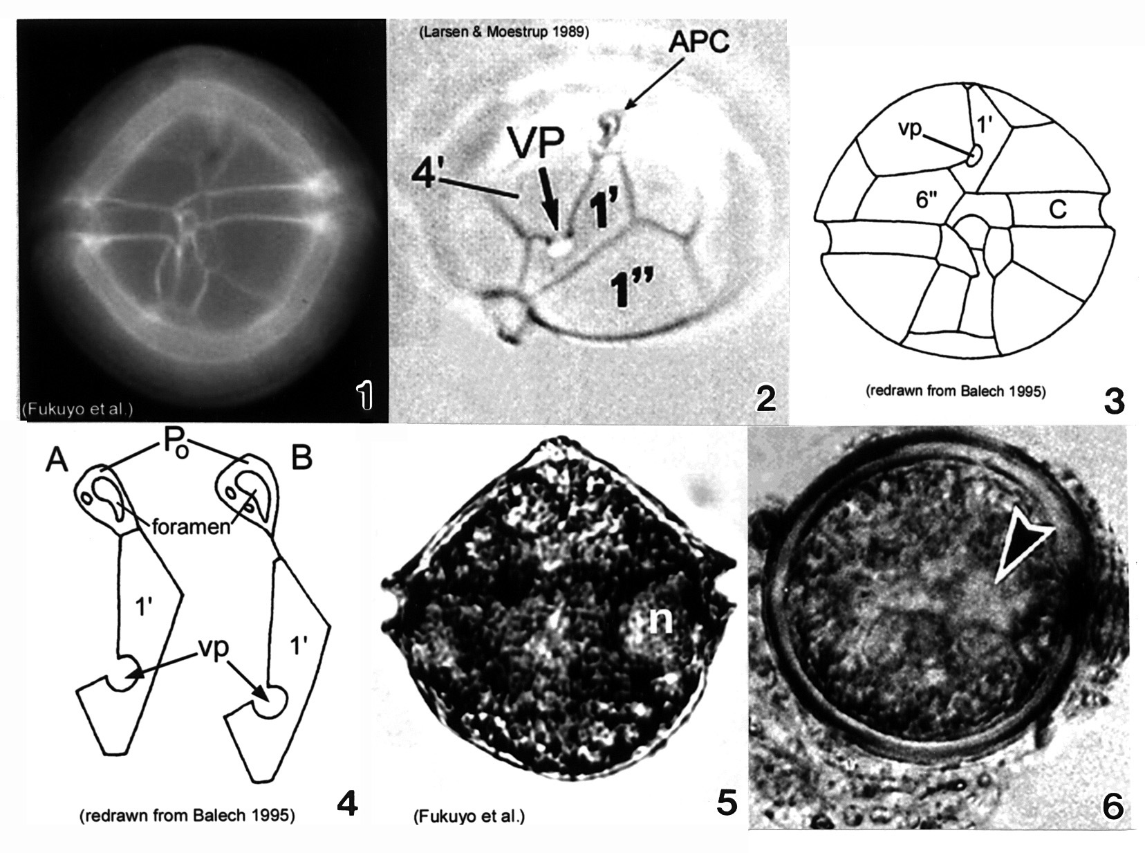
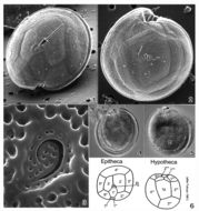
Plate 5. Alexandrium ostenfeldii. Figs. 1-3. LM. Fig. 1. Ventral view. Cell large and nearly spherical. Cingulum deeply excavated. Epitheca broad and convex-conical. Hypotheca hemispherical with an obliquely flattened antapex. Fig. 2. Epitheca: apical view. Ventral pore (vp) large and distinct. First apical plate (1') forms a 90 degree angle at the point where vp and 4' plate come in contact. Apical pore complex (APC) with comma-shaped foramen. Figs. 3-4. Line drawings. Fig. 3. Ventral view: 6'' plate wider than high. Cingulum (C) slightly excavated. Fig. 4. APC and 1' plate: a. Po in direct contact with 1'; b. Po in indirect contact with 1' via thin suture. Fig. 5. LM: vegetative cell. Small equatorial nucleus (n). Fig. 6. LM: temporary cyst large and spherical, covered in mucilage. Nucleus visible (arrowhead)(Mackenzie et al. 1996).
-
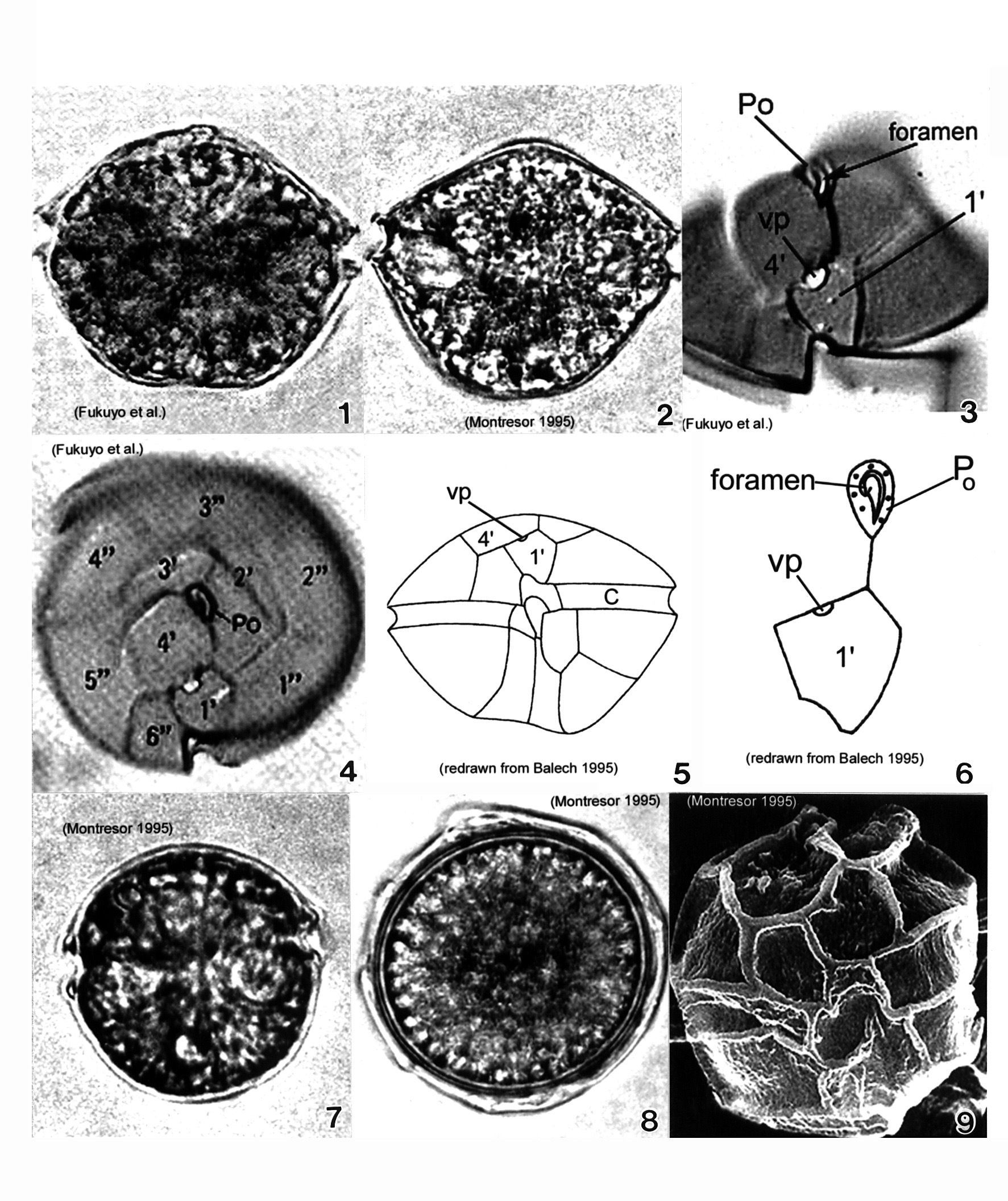
Plate 6. Alexandrium pseudogonyaulax. Figs. 1-4. LM. Fig. 1. Ventral view. Cell broadly pentagonal; wider than long. Epitheca short and dome-shaped. Hypotheca longer than epitheca. Cingulum shallow and barely displaced. Fig. 2. Dorsal view. Antapex obliquely concave. Fig. 3. Epitheca: ventral view. Apical pore plate (Po) with comma-shaped foramen. 1' plate pentagonal with large wide ventral pore (vp) on 4' plate margin. Fig. 4. Epitheca: apical view. 1' plate does not come in contact with Po. Po oval and longitudinal on apex. Figs. 5-6. Line drawings. Fig. 6. Po and 1' plate not in contact. Fig. 7. LM: isogamous gametes smaller and rounder than vegetative cells. Fig. 8. LM: round resting cyst. Fig. 9. SEM: paratabulate cyst.
-
Plate 7. Alexandrium tamarense. Fig. 1. LM. Two cell chain: cells small to medium; slightly longer than wide, nearly spherical. Cingulum (C) deeply escavated and lipped. Left hypothcal lobe slightly larger than right. Nucleus (n) visible. Figs. 2-4. SEM. Fig. 2. Two cell chain: cingulum displaced 1X its width. Deep sulcus (s) widens posteriorly. Fig. 3. Epitheca: apical view. Apical pore plate (Po) rectangular; narrows ventrally. Po and first apical plate (1') in direct contact. Small ventral pore present on 1' plate. Fig. 4. Apical pore complex (APC): foramen large and fishhook shaped. Small round anterior attachment pore (aap) present (Hallegraeff 1991). Fig. 5. Line drawing. Fig. 6. LM. Oblong resting cyst with rounded ends, reddish lipid bodies; covered in mucilage.
-
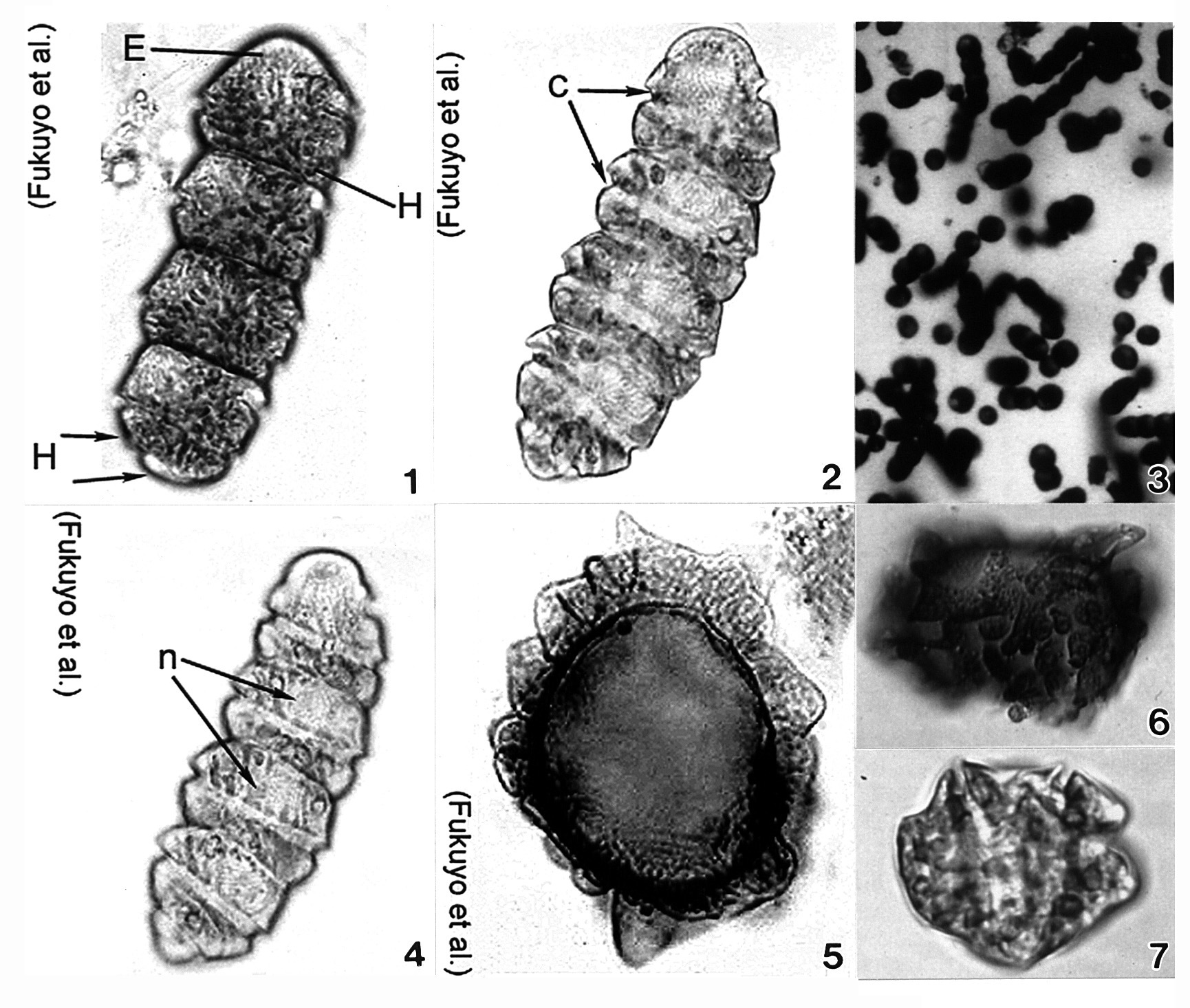
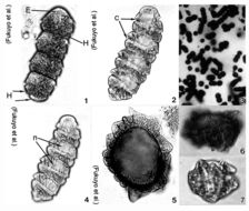
Plate 9. Cochlodinium polykrikoides. Figs. 1-7. LM. Fig. 1. Four cell chain. Single cell small and ellipsoid. Epitheca (E) rounded and conical. Hypotheca (H) divided into two posterior lobes (arrows). Numerous rod-shaped chloroplasts. Fig. 2. Cingulum (c) deeply excavated; circles cell 1.8-1.9 times. Fig. 3. Colony of single and chained cells. Fig. 4. Large nucleus (n) in epitheca. Figs. 5-7. Cysts. (Figs. 3,6,7 by Matsuoka & Fukuyo)
-
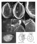
Plate 10. Coolia monotis: Figs. 1-5. SEM. Fig. 1. Ventral view: spherical shape. Cingulum lipped and equatorial. Sulcus with flexible lists (arrowheads). Ventral pore present (arrow). Fig. 2. Dorsal view: apical pore plate (arrow), Po, located off-center on epitheca. Fig. 3. Antapical view: hypothecal plates. Fig. 4. Smooth edged thecal pores unevenly distributed. Fig. 5. Po about 12 _ long, slightly curved and narrow with a slit-like apical pore. Two supporting rib-like costae (arrows) and evenly spaced round pores surround the pore. Figs. 6,7. LM. Fig. 6. Ventral view of lipped cingulum and sulcus. Fig. 7. Planozygote with two longitudinal flagella (arrows). Fig. 8. Line drawing: thecal plate arrangement.
-
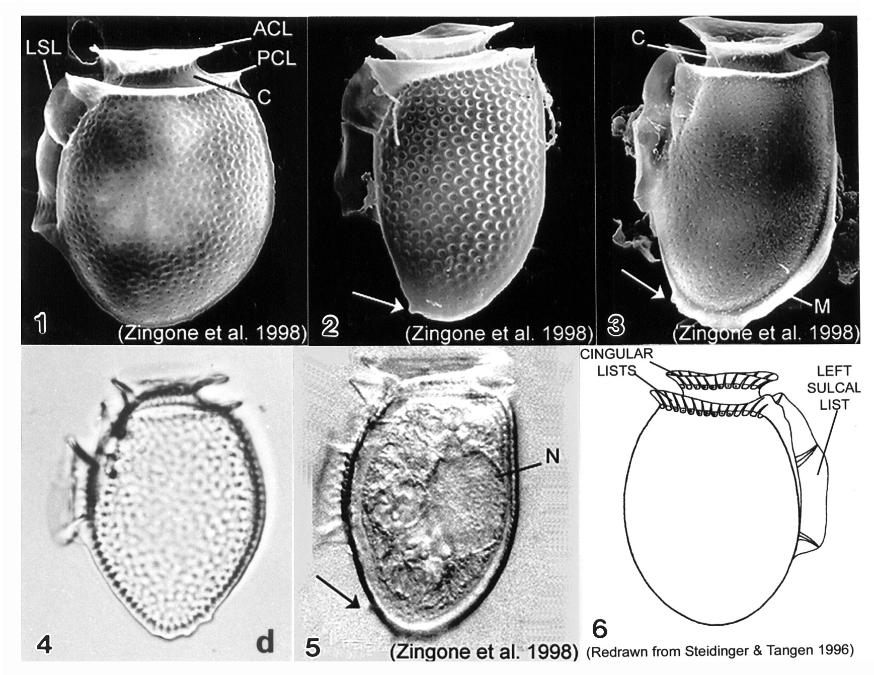
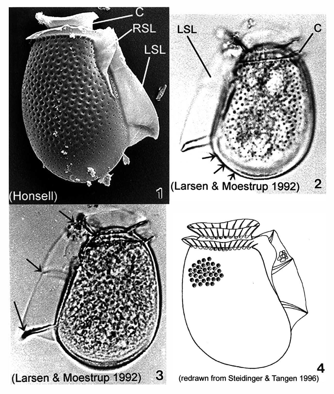
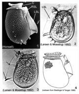
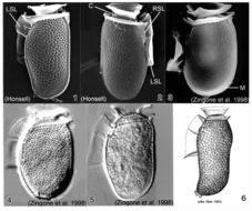
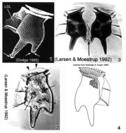
Plate 11. Dinophysis acuminata. Figs. 1-5. SEM: lateral view. Fig. 1. Cell oval and rotund; thecal surface with shallow depressions and scattered pores. Left sulcal list (LSL) extends beyond midpoint of cell. Well-developed cingular lists: anterior cingular list (ACL); posterior cingular list (PCL). C=cingulum. Fig. 2. Long and narrow cell with prominent surface areolae, each with a pore. Antapex tapered and ventrally off-center. Small posterior protrusion present (arrow). Fig. 3. Long and narrow cell. Thecal surface smooth with small scattered pores. Megacytic zone (M) void of pores. Posterior protrusions on antapex (arrow). Figs. 4-5. LM: lateral view. Fig. 4. Surface areolae and tapered antapex (from Larsen & Moestrup 1992: fig. 1d). Fig. 5. Large dorsal nucleus (N). Small, blunt projections on tapered antapex (arrow). Fig. 6. Line drawing.
-
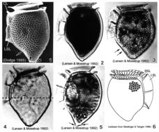
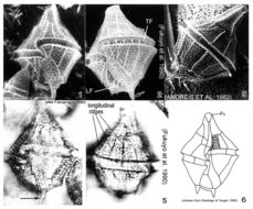
Plate 12. Dinophysis acuta. Fig. 1. SEM: lateral view. Cell oblong and robust; theca heavily areolated. Well developed cingular lists (CL) and left sulcal list (LSL). Pointed antapex. Figs. 2-3. LM: lateral view (from Larsen & Moestrup 1992: fig.s 2a,d; scale bars=20 _). Fig. 2. Large areolae, each with a pore (arrows). Fig. 3. Widest point below mid-section (dashed line) aligned with third sulcal rib (arrow). Fig. 4. Line drawing.
-
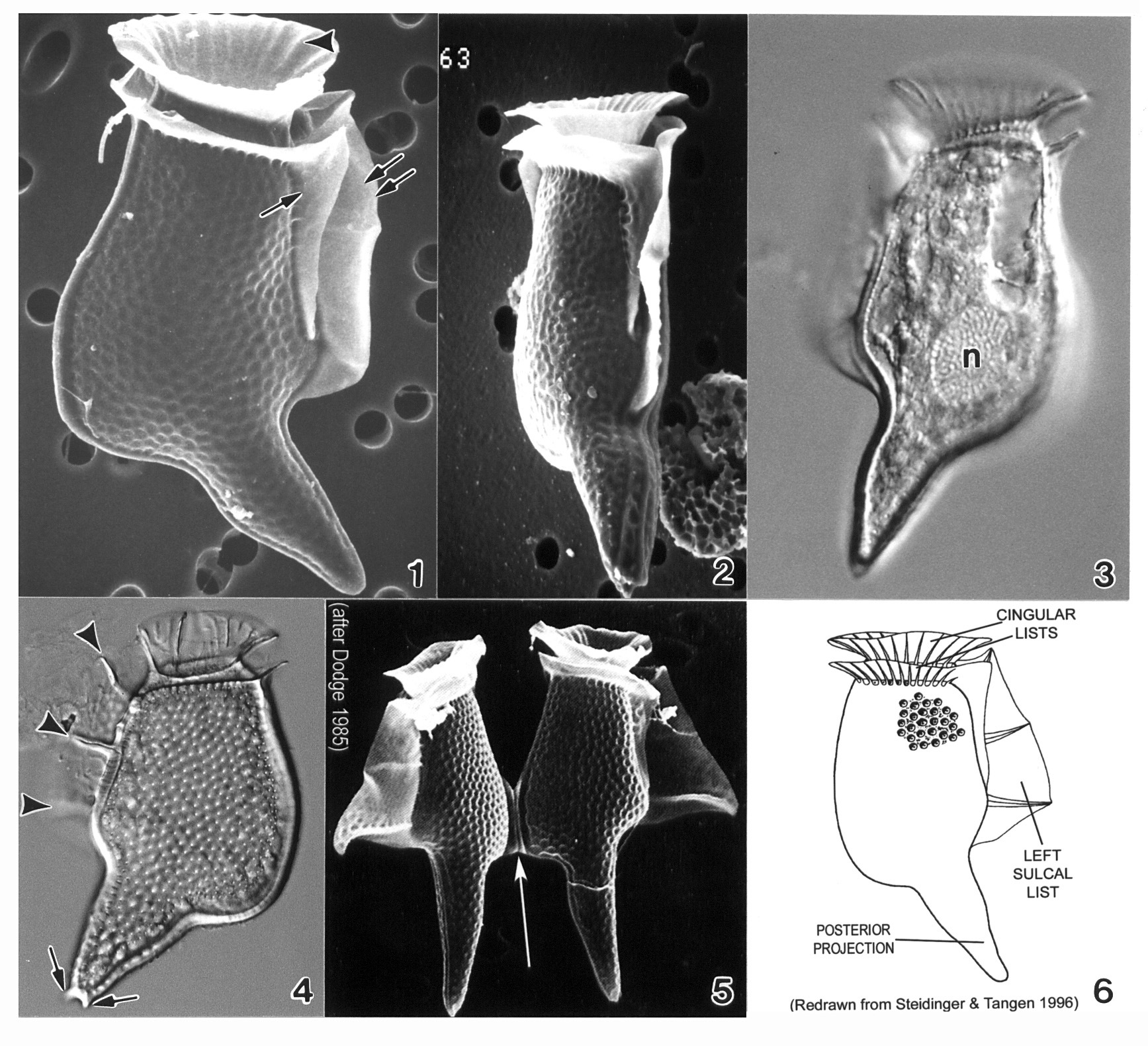
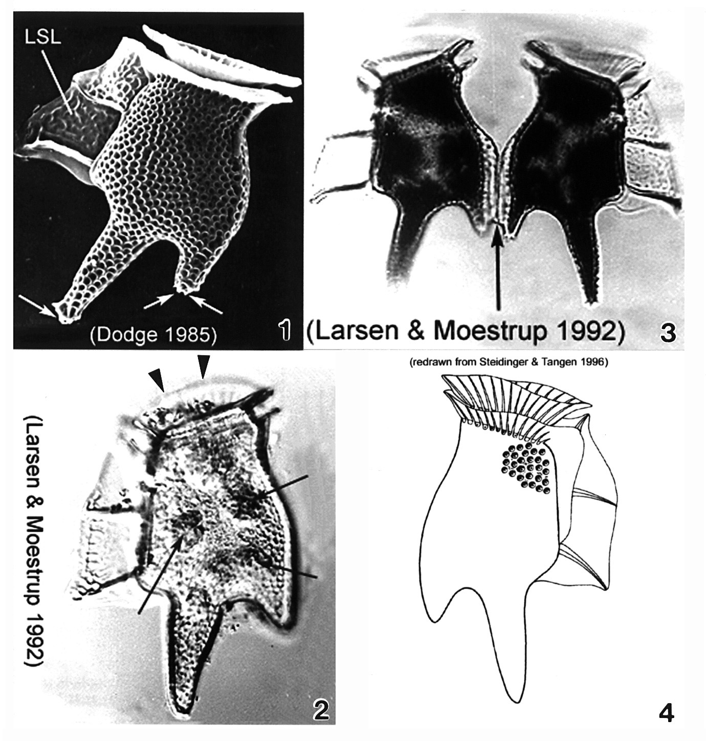
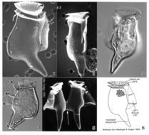
Plate 13. Dinophysis caudata. Figs. 1-2. SEM. Fig. 1. Large, long and distinctive cell with extended ventral hypothecal process. Cingulum narrow; lists supported by ribs (arrowhead). Strong left sulcal list (double arrows). Right sulcal list present (single arrow). Fig. 2. Ventral view: cell compressed laterally. Figs. 3-4. LM. Fig. 3. Large posterior nucleus (n). Fig. 4. Left sulcal list with three supporting ribs (arrowheads); posterior projection with small knob-like spines (arrows). Surface areolae evident. Fig. 5. SEM. Paired cells joined at dorsal expansion (arrow). Fig. 6. Line drawing.
-
Plate 14. Dinophysis fortii. Fig. 1. SEM: lateral view. Left sulcul list (LSL) long and well-developed. Right sulcal list (RSL) present. Cingulum (C) obscures low and small epitheca. Thecal surface covered with areolae. Figs. 2-3. LM: lateral view. Fig. 2. Cell subovate with a wide round posterior bottom (dorsal bulge)(arrows). Fig. 3. LSL supported by three strong ribs (arrows). Smoothly convex dorsal margin. Fig. 4. Line drawing.
-
Plate 16. Dinophysis norvegica. Fig. 1. SEM: lateral view. Cell heavily areolated with pointed antapex and posterior protrusions (arrowheads). Ventral margin concave below left sulcal list (LSL)(arrow). Well developed cingular lists (CL) and LSL. Figs. 2-5. LM: lateral view. Fig. 2. Cell less robust than in Fig. 1; pointed antapex. Fig. 3. Robust cell with rounded antapex. Heavily areolated. Ventral margin straight below LSL (arrows). Fig. 4. Deepest point of cell through mid-point (dashed line), just above third rib of LSL. Fig. 5. Large posterior nucleus (n). Pointed antapex with posterior projections (arrows). Fig. 6. Line drawing. Right sulcal list depicted (RSL).
-
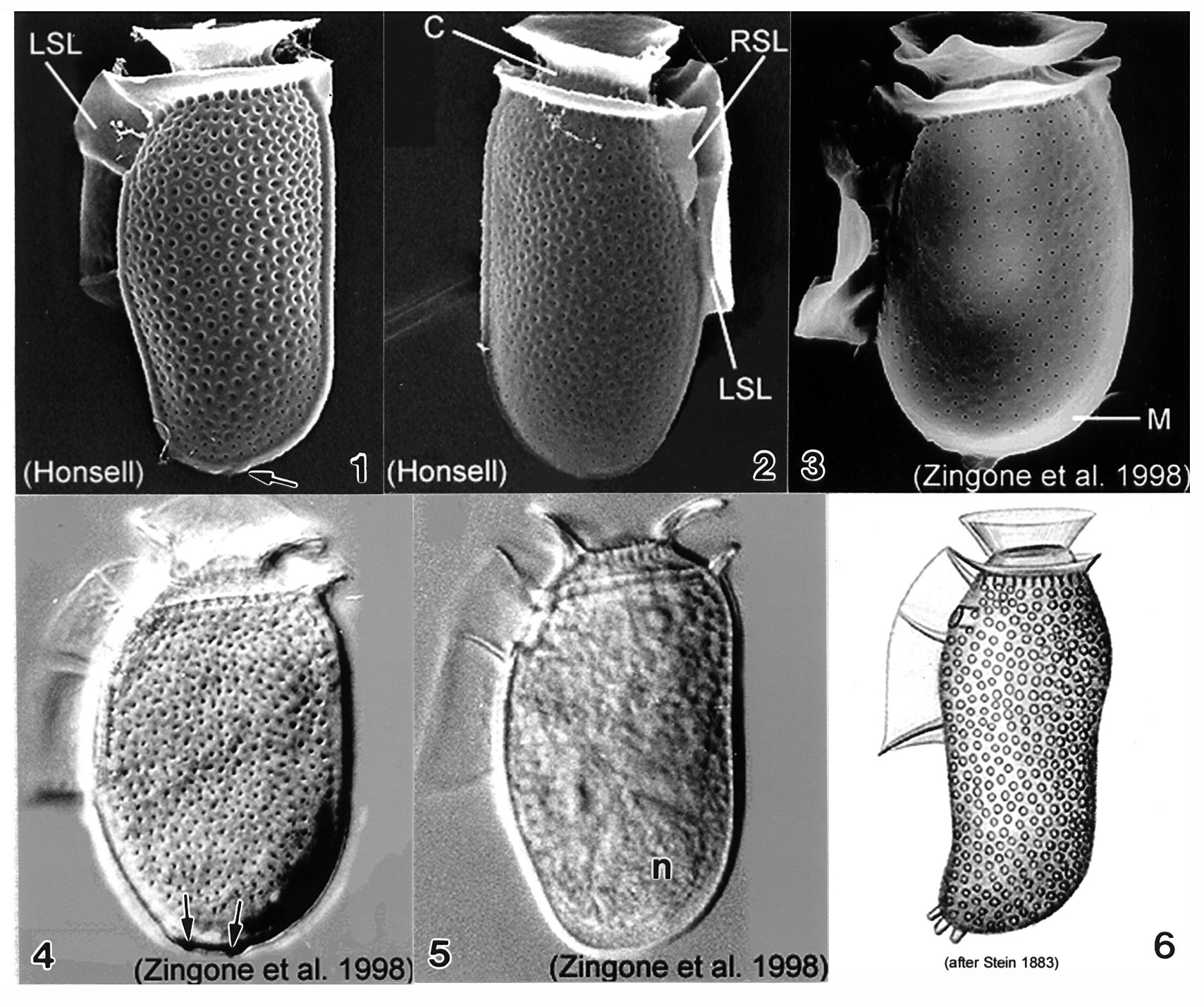
Plate 18. Dinophysis sacculus. Figs. 1-3. SEM: lateral view. Fig. 1. Cell oblong with rounded posterior. Hypotheca long, margins undulate. Thecal surface coarsely areolated. Short left sulcal list (LSL). Cingulum with two well developed lists. Small blunt posterior projections (arrow). Fig. 2. Cingulum lined with pores. Right sulcal list (RSL) visible. Fig. 3. Smooth thecal surface with pores. Metacytic zone (M) devoid of pores. Figs. 4-5. LM: lateral view. Fig. 4. Hypotheca sack-like with deep thecal pores. Posterior end with two blunt projections (arrows). Fig. 5. Large posterior nucleus (n). Fig. 6. Line drawing: morphotype from Stein (1883).
-
Plate 19. Dinophysis tripos. Fig. 1. SEM: lateral view. Cell large, oblong and heavily areolated. Hypothecal projections with toothed posterior ends (arrows). Left sulcal list (LSL) large, wide and reticulated. Figs. 2,3. LM: lateral view. Fig. 2. Anterior cingular list (ACL) projected anteriorly obscuring low epitheca (arrowheads). Narrow cingulum. Chloroplasts visible (arrows). Fig. 3. Paired cells. Hypothecal projection on dorsal margin sometimes seen with a narrow list (arrow) connecting two daughter cells during cell division. Fig. 4. Line drawing.
-
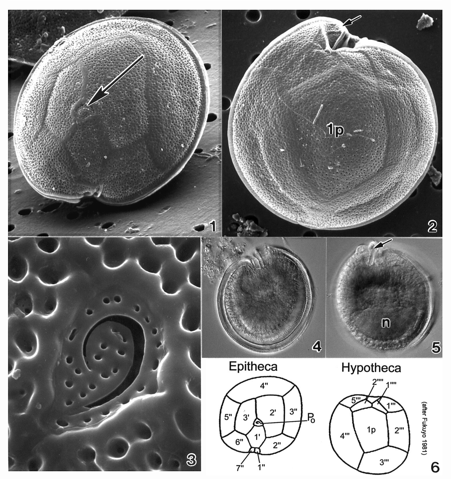
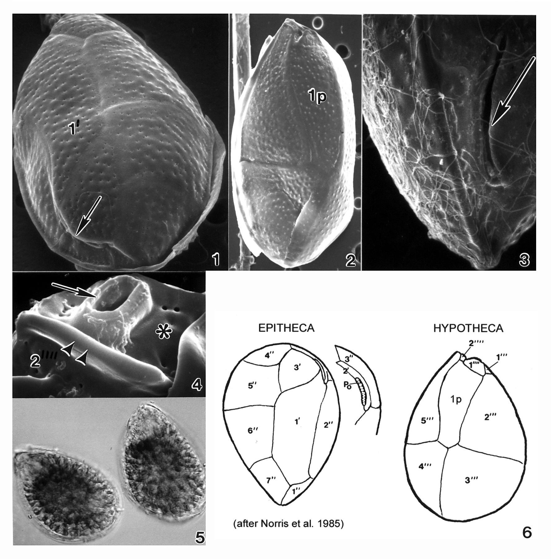
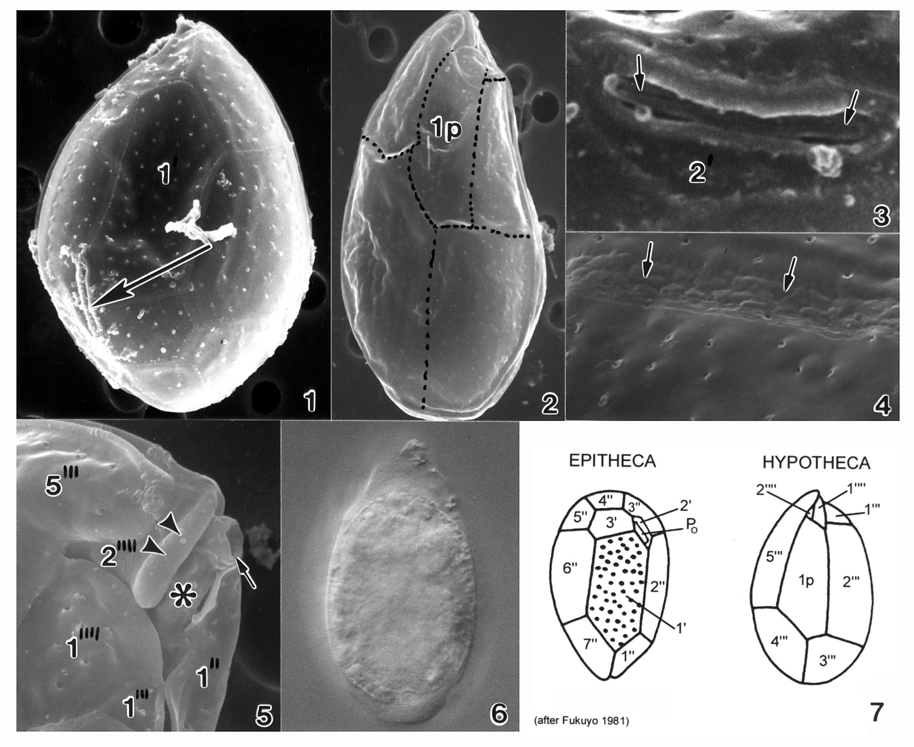
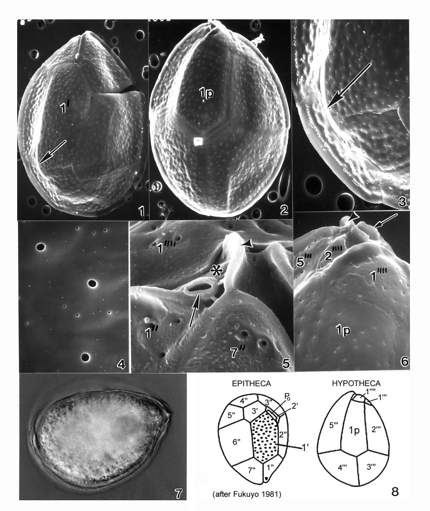
Plate 20. Gambierdiscus toxicus. Figs. 1-3. SEM. Fig. 1. Epitheca: cell round to ellipsoid; anterior-posteriorly compressed. Cell surface smooth with small scattered pores. Apical pore complex located at the apex (arrow). Fig. 2. Hypotheca: 1p plate large and pentagonal. Sulcal region deeply excavated (arrow). Fig. 3. Apical pore plate with characteristic fishhook shaped apical pore. Fig. 4. LM. Epitheca: cingulum and sulcal region in focus. Fig. 5. LM. Hypotheca: sulcal ridge (arrow); large nucleus (n). Fig. 6. Line drawing: thecal plate arrangement.
-
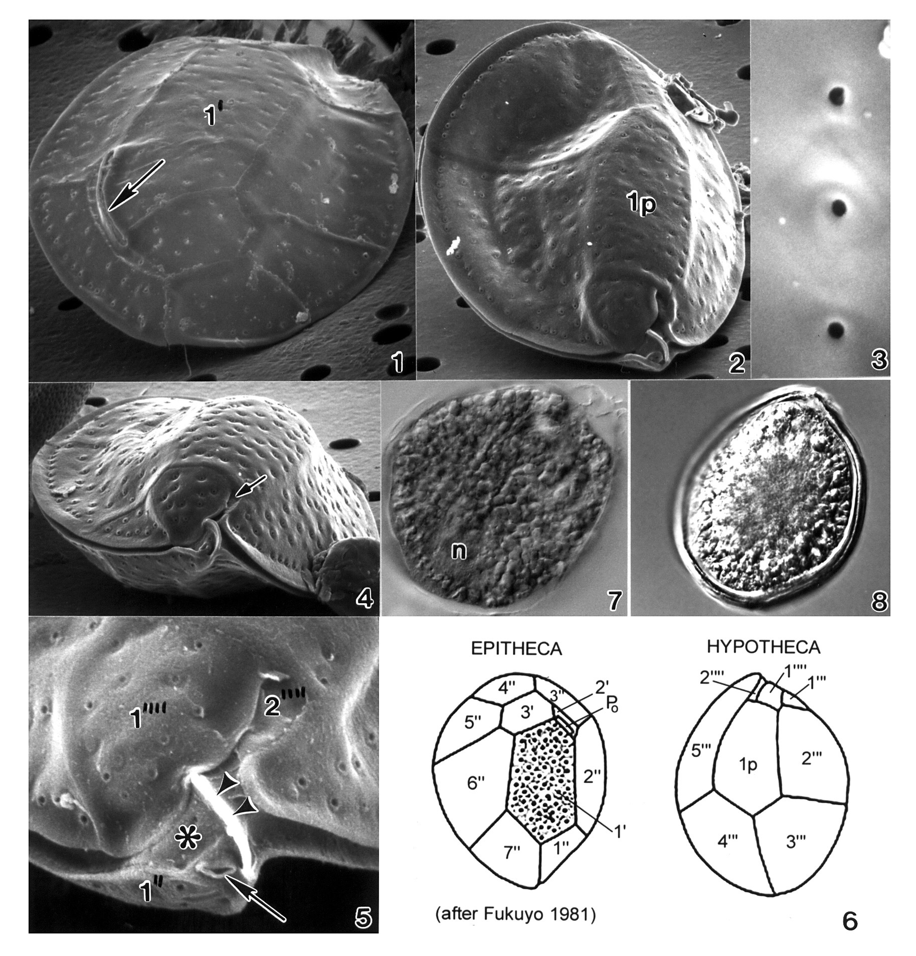
Plate 21. Gonyaulax polygramma. Figs. 1-3. SEM. Fig. 1. Ventral view: cell large, elongate and quadrilateral. Epitheca with prominent apical horn (arrow). Cingulum left-handed, displaced 1.5 X its width; sulcus widens posteriorly. Longitudinal ridges on thecal surface with reticulations in between. Fig. 2. Lateral ventral view: transverse (TF) and longitudinal (LF) flagella present. One antapical spine (arrow). Fig. 3. Dorsal view: hypotheca truncate with straight sides. Three antapical spines (arrows): one large and two small. Figs. 4-5. LM. Fig. 4. Ventral view: reticulations evident; one long antapical spine (arrow). Fig. 5. Dorsal view: prominent longitudinal ridges. Fig. 6. Line drawing.
-
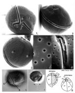
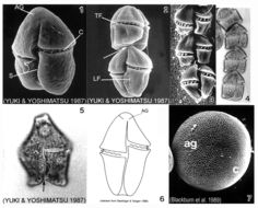
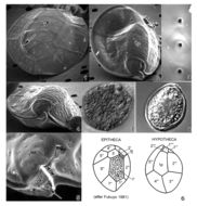
Plate 23. Gymnodinium catenatum. Figs. 1-3. SEM: ventral view. Fig. 1. Cell small, elongate-ovoid with slight dorso-ventral compression. Conical apex; rounded and notched antapex. Cingulum (C) excavated; sulcus (S) long. Distinctive horse-shoe shaped apical groove (AG) encircles apex. Fig. 2. Two cell chain; attachment point visible (arrow). Premedian cingulum displaced 2X its width. Longitudinal (LF) and transverse (TF) flagella visible. Fig. 3. Chain cells with anterior-posterior compression. Terminal cell slightly longer. Thecal surface rugose to smooth (Blackburn et al. 1989). Figs. 4-5. LM. Fig. 4. Chain-formation (Yuki and Yoshimatsu 1987). Fig. 5. Single cell. Conical epitheca with concave to flat apex. Bilobed hypotheca (arrow). Fig. 6. Line drawing. Fig. 7. SEM: cyst with microreticulations. ag=apical groove; c=cingulum
-
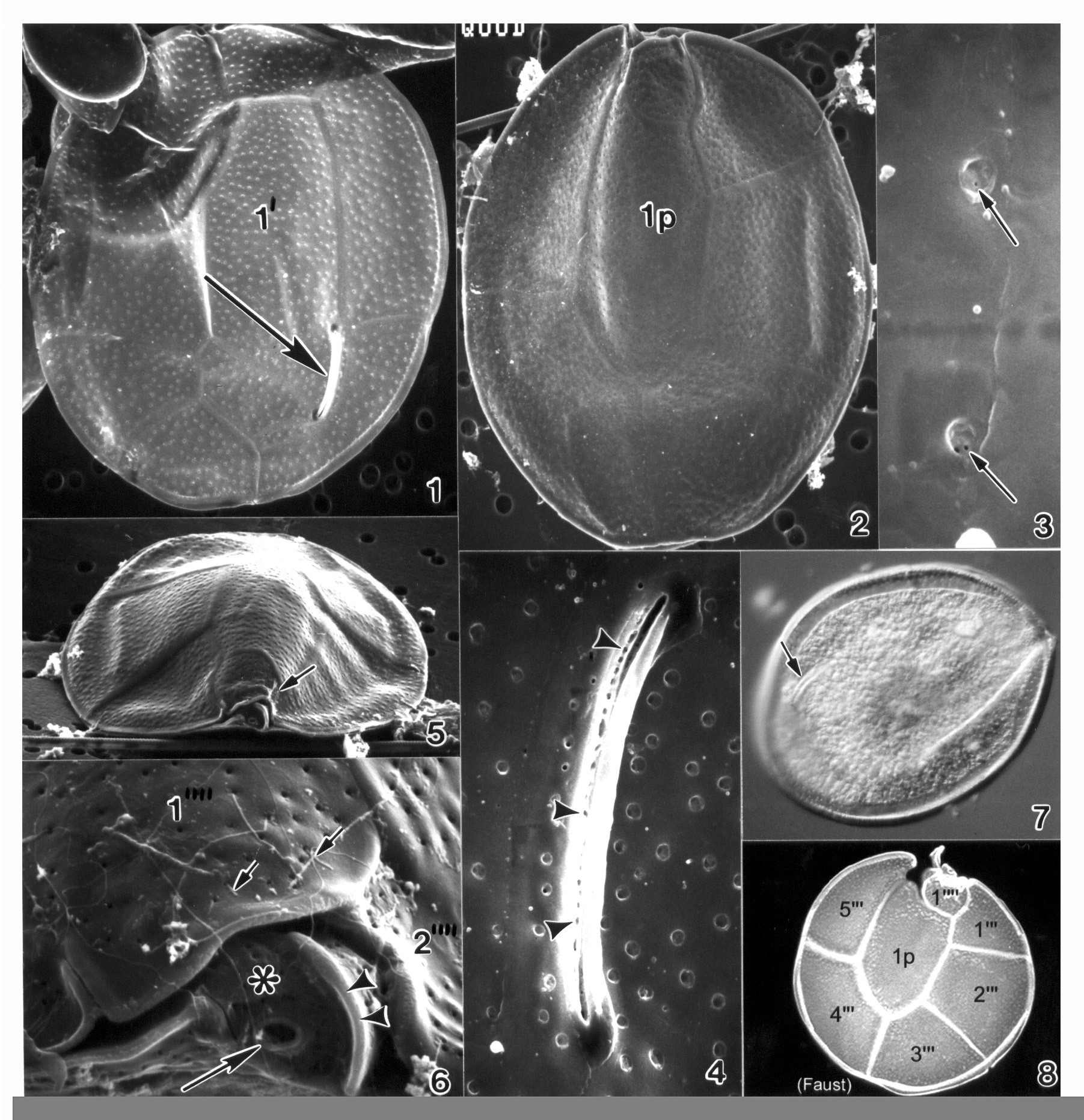
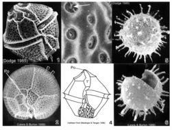
Plate 29. Lingulodinium polyedrum. Figs. 1-3. SEM. Fig. 1. Ventral view: cells angular and polyhedral-shaped. Thick plates well defined and coarsely areolate. Epitheca with shoulders and nearly flattened apex. Hypotheca with straight sides and flattened antapex (arrow). Cingulum deep and displaced 1-2 X its width. Sulcus widens posteriorly. Fig. 2. Apical view: first apical plate (1') long and narrow. Apical pore plate (Po) with raised inner elliptical ridge. Cingulum with lists (arrowheads). Strong ridges along sutures outline thecal plates. Fig. 3. Thecal areolae with large trichocysts (arrow)(Lewis and Burton 1988). Fig. 4. Line drawing. Figs. 5-6. SEM: resting cysts. Fig. 5. Cyst sperical with numerous tapering spines. Fig. 6. Cyst theca after excystment.
-
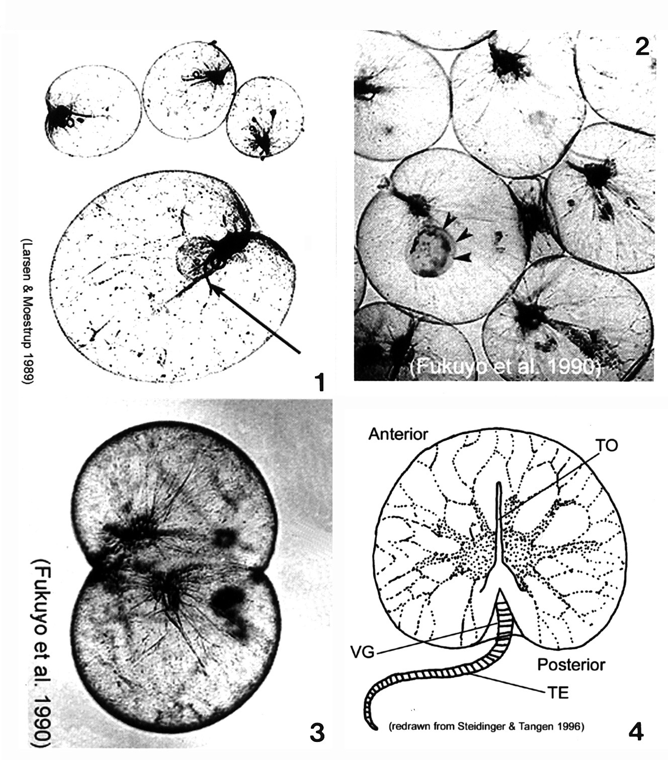
Plate 30. Noctiluca scintillans. Figs. 1-3. LM. Fig. 1. Cells large, balloon-shaped, nearly spherical, and colorless. A single flagellum housed in the ventral groove (arrow). Fig. 2. Cytoplasmic strands extend from nucleus (near the groove) to cell perifery. Engulfed cell (arrowheads). Fig. 3. Asexually dividing cell. Fig. 4. Line drawing. Deep and wide ventral groove (VG) houses the tooth (TO), an extension of the cell wall. Striated tentacle (TE).
-
Plate 31. Ostreopsis heptagona. Figs. 1-4. SEM. Fig. 1. Epithecal view: cells broadly oval, oblong and pointed. Long curved apical pore plate, Po, off-center (arrow). Plate 1' heptagonal and distinctive. Fig. 2. Hypothecal view: plate 1p pentagonal and dorso-ventrally elongate. Fig. 3. Po long, narrow and curved. Narrow mucilage strands cover cell surface. Fig. 4. Ventral view: location of ventral opening (arrow), ventral plate (asterisk), and rigid plate (asterisk) within cingulum. Fig. 5. LM. Two cells. Fig. 6. Line drawing: thecal plate arrangement.
-
Plate 32. Ostreopsis lenticularis. Figs. 1-5. SEM. Fig. 1. Epithecal view: cell lenticulate to broadly oval. Curved off-center apical pore plate with a slit-like apical pore (arrow). Plate 1' irregularly pentagonal. Fig. 2. Hypothecal view: plate 1p central and pentagonal. Fig. 3. Smooth thecal surface. Round pores with smooth raised edges. Fig. 4. Hypothecal ventral view: cell anterio-posteriorly compressed. Shallow cingulum with smooth edge. Small sulcus hidden (arrow). Fig. 5. Location of ventral opening (arrow), ventral plate (asterisk), and rigid plate (arrowheads) within cingulum. Fig. 6. Line drawing: thecal plate arrangement. Figs. 7,8. LM. Fig. 7. Cytoplasma granulated; posterior nucleus (n). Fig. 8. Distinct cingular list.
-
Plate 33. Ostreopsis mascarenensis. Figs. 1-5. SEM. Fig. 1. Epitheca: inner thecal surface. Cell very large, broadly ovate, large plates. Plate 1' elongate and hexagonal. Apical pore plate (Po) nearly straight. Fig. 2. Hypotheca: plate 1p long and wide. Fig. 3. Smooth cell surface with round pores; pores with two small openings (arrows). Fig. 4. Po with long narrow apical pore; small pores line the opening (arrowheads). Figs. 5-6. Ventral view of epitheca. Fig. 5. Cell compressed anterio-posteriorly; cingulum narrow with smooth edge. Small sulcus hidden (arrow). Fig. 6. Location of ventral opening (large arrow), ventral plate (asterisk), and rigid plate (arrowheads) within cingulum. Pores with ejected trichocysts (small arrows). Fig. 7. LM. Epitheca: Po (arrow) and cingulum in focus. Fig. 8. Line drawing: hypotheca plate arrangement.
-
Plate 34. Ostreopsis ovata. Figs. 1-5. SEM. Fig. 1. Epithecal view: cell slender and tear-shaped. Apical pore plate (Po) off-center (arrow). Plate 1' large and hexagonal. Cingulum wide with narrow lists. Fig. 2. Hypothecal view: plates delicate. Plate 1p long and narrow. Fig. 3. Po: short and straight, adjacent to plate 2'. Fig. 4. Thecal surface smooth with scattered small pores. Suture line uneven and bumpy (arrows). Fig. 5. Hypothecal view: ventral opening (arrow), ventral plate (asterisk), and rigid plate (arrowhead) on cingulum. Fig. 6. LM. Large posterior nucleus. Fig. 7. Line drawing: thecal plate arrangement.
-
Plate 35. Ostreopsis siamensis. Figs. 1-6. SEM. Fig. 1. Epithecal view: cell broad and tear-shaped. Thecal surface smooth with scattered pores. Apical pore plate (Po) off-center (arrow). Narrow cingulum with smooth edge. Plate 1' narrow and pentagonal. Fig. 2. Hypothecal view: plate 1p long and pentagonal. Fig. 3. Po: long, curved and narrow. Fig. 4. Large and small pores on thecal surface. Fig. 5. Ventral view: location of ventral opening (arrow), ventral plate (asterisk), and rigid plate (arrowhead) on cingulum. Fig. 6. Hypothecal view: Vo (arrow) and Rp (arrowhead). Fig. 7. LM. Hypotheca. Fig. 8. Line drawing: thecal plate arrangement.