Plate 20
Description:
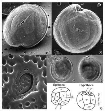
Plate 20. Gambierdiscus toxicus. Figs. 1-3. SEM. Fig. 1. Epitheca: cell round to ellipsoid; anterior-posteriorly compressed. Cell surface smooth with small scattered pores. Apical pore complex located at the apex (arrow). Fig. 2. Hypotheca: 1p plate large and pentagonal. Sulcal region deeply excavated (arrow). Fig. 3. Apical pore plate with characteristic fishhook shaped apical pore. Fig. 4. LM. Epitheca: cingulum and sulcal region in focus. Fig. 5. LM. Hypotheca: sulcal ridge (arrow); large nucleus (n). Fig. 6. Line drawing: thecal plate arrangement.
Included On The Following Pages:
- Life (creatures)
- Cellular (cellular organisms)
- Eukaryota (eukaryotes)
- SAR (Stramenopiles, Alveolates, Rhizaria)
- Alveolata (alveolates)
- Dinophyceae
- Gonyaulacales
- Goniodomataceae
- Gambierdiscus
- Gambierdiscus toxicus
- Dinoflagellata (dinoflagellates)
This image is not featured in any collections.
Source Information
- license
- cc-publicdomain
- bibliographic citation
- Faust, Maria A. and Rose A. Gulledge. Identifying Harmful Marine Dinoflagellates. Smithsonian Contributions from the United States National Herbarium, volume 42: 1-144 (including 48 plates, 1 figure and 1 table).
- original
- original media file
- visit source
- partner site
- NMNH Marine Dinoflagellates
- ID