-
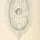
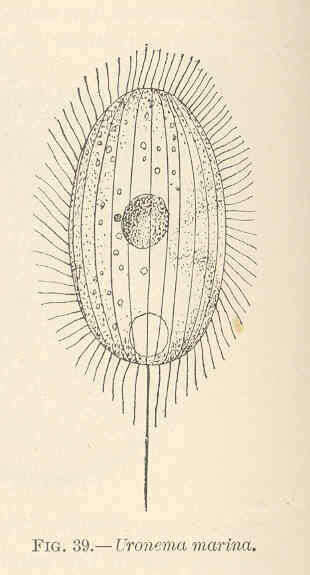
Uronema marina.
-
-
-
-
-
-
-
-
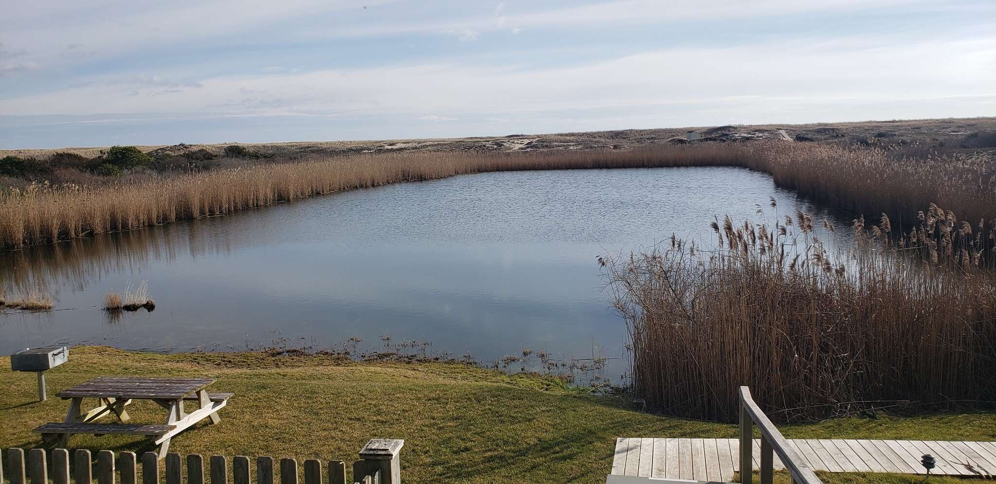
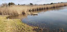
Peniscola, Valencia, Spain
-
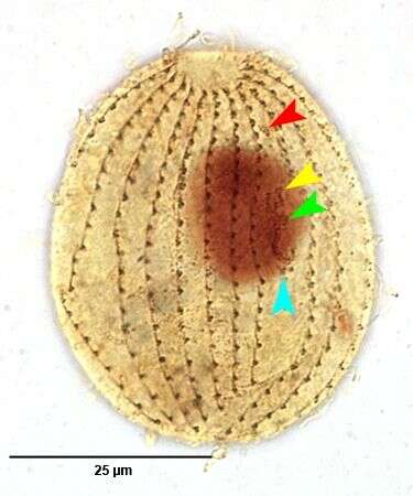
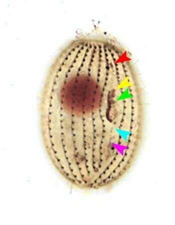
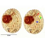
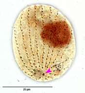
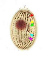
Infraciliature of the common marine scuticolciliate, Uronema marinum (DUJARDIN,1841). The red,yellow and green arrowheads indicate the 1st,2nd and 3rd adoral membranelle respectively. the blue arrowhead indicates the right paraoral membrane. Collected from effluent of the protein skimmer of a commercial marine aquarium in Boise, Idaho. January 2007.Stained by a silver carbonate technique for marine ciliates. (Ma,H. et al.An Improved Silver Carbonate Impregnation for Marine Ciliated Protozoa.Acta Protozool. 42: 161 â 164;2003).Brightfield.
-
Infraciliature of the common marine scuticolciliate, Uronema marinum (DUJARDIN,1841). The red,yellow and green arrowheads indicate the 1st,2nd and 3rd adoral membranelle respectively. The light blue arrowhead indicates the scutica. Collected from effluent of the protein skimmer of a commercial marine aquarium in Boise, Idaho. January 2007.Stained by a silver carbonate technique for marine ciliates. (Ma,H. et al.An Improved Silver Carbonate Impregnation for Marine Ciliated Protozoa.Acta Protozool. 42: 161 â 164;2003).Brightfield.
-
Infraciliature of the common marine scuticolciliate, Uronema marinum (DUJARDIN,1841). The pink arrowhead indicates the kinetosome of the long posterior cilium. Collected from effluent of the protein skimmer of a commercial marine aquarium in Boise, Idaho. January 2007.Stained by a silver carbonate technique for marine ciliates. (Ma,H. et al.An Improved Silver Carbonate Impregnation for Marine Ciliated Protozoa.Acta Protozool. 42: 161 â 164;2003).Brightfield.
-
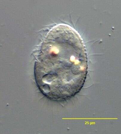
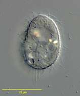
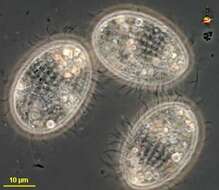
Dorsal surface view of the common marine scuticolciliate, Uronema marinum (DUJARDIN,1841). Collected from effluent of the protein skimmer of a commercial marine aquarium in Boise, Idaho. January 2007. DIC.
-
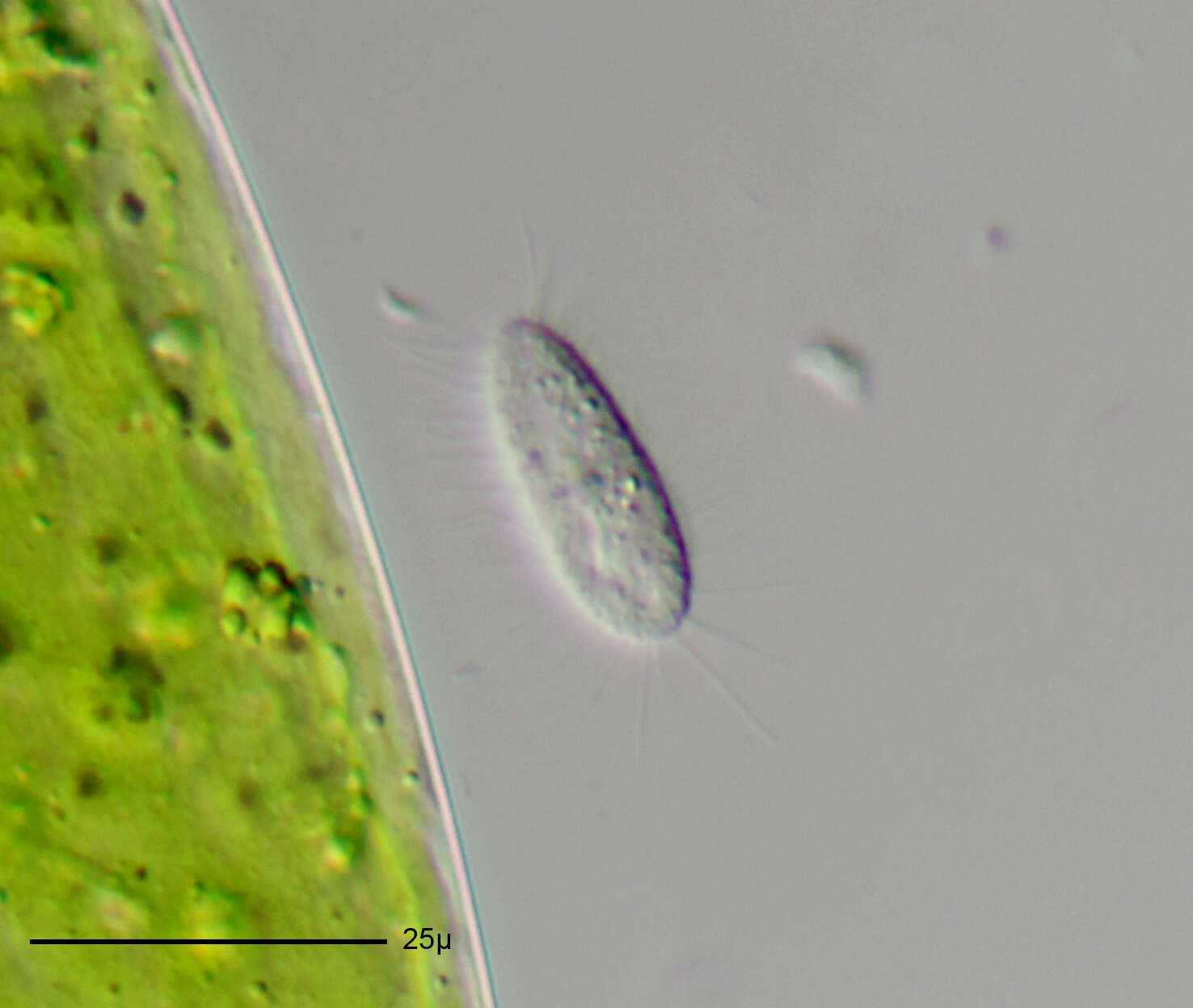
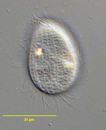
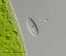
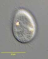
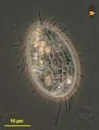
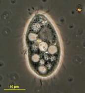
In vivo portrait of the common marine scuticolciliate, Uronema marinum (DUJARDIN,1841). Collected from effluent of the protein skimmer of a commercial marine aquarium in Boise, Idaho. January 2007. DIC.
-
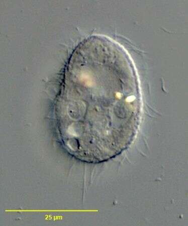
Infraciliature of the common marine scuticolciliate, Uronema marinum (DUJARDIN,1841). Collected from effluent of the protein skimmer of a commercial marine aquarium in Boise, Idaho. January 2007. DIC.
-
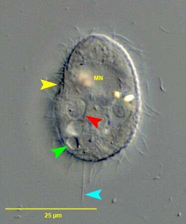
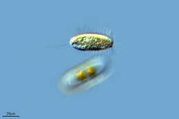
Portrait of the common marine scuticociliate, Uronema marinum (DUJARDIN,1841). The large spherical macronucleus (MN) is just anterior to the cell center. The oral apperatus is indicated by the yellow arrowhead. The small subterminal contractile vacuole is indicated by the green arrowhead. A food vacuole is indicated by the red arrowhead. The long terminal cilium is indicated by the light blue arrowhead. Collected from effluent of the protein skimmer of a commercial marine aquarium in Boise, Idaho. January 2007. DIC.
-
Infraciliature of the common marine scuticolciliate, Uronema marinum (DUJARDIN,1841). The red,yellow and green arrowheads indicate the 1st,2nd and 3rd adoral membranelle respectively. The light blue arrowhead indicates the unciliated kinetosomes of the scutica. The pink arrowhead indicates the linear cytopyge posterior to the scutica. Collected from effluent of the protein skimmer of a commercial marine aquarium in Boise, Idaho. January 2007.Stained by a silver carbonate technique for marine ciliates. (Ma,H. et al.An Improved Silver Carbonate Impregnation for Marine Ciliated Protozoa.Acta Protozool. 42: 161 â 164;2003).Brightfield.
-
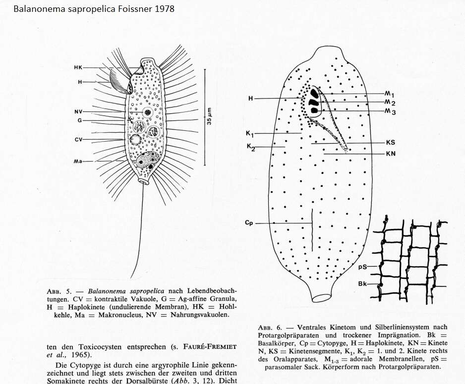
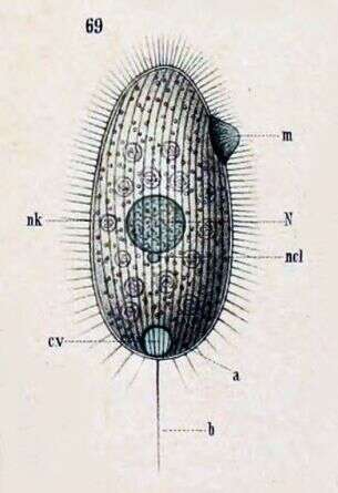
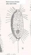
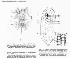
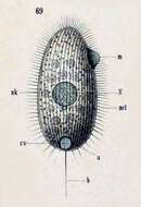
(Synonym: Uronema marinum) Key to Schewiakoff's abbreviations a--Anus b--Sensory bristle cv--Contractile vacuole m--Undulating membrane N--Macronucleus ncl--Micronucleus nk--Food particle
-
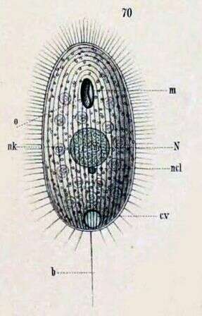
(Synonym: Uronema marina) Ventral view. Key to Schewiakoff's abbreviations b--Sensory bristle cv--Contractile vacuole m--Undulating membrane N--Macronucleus ncl--Micronucleus nk--Food particle o -- Mouth
-
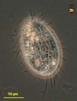
Urocyclon (your-owe-sigh-clone) a small scuticociliate, mouth small and located about mid-way down the body. Phase contrast micrograph.
-
Urocyclon (your-owe-sigh-clone) a small scuticociliate, mouth small and located about mid-way down the body. These cells are in focus on the surface, and elements of the infraciliature - the skeletal network associated with the bases of the cilia) are evident. Phase contrast micrograph.
-
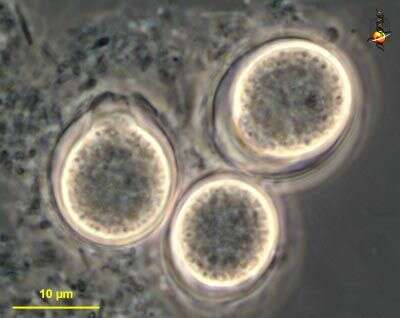
Homalogastra (hom-al-owe-gast-ra) small bacterivorous ciliate. This image shows cysts. Phase contrast micrograph.
-
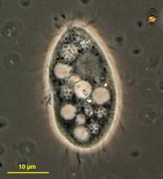
Homalogastra (hom-al-owe-gast-ra) small bacterivorous ciliate. The lighter vacuoles are food vacuoles, the grey area anterior right is the macronucleus. Phase contrast micrograph.
-
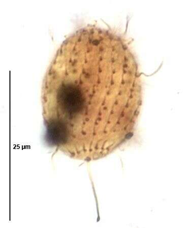
Dorsal infraciliature of the scuticociliate, Homalogastra setosa (KAHL,1926).From non-flooded petri dish culture of topsoil from a public park in Boise,Idaho. Stained by the silver carbonate technique (see Foissner, W.Europ. J. Protistol.27:313-330;1991).Brightfield.