-
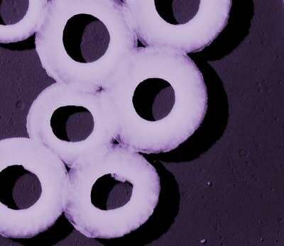
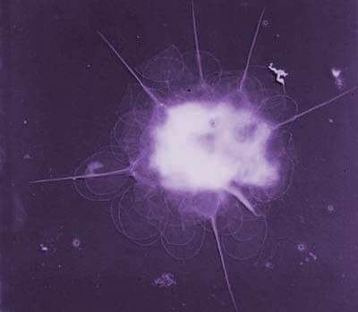
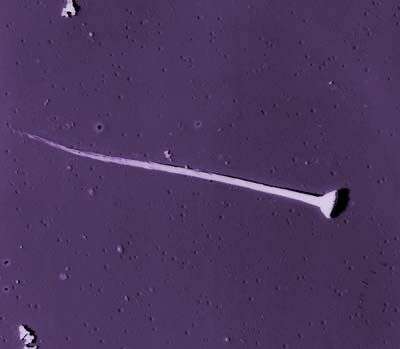
This image was made from samples taken during a scientific cruise in the Pacific. Water was filtered to concentrate the organisms that were present, then dried onto a thin sheet of plastic and then shadowed with a fine layer of metal to provide contrast. The preparation was then observed with an electron-microscope. This technique has been used to document the diversity of marine microbes, especially, protists in the oceans.
-
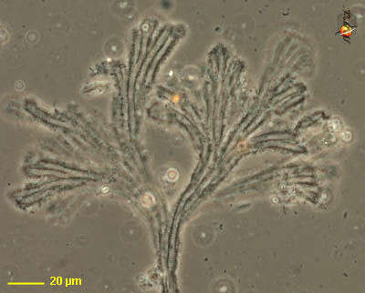
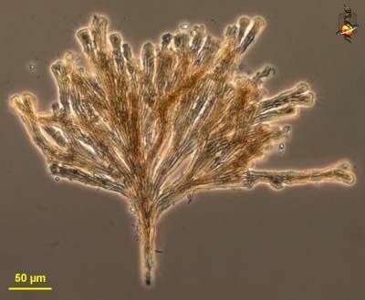
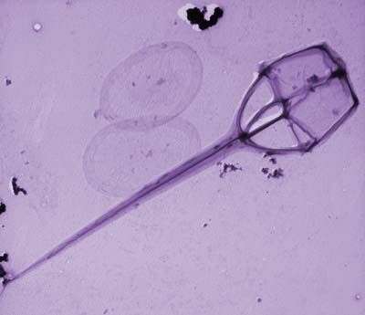
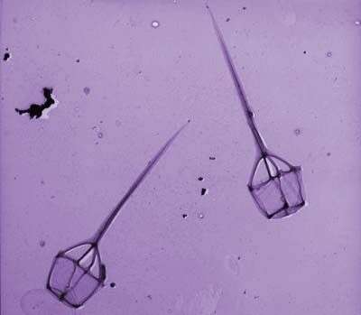
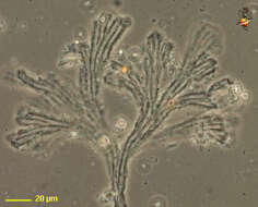
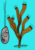
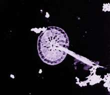
Rhipidodendron (rip-ee-doe-dend-ron, is a colonial spongomonad flagellate, in which the cells are located at the end of a branching (aborescent) colony. The matrix of the colony is formed from adhering small globules of mucilage. The branches are flat, with several channels in each blade. One cell is located at the end of each channel (many of the cells were dislodged from this preparation). Phase contrast.
-
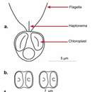
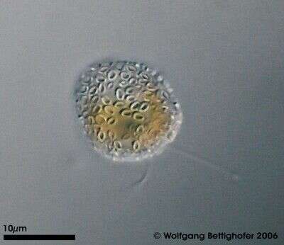
Chrysochromulina (cry-so-crumb-you-line-a) ericina a single-celled haptophyte, with two similar flagella, long anterior haptonema and a golden colour from two yellow-brown chloroplasts. Small scales lie on the surface of the cell but these are not evident in this image. Phase contrast microscopy.
data on this strain.
-
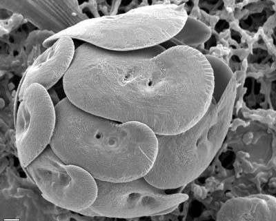
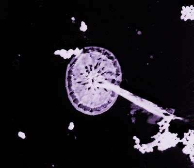
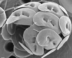
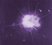
This image was made from samples taken during a scientific cruise in the Pacific. Water was filtered to concentrate the organisms that were present, then dried onto a thin sheet of plastic and then shadowed with a fine layer of metal to provide contrast. The preparation was then observed with an electron-microscope. This technique has been used to document the diversity of marine microbes, especially, protists in the oceans. According to Jeremy Young, this is a fragment of a coccosphere of Umbilicosphaera sibogae.
-
Rhipidodendron (rip-ee-doe-dend-ron, is a colonial spongomonad flagellate, in which the cells are located at the end of a branching (aborescent) colony. The matrix of the colony is formed from adhering small globules of mucilage. The branches are flat, with several channels in each blade. One cell is located at the end of each channel (many of the cells were dislodged from this preparation). Phase contrast.
-
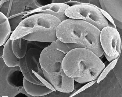
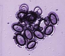
SEM of coccosphere. Flagellar opening is toward top right. Note considerable variation in coccolith size and wing development
-
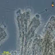
Rhipidodendron (rip-ee-doe-dend-ron, is a colonial spongomonad flagellate, in which the cells are located at the end of a branching (aborescent) colony. The matrix of the colony is formed from adhering small globules of mucilage. The branches are flat, with several channels in each blade. One cell is located at the end of each channel and the cells have two flagella. Phase contrast.
-
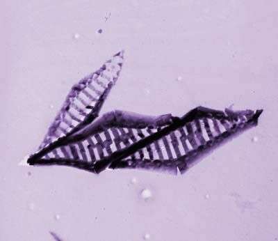
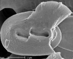
Broken coccolith of H. wallichii in distal view, breakage shows the proximal shield.
-
-
Helicosphaera wallichii (Lohmann 1902) Okada & McIntyre 1977 [Coccolithophora] Like H. carteri but: central-area with oblique twisted slits; bridge typically better developed; and liths perhaps slightly larger. NB Slits obliquity: In distal view the slits are rotated about 10-20° clockwise (and so away from the wing), this is the ânormalâ sense of obliquity in Helicosphaera, as shown by many fossil species. The Pleistocene species H. inversa is similar but shows the opposite sense of obliquity. HOL phase - unknown but H. wallichii often co-occurs with Syracolithus dalmaticus in our samples (Geisen et al., 2004).
-
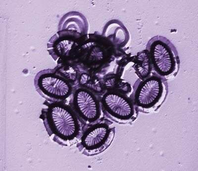
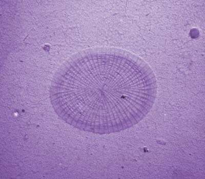
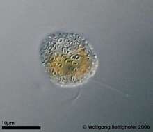
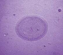
This tiny prymnesiomonad belongs to group of Coccolithophoraceae. In a mucous cover they wear lots of calcified scales (coccoliths). The depth of focus picture shows coccoliths covering the monad cell. Picture generated from 7 shots using CombineZ by Alan Hadley. For details see ZIP archive. Collected from littoral region (stand of Phragmites) of a rain storage reservoir in Kiel (Schleswig-Holstein, Germany). Images were taken using Zeiss Universal with Olympus C7070 CCD camera.
-
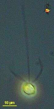
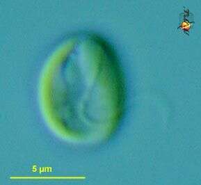
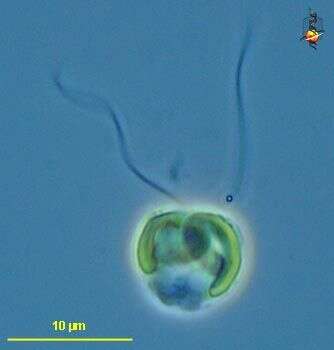
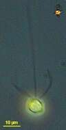
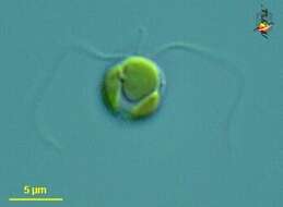
Pavlova (pav-low-va) small atypical haptophyte (prymnesiophyte, pavlovophyte) alga, two plastids, two smooth flagella. Differential interference microscopy.
data on this strain.
-
Pavlova (pav-low-va) small atypical haptophyte (prymnesiophyte, pavlovophyte) alga, two plastids, two smooth flagella. Differential interference microscopy.
data on this strain.
-
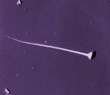
This image was made from samples taken during a scientific cruise in the Pacific. Water was filtered to concentrate the organisms that were present, then dried onto a thin sheet of plastic and then shadowed with a fine layer of metal to provide contrast. The preparation was then observed with an electron-microscope. This technique has been used to document the diversity of marine microbes, especially, protists in the oceans.
-
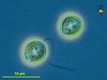
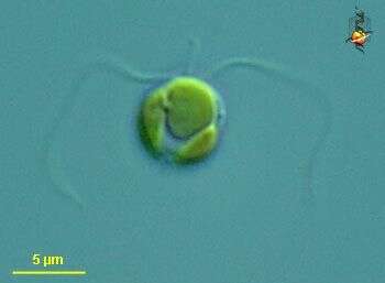
Chrysochromulina (cry-so-crumb-you-line-a) a single-celled haptophyte, with two similar flagella, a short haptonema lying between the flagella, and golden plastids. This may be C. herdlensis. Differential interference microscopy.
data on this strain.
-
Chrysochromulina (cry-so-crumb-you-line-a) a single-celled haptophyte, with two similar flagella, a short haptonema lying between the flagella, and two golden plastids. This may be C. herdlensis. Differential interference microscopy.
data on this strain.
-
This image was made from samples taken during a scientific cruise in the Pacific. Water was filtered to concentrate the organisms that were present, then dried onto a thin sheet of plastic and then shadowed with a fine layer of metal to provide contrast. The preparation was then observed with an electron-microscope. This technique has been used to document the diversity of marine microbes, especially, protists in the oceans.
-
This image was made from samples taken during a scientific cruise in the Pacific. Water was filtered to concentrate the organisms that were present, then dried onto a thin sheet of plastic and then shadowed with a fine layer of metal to provide contrast. The preparation was then observed with an electron-microscope. This technique has been used to document the diversity of marine microbes, especially, protists in the oceans.