Description
provided by Zookeys
Body (Figure 5A) 3.44 long, composed of elongate, slender prosome with 3 pairs of long lateral processes and small 2-segmented urosome. Prosome (Figure 5A, B) composed of anterior region, cephalosome, middle region comprising first to second pedigerous somites, and posterior region as third pedigerous somite. Cephalosome (Figure 5B, F) elongate, bent ventrally, with projecting rostral area. Middle region (Figure 5A, B) large, constricted posterior to base of anterior lateral processes with paired and single dorsal protrusions. Posterior region (Figure 5A–C) broad, without armature. Lateral processes (Figure 5A) long and slender, distinctly longer than body length. Urosome (Figure 5C, D) small; genito-abdomen narrower posteriorly with paired posterolateral lobes; unarmed opercula and genital aperture located on ventral. Caudal rami (Figure 5E) small, about twice as long as wide, bearing 6 setae and 1 dorsal spiniform process; apical seta long, styliform.
Antennule (Figures 5F, 6A) 2-segmented; terminal segment bearing 2 constrictions making it appearing as original segmentation; proximal segment bearing 2 blunt spines; terminal segment bearing 2 blunt spines and 1 seta in proximal part, 3 setae and 1 aesthetasc in middle part, and 9 setae and 2 aesthetascs in distal part. Antenna (Figures 5F, 6B) 3-segmented; coxo-basis broad, bearing 1 inner spine with spiniform tip; proximal segment of endopod bearing 1 inner spine; terminal segment of endopod tapering into strong apical claw, with 2 spines and 2 setal elements. Labrum (Figure 6B) bilobate, bearing flat surface. Mandible (Figure 6C) spatulate, tapering into single curved blade with 2 dentiform processes giving trifid appearance. Labium (Figure 6B) developed with paired spinulose patches. Maxillule not observed. Paragnath (Figure 6B) developed, represented by pinnate lobe. Maxilla (Figure 6B, D) 2-segmented; syncoxa unarmed; allobasis tapering into spiniform element, with seta. Maxilliped absent.
Leg 1(Figures 5B, 6E) unsegmented, weakly sclerotized and drawn out into elongate exopod and small endopod; protopod bearing outer basal seta; exopod drawn out into spiniform lobe bearing multiple constrictions, wrinkly surface, 3 outer and 1 inner setal elements; endopod a small lobe tipped with seta. Leg 2 (Figures 5B, 6F) unsegmented, weakly sclerotized; protopod drawn out into long exopod and small, cylindrical endopod; protopod bearing outer basal seta; exopod tapering into a pointed process with three outer and 1 inner small element; endopodal lobe bearing small apical seta. Leg 3 (Figures 5C, 6G) represented by conical process with apical seta, located near posterolateral corner on ventral side of prosome.
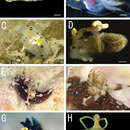
Egg sacs (Figure 5A) bilobate, bearing curved side and swollen side; color in life cream (Figure 1C, D).
Variation of female morphology. The morphology of female paratypes is as in the holotype. The specimens from type series (n = 3) range from 2.81–4.47 (3.57 ± 0.83) BL.
- license
- cc-by-3.0
- copyright
- Daisuke Uyeno, Kazuya Nagasawa
- bibliographic citation
- Uyeno D, Nagasawa K (2012) Four new species of splanchnotrophid copepods (Poecilostomatoida) parasitic on doridacean nudibranchs (Gastropoda, Opistobranchia) from Japan, with proposition of one new genus ZooKeys 247: 1–29
- author
- Daisuke Uyeno
- author
- Kazuya Nagasawa