-
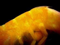
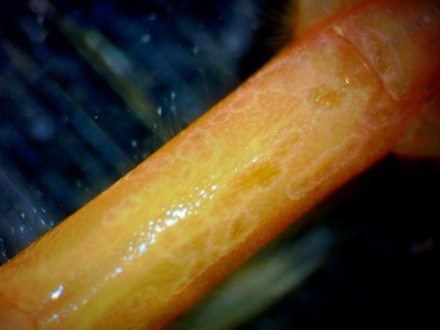
This photograph of the left side of the shrimp shows pereopods 3-5 (#5 is to the right). The endopods are robust and have reddish setae. The ventral edge of the carapace is visible at the top of the photo. The epipod on pereopod 4 can be seen must below the edge of the carapace, branching from the coxa. The exopods, which look like long flexible yellowish extensions, can be seen on pereopods 3-5, branching from the basis. Note that the exopods are of similar length to each other.
-
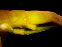
A side view of abdominal segments 1-3. Note that segment 2 has no posterior dorsal spine, while segment 3 is dorsally carinate and ends with a long medial dorsal spine. This view is of the left side of the shrimp, anterior is to the left.
-
A side view of abdominal segments 4-6 (4 is to the left). Note the small posterior medial dorsal spines on segments 4,5 and the small spines lateral to them. Note also the tooth on the posterior margin of the pleuron of segment 5. Anterior is to the left.
-
This dorsal view of abdominal segments 5-6 shows the small posterior medial dorsal spine on segment 5 and the small spines lateral to it. Anterior is to the left.
-
This lateral closeup view of abdominal pleuron 5 shows the sharp tooth along the posterior margin. Anterior is to the left.
-
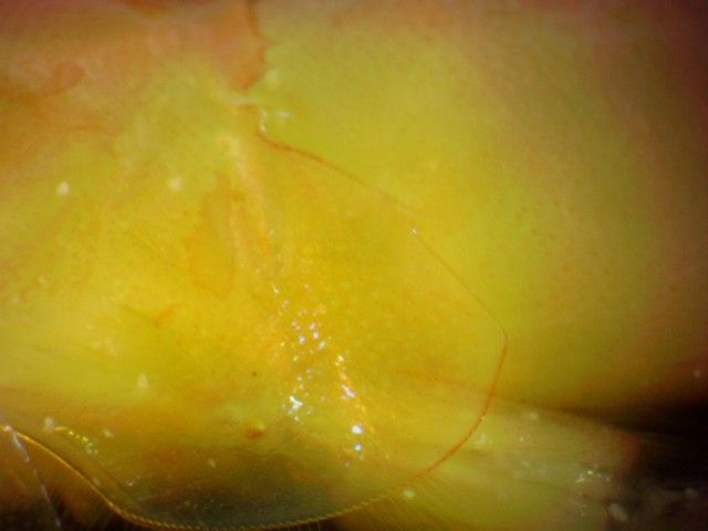
This dorsal view of abdominal segment 6 shows that it rounded and has no dorsal carina (ridge). Anterior is to the left and the base of the uropods and telson are on the upper right.
-
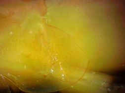
This dorsal view of the pleopods and telson show that the telson has a shallow dorsal groove (sulcus). It also has a spiny endpiece flanked by two spines, and two dorsolateral rows of 4-8 spines.
-
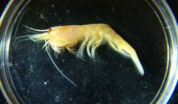
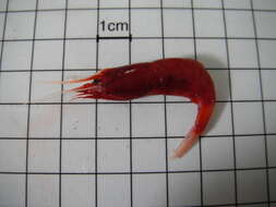
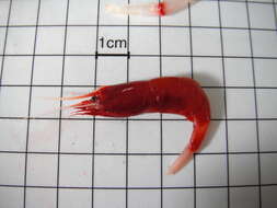
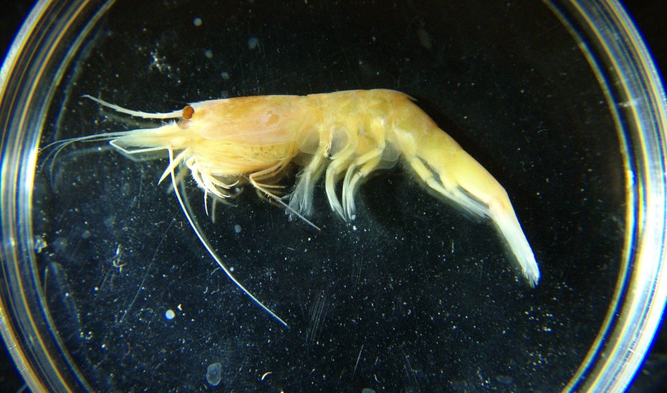
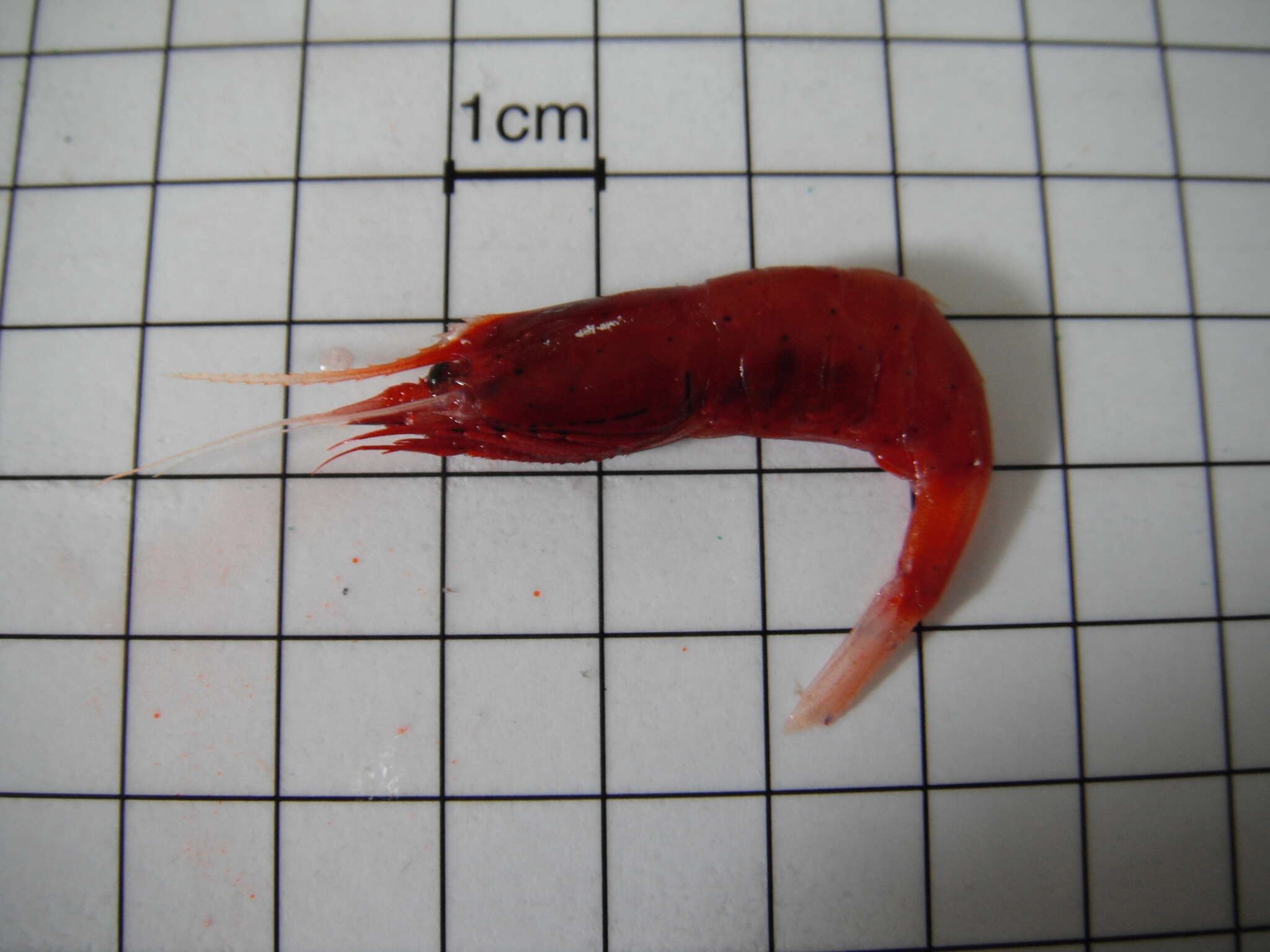
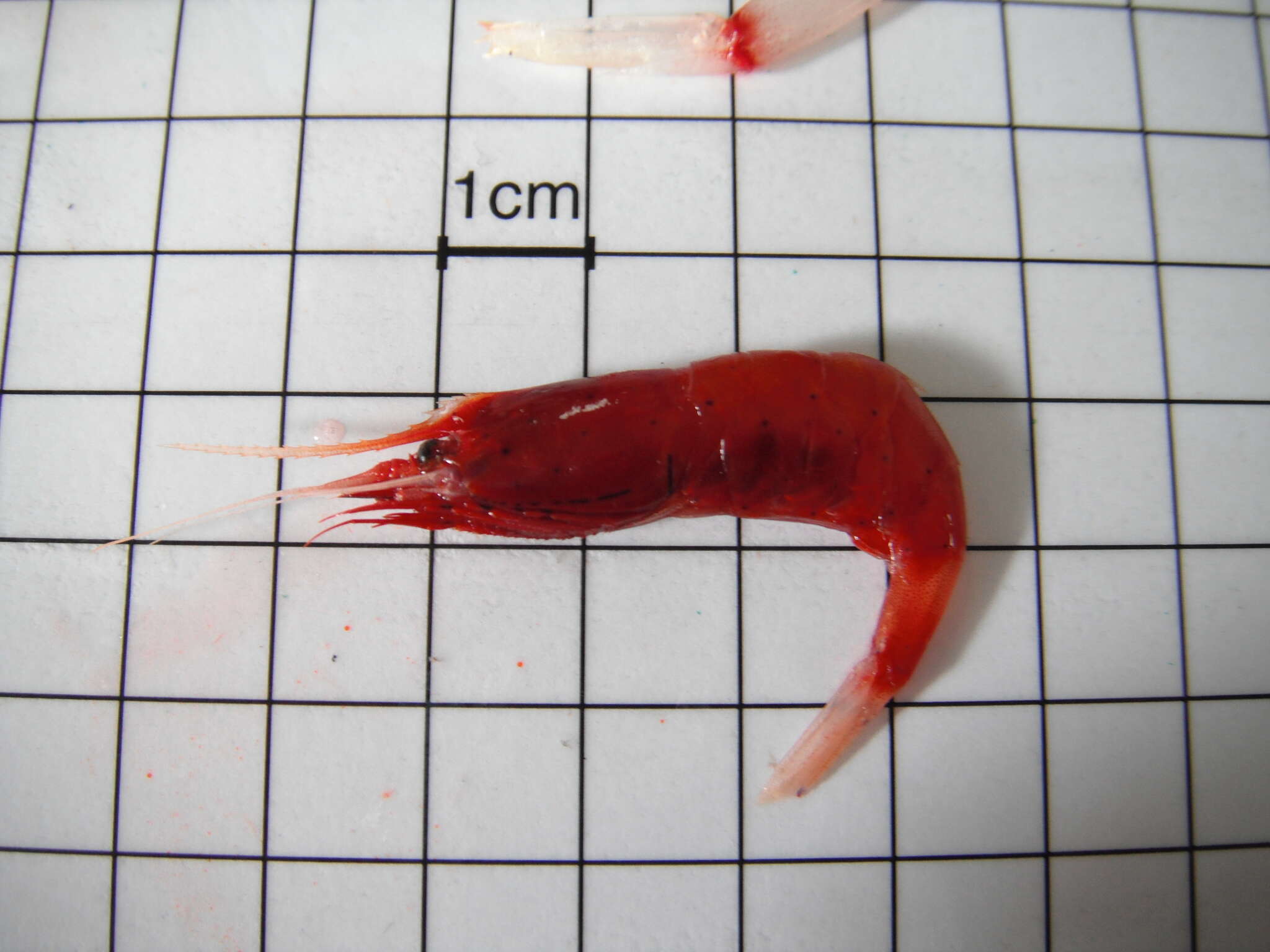
Systellaspis debilis, preserved specimen captured in midwater off Oahu, Hawaii in 1996. Compare petri dish for size. In life the shrimp would be bright red. (Photo by: Dave Cowles)
-
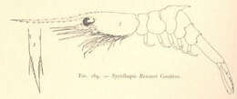
Systellaspis Bouvieri Coutiere.
-
-