“Spikebuccinum stephaniae new species
(Figures 1-23, Table 1)
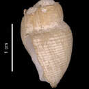
Description: Shell (Figures 1-3, 5-7, 9, 10) small (to 19.9 mm), very thin, translucent, ovate, with rounded anterior, eroded spire. Protoconch (Figure 11), known from a single juvenile (Paratype 12), increasing in diameter from 0.4 mm to 3.5 mm, in 2 ⅔ smooth, evenly rounded, pitted whorls. Transition to teleoconch distinguished by slight change in color, from cream to white, and by abrupt transition from coarse, irregular axial growth striae, to finer, regular growth lines. Protoconch and upper whorls eroded on all other specimens. Extrapolation from growth series suggests that teleoconch may reach 5-6 whorls, of which all but last 2-2 ½ whorls eroded. Whorls evenly rounded, with indistinct shoulder, abutting suture. Axial sculpture limited to very fine, straight, strongly prosocline growth lines. Spiral sculpture of fine, sharp, uniform, evenly spaced cords (21-29 on final whorl, 11-14 on penultimate whorl). Aperture large (AL/SL = 0.60-0.67 when using length of eroded shell; AL/SL = 0.50-0.55, as estimated by linear projections of apex), broadly oval, deflected from shell axis by 22-25°. Outer lip very thin, not reflected, evenly rounded from suture to siphonal notch. Inner lip consists of a long, straight parietal region that meets the shorter, concavely indented axial portion of columella, ending in strong siphonal fold. Columella shorter than aperture, giving rise to a broad siphonal notch. Parietal callus uniformly narrow from suture to siphonal fold. Short, weak, siphonal fasciole and pseudoumbilicus present, often obscured by erosion. Shell color uniformly white. Periostracum very thin, straw yellow in color, with densely spaced axial lamellae, producing short, fine hairs at intersection with spiral cords, giving shell a finely hirsute appearance. Operculum (Figures 4, 8) small, spanning ~0.36 AL, yellowish brown, broadly ovate, paucispiral, with subterminal nucleus rotated relative to opercular axis.
Anatomy (Holotype): Soft tissues (Figures 14-22) comprise approximately 2 ¼ whorls. Mantle cavity spans just under ⅓ whorl, kidney ¼ whorl, digestive gland and gonad just under 1 ½ whorl. Columellar muscle short, broad, comprising slightly more than one whorl, attached to shell at rear of mantle cavity. Foot large, broadly rectangular (L/W ≈ 1). Body color yellowish tan, without pigmentation. Head large with long, thin tapering tentacles (Figure 15, tn), without neck. Eyes absent. Nephridium with semi-transparent walls that clearly reveal folds. Nephridial gland (Figure 14, ng) small, very narrow. Pericardium oriented antero-ventrally. Digestive gland lobes (Figure 19, adg, pdg) not fused, separated by ovary (Figure 19, ov).
Mantle Cavity (Figure 20): Mantle cavity of medium width (L/W ~0.8), mantle edge slightly serrated. Siphon long (0.42 AL), free, muscular, extending substantially beyond mantle edge. Osphradium (Figure 20, os) small (~0.4 mantle cavity length) yellowish, bipectinate, with narrow osphradial nerve. Ctenidium (Figure 20, et) large, very wide, spans about ¾ of mantle cavity length. The ctenidial lamellae are low. Hypobranchial gland lacks distinct folds, covered by thick layer of mucus.
Alimentary System (Figures 14-19): Proboscis (Figures 16-18, pr) of moderate length when retracted (~0.36 SL, 0.54 AL), thick (L/D ~3.3), smooth, non-pigmented. Proboscis retractor muscles (Figures 16-18, prr) not numerous, but thick, powerful, attached to proboscis sheath at mid-length when proboscis retracted. Proboscis sheath thin-walled, translucent along anterior half, thickened posteriorly, but thinner than proboscis wall. Proboscis wall thick, comprising ~1/10 of retracted proboscis diameter. Mouth opening dorso-ventrally compressed slit. Buccal mass muscular, large, filling retracted proboscis and protruding significantly beyond its rear. Odontophoral cartilages paired, fused anteriorly, slightly longer, than retracted proboscis. Radular ribbon longer than retracted proboscis, 5.4 mm long (0.41 AL), about 480 μm wide (0.037 AL), triserial (Figures 12-13), consisting of 58 rows of teeth, posteriormost 6 rows nascent. Rachidian teeth with 3 short cusps emanating from central portion of broad, anteriorly deeply arched basal plate. Central cusp slightly shorter, narrower than lateral cusps. Lateral teeth with long basal plate flanked by two main cusps, outermost nearly twice as long as innermost, 3-4 smaller, intermediate denticles vary in size and position from row to row, innermost denticle often abutting inner cusp.
Salivary glands (Figures 16-18, rsg, lsg) small, acinous, yellowish, not fused. Right salivary gland (rsg) dorsal to nerve ring, enveloping most of valve of Leiblein. Left salivary gland (lsg) lateral to, partially covering nerve ring. Salivary ducts (Figures 17-18, sd) thick, attached both to oesophagus and proboscis sheath by numerous connective tissue fibers. Salivary ducts pass along both sides of oesophagus, become "embedded" into oesophagus walls immediately after entering the proboscis. Valve of Leiblein (Figure 18, vL) well defined, large, pyriform, with whitish glandular pad containing ciliary cone visible through walls of valve.
Gland of Leiblein (Figures 16-18, gL) large, massive, glandular anteriorly; flaccid, transparent, lacking glandular tissue posteriorly; opens, via short duct, into oesophagus just posterior to nerve ring. Gland yellowish, only slightly darker than other organs of the cephalic haem ocoel.
Oesophagus thick anterior to nerve ring, becoming thin-walled, flattened posterior to nerve ring. Oesophagus widens to form a "crop" (Figure 19, poe) before entering stomach.
Stomach (Figures 14, 15, 19, st) relatively large, broadly U-shaped, without posterior mixing area. Due to poor preservation of holotype it was impossible to examine the internal morphology of the stomach in detail. Digestive gland ducts paired. Arrangement of stomach similar to Lusitromina abyssorum (Figure 132) but differs in having well developed, highly cuticularized gastric shield, with crescent-shaped dorsal side that is lifted and significantly protruded into stomach lumen.
Female Reproductive System: Female reproductive system typically buccinoidean, with small albumen gland partially overlapping posterior portion of capsule gland dorsally. Ingesting gland small, opening between albumen and long, broad capsule gland. Small bursa copulatrix situated anterior to capsule gland, tapering anteriorly to form female opening.
Male Reproductive System: Paratype 1, mature male. Seminal vesicle (Figure 22) of medium size, spans > ½ whorl, formed of numerous loops. Penis (Figure 21) long, narrow, non-pigmented, with slightly folded walls. Penial papilla long, cylindrical, surrounded by circular fold around base.
Type Locality: Off South Georgia Island, 53°02' S, 37°40' W, in 3056-3102 m [R/V ELTANIN cruise 9, sta. 735, 13 Sep 1963].
Type Material: Holotype, ♀, USNM 896368; Paratypes 1-4, USNM 1010626, all from the type locality. Paratypes 5-7, USNM 1010628, off South Georgia Island, 53°26.7' S, 36°32.6' W, in 1967-2186 m, [R/V ISLAS ORCADAS sta. 28, 12 May 1976]; Paratypes 8-11, USNM 1010629, S of Southern Georgia Island, 58°04' S, 37°50' W, in 3255-3166 m, [R/V ELTANIN cruise 9, sta. 699, 30 Aug 1963]; Paratype 12, ZSM 20021125, E of South Sandwich Islands, 58°24.98' S, 25° 1.00' W, in 2285.5 m, [R/V POLARSTERN cruise ANTXIX, sta. PS61/141-8, 22 Mar 2002].
Table 1. Spikebuccinum stephaniae new species. Measurements of shell characters. Linear measurements in mm. (n = 7, including holotype. Juvenile specimens excluded).
Character
Mean
σ
Range
Holotype
Shell length (SL)
15.6
2.2
13.4-19.9
19.9
Final whorl length (FWL)
13.9
2.2
11.9-18.0
18.0
Aperture length (AL)
10.3
1.7
8.3-13.2
13.2
Shell width (SW)
10.1
1.6
8.6-13.2
13.2
FWL/SL
0.89
0.04
0.81-0.91
0.90
AL/SL
0.65
0.03
0.60-0.68
0.66
SW/SL
0.64
0.02
0.59-0.66
0.66
Number of spiral cords on penultimate whorl
12.4
1.0
11-14
14
Number of spiral cords on final whorl
24.6
2.9
21-29
29
Other Material Examined: USNM 896337, E of South Sandwich Islands, 54°51' S, 14°54' W, in 3947-4063 m, [R/V ELTANIN sta. 1571, 28 Feb 1966], 1 specimen; USNM 1010630, E of South Sandwich Islands, 58°27' S, 22°22' W, in 4643-4645 m, [R/V ELTANIN cruise 9, sta. 603, 5 May 1963], 1 specimen + 1 shell.
Distribution (Figure 23): The species is found in the Scotia Sea and adjacent abyssal plains at depths of 1967-4645 m.
Etymology: This species is named for the senior author's elder daughter, Stephanie Alexandra Harasewych.
Remarks: The shell morphology of Spikebuccinum stephaniae superficially resembles that of several species of Chlanidota, especially C. signeyana Powell, 1971 and C. (Pfefferia) invenusta Harasewych and Kantor 1999, and, to a lesser extent, an eroded Neobuccinum eatoni (Smith, 1875). However, it can readily be distinguished from these taxa by its very short columella, an inconspicuous siphonal fasciole, as well as by having a rachidian tooth in which the central cusp is shorter and narrower, rather than longer and stouter than the outer cusps. The lateral teeth of all species of Chlanidota and the monotypic Neobuccinum have a strong, single intermediate cusp, rather than the multiple denticles of Spikebuccinum. The shell of Spikebuccinum stephaniae is also somewhat similar to that of Antarctodomus okutanii Numanami, 1996, which has a clearly cominelline radula, with tricuspid rachidian and bicuspid lateral teeth.
The radular morphology of Spikebuccinum stephaniae is distinctive, and suffices to distinguish it from all Antarctic and Magellanic buccinoideans. The presence of multiple denticles between the flanking cusps of the lateral teeth would appear to preclude the inclusion of this genus in the subfamily Buccinulinae, which is defined on the basis of having tricuspid rachidian and lateral teeth (Powell, 1951). However, Powell (1951:131) expanded this criterion to include Bathydomus Thiele, 1912, within Buccinulinae, citing the conchological affinities of Bathydomus to Chlanidota. We questionably include Spikebuccinum within the subfamily Buccinulinae, noting the possibly pleisiomorphic similarity of its radula to such boreal and temperate taxa as Neptunea, some Buccinum, Cantharus and certain Busycotypus.
The strongly paucispiral operculum of Spikebuccinum stephaniae is a feature it shares with a number of genera, among them Neobuccinum, Parficulina Powell, 1958, Falsitromina Dell, 1990, Parabuccinum Harasewych, Kantor and Linse, 2000, and such boreal genera as Mohnia Friele in Kobelt, 1878, and Pararetifusus Kosuge, 1976. While operculum morphology is undoubtedly useful for distinguishing genera, its utility for discerning phylogenetic relationships among supraspecific taxa is less clear.
Comparative anatomical data is available for only a very few buccinulid taxa, among them Chlanidota (Harasewych and Kantor, 1999) and Parabuccinum (Harasewych, Kantor and Linse, 2000). Of these, Spikebuccinum appears most similar anatomically to Chlanidota, but differs in having proportionally longer odontophoral cartilages, salivary glands that are not fused, a valve of Leiblein with a ciliary cone, a gland of Leiblein that opens to the mid-oesophagus via a narrow rather than broad duct, a broader stomach with a well-defined gastric shield, and a tapering rather than hemispherical bursa copulatrix.”
(Harasewych & Kantor, 2004:2-7)
Spikebuccinum stephaniae is a species of sea snail, a marine gastropod mollusk in the family Buccinidae.[1]
Spikebuccinum stephaniae is a species of sea snail, a marine gastropod mollusk in the family Buccinidae.