DeeAnn M. Reeder, Kristofer M. Helgen, Megan E. Vodzak, Darrin P. Lunde, Imran Ejotre
Zookeys
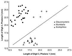
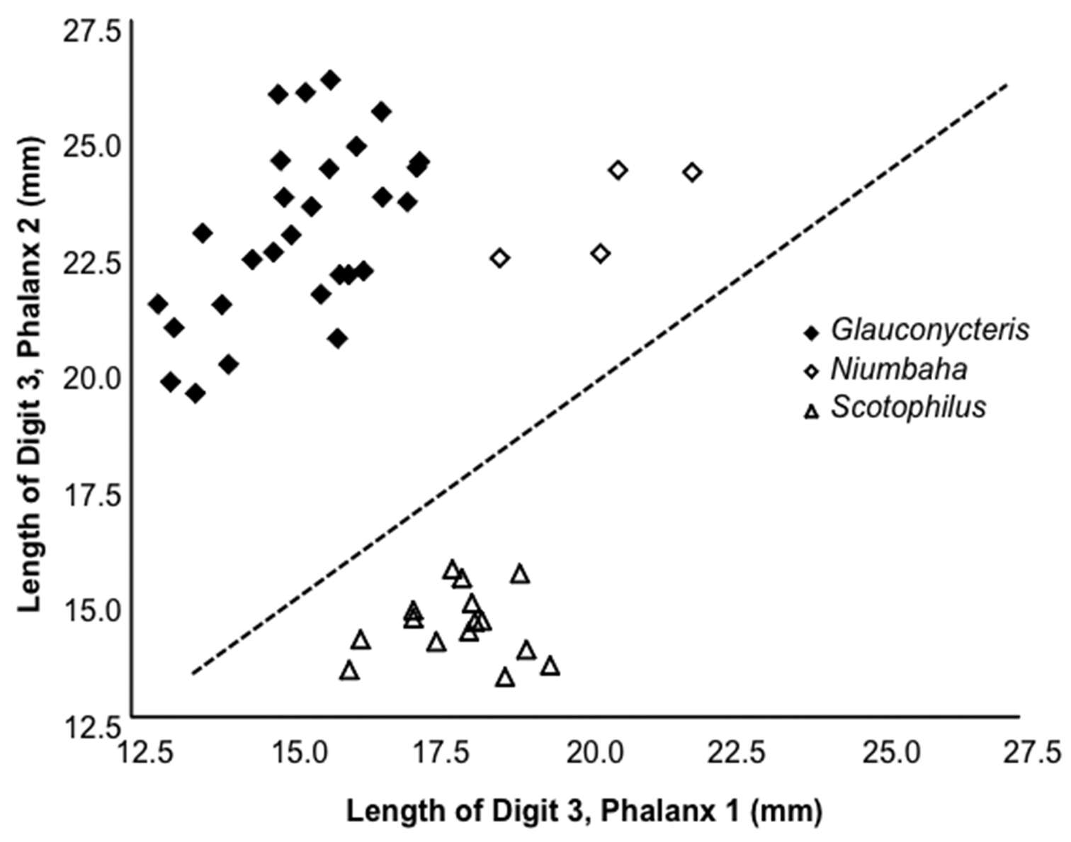
Figure 4.Length of the 2nd phalanx (2PL) of the 3rd digit vs. the 1st phalanx (1PL) of the 3rd digit. Several species of Glauconycteris are shown (closed diamond), as is Niumbaha superba (open diamond), and for comparison, two species of Scotophilus (open triangle; a ‘typical’ African vespertilionid bat). The ratio of 2PL/1PL is significantly greater in Glauconycteris than in Niumbaha (with a theoretical 1:1 ratio indicated by the dashed line). Data as reported in Table 2.
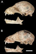
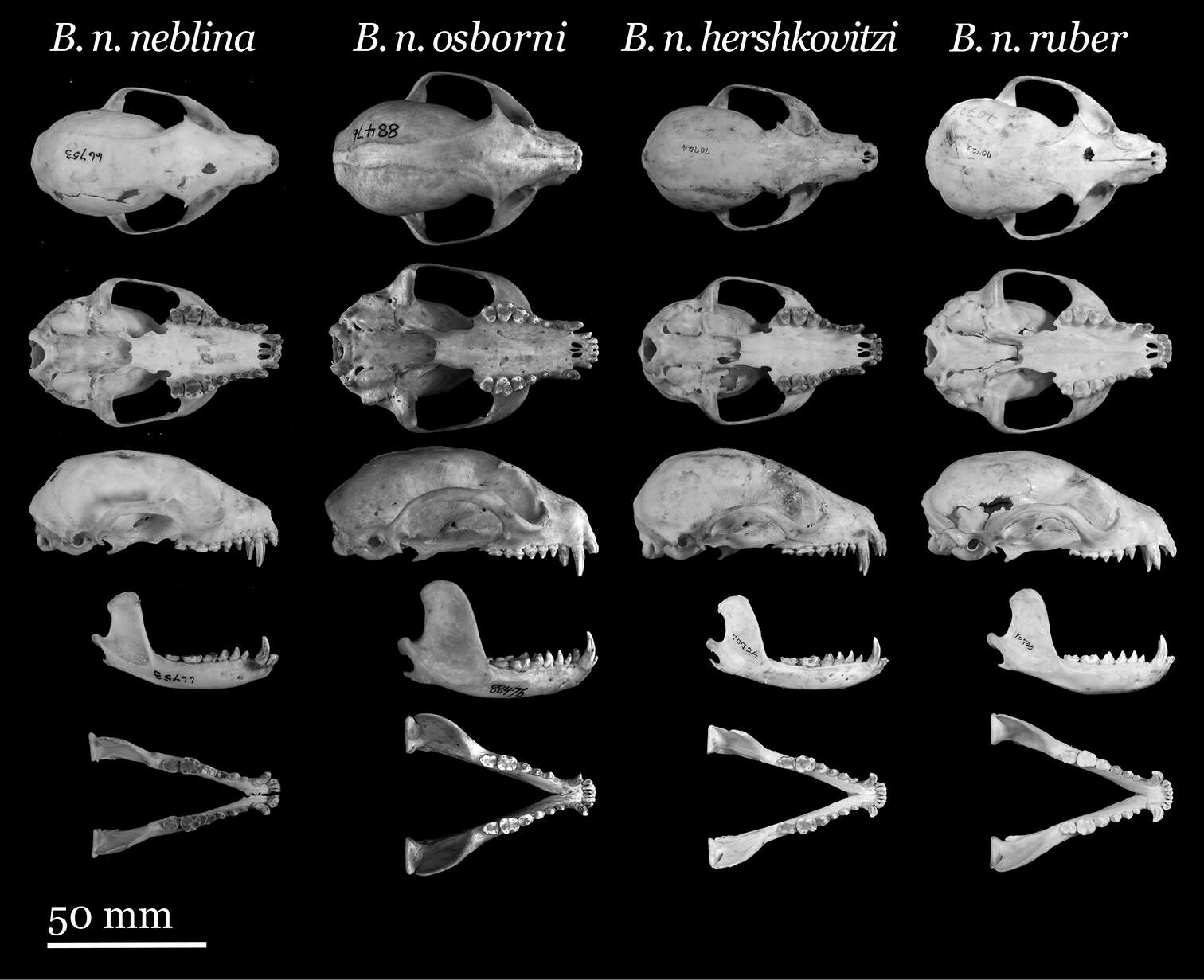
Figure 4.Lateral views of the cranium and mandible of A Sturnira bakeri (QCAZ 14635 ♀). Lateral views of the cranium and mandible of B Sturnira burtonlimi (ROM 104294 ♂). Scale bar = 5 mm.
DeeAnn M. Reeder, Kristofer M. Helgen, Megan E. Vodzak, Darrin P. Lunde, Imran Ejotre
Zookeys
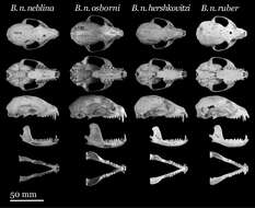
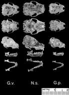
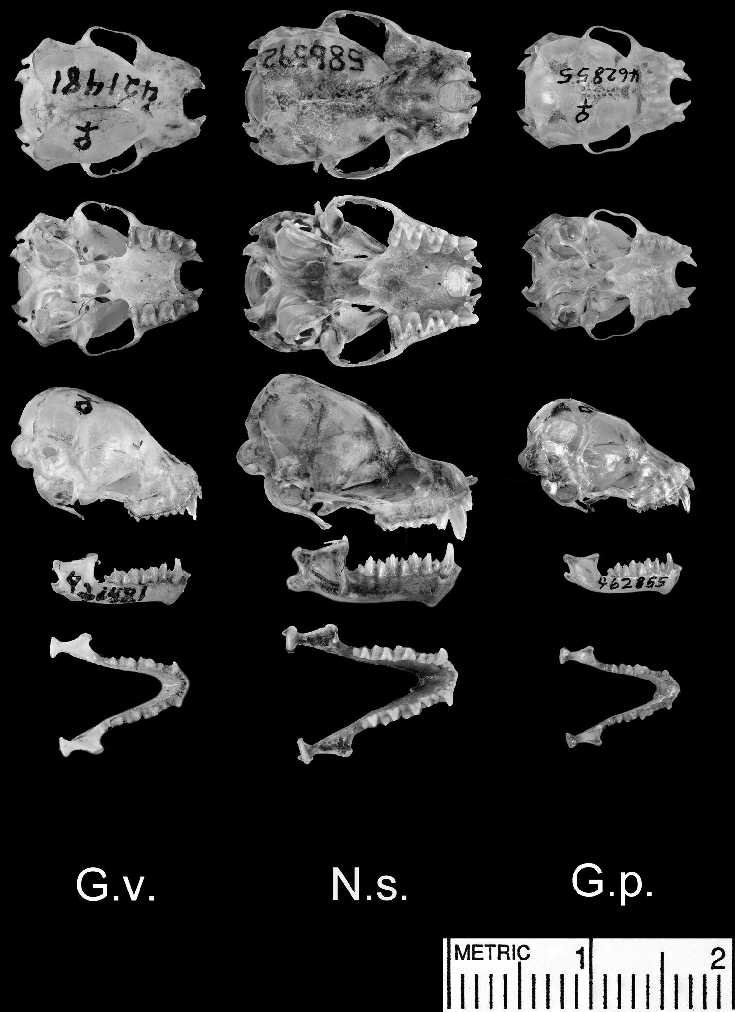
Figure 5.Dorsal and ventral views of the cranium, lateral views of the cranium and mandible, and dorsal view of the mandible. Species shown include Glauconycteris variegata (G.v.; a relatively large species of Glauconycteris, which nearly matches Niumbaha superba in linear body size, but not in skull size); Niumbaha superba (N.s.; the type species of Niumbaha), and Glauconycteris poensis (G.p., the type species of Glauconycteris).
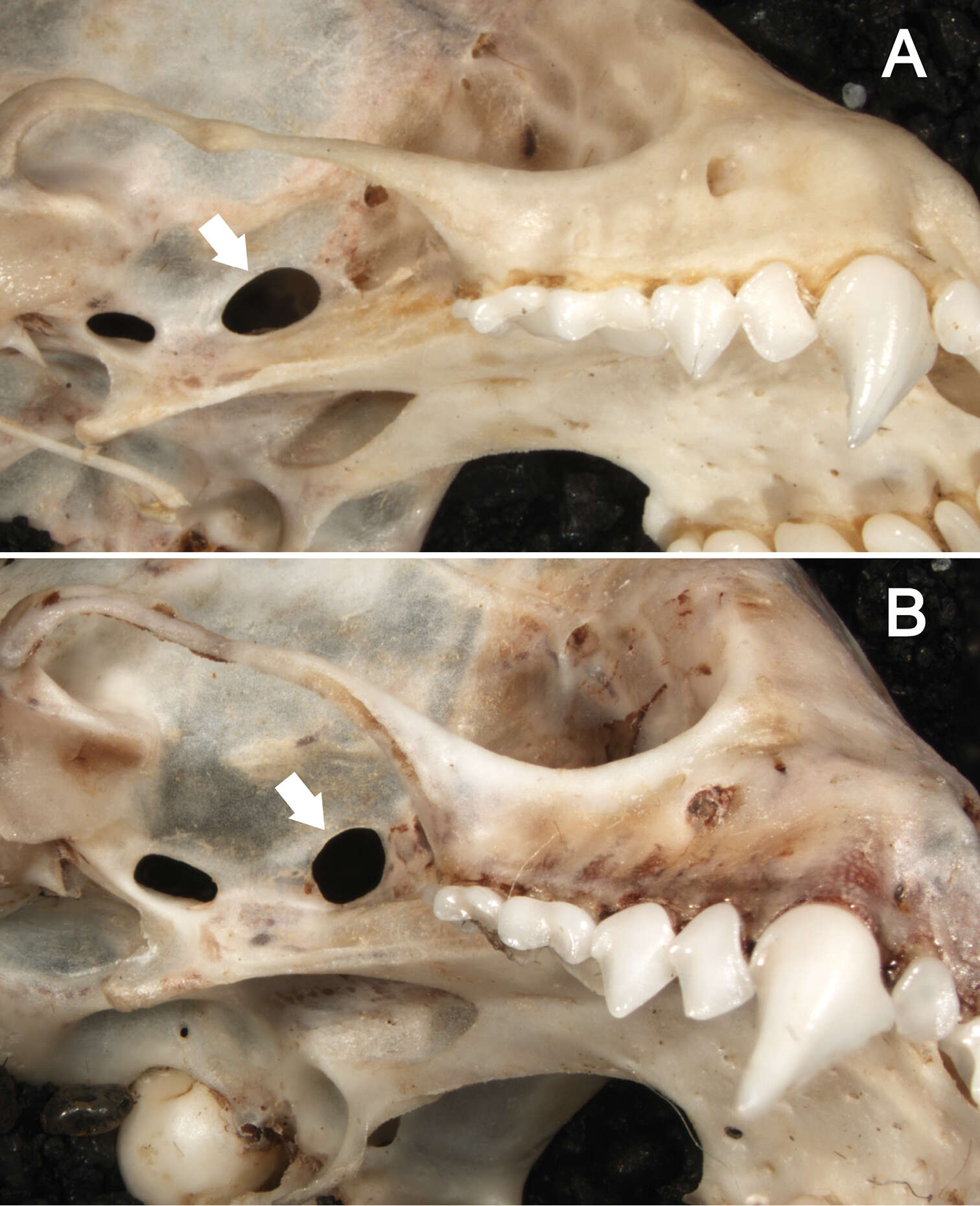
Figure 5.Ventrolateral views of the right orbital region in Sturnira bakeri (A, QCAZ 14635 ♀) and Sturnira luisi (B, ROM 104204 ♂) illustrating taxonomic differences in the shape of the sphenorbital fissure. In Sturnira bakeri, the sphenorbital fissure is oval (arrow). In Sturnira luisi, however, the sphenorbital fissure is semicircular (arrow).
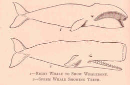
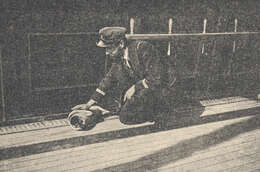
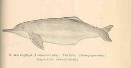
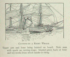
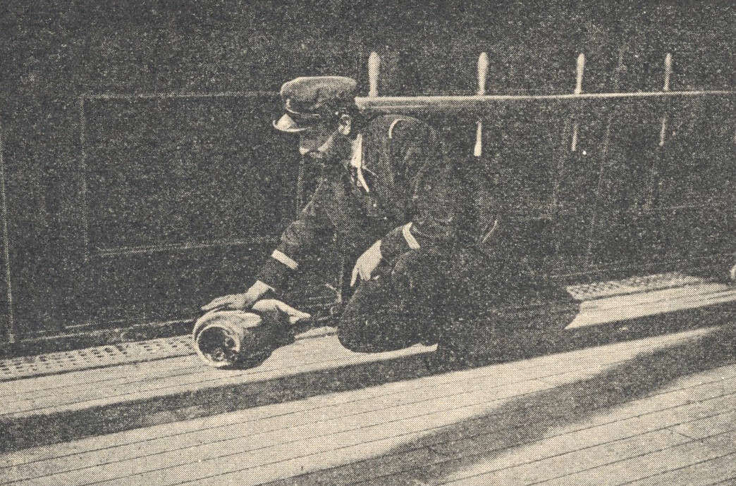
Cutting-In a Right Whale. Upper jaw and bone being hoisted on board. Note man with spade on cutting-stage; blanket-piece back of bone and try-works from which smoke is rising..