Comprehensive Description
provided by Smithsonian Contributions to Zoology
Cranchia scabra Leach, 1817
Cranchia scabra Leach, 1817, p.140.—Hoyle, 1904, p.43, pl. 10, fig. ll.—Pfeffer, 1912, p.679, pl.48, figs. 22–28.— Sasaki, 929, p.329, pl.26, figs. 13–15, text – figs. 151–153.— Voss, 1963, p. 142, text fig. 31.—Pearcy, 1965, p.261.
Cranchia tenuitentaculata Pfeffer, 1884, p.26, pl.3, fig. 36.
Cranchia hispida Pfeffer, 1884, p.27, pl.3, fig.37.
DESCRIPTION.—The mantle is saclike, although it tapers abruptly to a point posteriorly. The mantle wall is very thin, but muscular. The anterior edge of the mantle is fused to the head in the nuchal region and to lateral corners of the funnel. From each of the latter points of fusion, two, short cartilaginous strips diverge to form a V-pattern. The entire surface of the mantle is covered with cartilaginous tubercules. The tubercules are somewhat variable, but generally have 3–4 well-separated cusps whose tips are usually subdivided into 2–3 additional cusps.
The fins are small and terminal (15–20% of the M.L. in length and 25–30% of the M.L. in total width). Each is nearly circular in outline and has free anterior and posterior lobes. The dorsal surface of the fins is covered with cartilaginous tubercules, but the ventral surface appears to have only small, rounded papillae.
The funnel reaches slightly past the bases of the arms. The dorsal pad of the funnel organ has roughly an inverted V-shape with a slender median ridge that is larger in its posterior portion; a flap occurs at the lateral edge of each diverging arm. The ventral pads are large and kidney-shaped, with the concave margin facing anteriorly. The surface of the funnel is covered with simple papillae; those on the ventral surface, immediately anterior to the mantle, are interconnected by low, narrow ridges that give a reticulated pattern. A funnel valve is present. The funnel is fused laterally to the head.
The head is short but broad and bears very large eyes that occupy the entire lateral sides of the head. Each eyelid has a small anterior sinus. Small, short “olfactory” papillae are present on either side of the head just lateral to the base of the funnel. Small, low papillae are scattered over the surface of the head.
The arms are short and in the order of III>IV=II>I. Arms I–III are joined basally by a web. The arms all have broad, trabeculate protective membranes on both dorsal and ventral borders. Well-developed aboral keels occur on arms III, but are barely detectable on arms I and II. Large lateral keels are present on arms IV. The arms have biserially arranged suckers except near the tips of arms III in males and on the hectocotylus. The suckers in the proximal halves of the arms each have about 5–7 low, rounded, irregular teeth on the distal portion of the inner chitinous ring. Distally, the broad teeth become more regular in arrangement and distinctly truncate. The teeth of the inner sucker rings in females seem to be more regularly arranged throughout the arm length, but are particularly so in the more distal suckers which have slender truncate teeth. Each arm III in males has the distal fifth modified by an increase to 4–5 slightly irregular longitudinal rows of suckers and by an abrupt decrease in the size of the suckers. The suckers become biserial again at the extreme tips of these arms.
The right arm IV is hectocotylized in males. It is considerably shorter than the left arm, and its tip is swollen and curled laterally. Proximally, the suckers of the hectocotylus are biserially arranged, but become tetraserial near the base of the curved tip. The curved tip has biserial suckers which are considerably enlarged proximally. Trabeculate protective membranes are present, but diminish on the curved portion. A large lateral keel extends to the tip of the arm. Each sucker on the proximal segment of the hectocotylus has about 6–7 broad truncate teeth on the distal margins of the inner chitinous ring. The largest suckers of the distal segment (curved portion) each have about 13–15 rounded teeth around the entire margin of the inner chitinous ring; the teeth on the distal margin are the largest.
The tentacles are short and carry small, compact clubs. Four longitudinal rows of suckers on the manus grade into 4 rows on the dactylus. Suckers of both manus and dactylus have a uniform size in a transverse series. There are 4–7 small, smooth-ringed suckers at the tip of the dactylus. The dentition of the inner rings of the manal suckers varies with the location of the sucker. The proximal suckers have 6–7 slender, pointed teeth on the distal half of the inner ring. On the more distal suckers, the number progressively increases and smaller teeth also develop on the proximal margin of the ring. At the distal end of the manus, the number of teeth per ring approaches 30. Pairs of alternating suckers and pads extend down the tentacular stalk for about two-thirds of its length; the total number of suckers varies from 33–36 in the 2 specimens in which accurate counts were possible.
The buccal membrane attaches to the dorsal borders of arms I and II and to the ventral borders of arms III and IV.
In preservation, the specimens have small, brown chromatophores scattered over the body, head, and arms. In life, with chromatophores expanded and head contracted into the mantle, the animal looks like a large orange.
Photophores are present only on the surface of the eyes. There is a proximal row consisting of 8 photophores in a U-shaped series passing from the anterior to the medial section to the posterior surface of the eye. A distal series of photophores lies near the lens and has 4 photophores in a cescent-shape row ventral to the lens and 2 photophores dorsal to the lens. The eye, therefore, carries a total of 14 photophores.
TYPE LOCALITY.—Off West Africa.
LOCATION OF TYPE.—British Museum (Natural History).
- bibliographic citation
- Young, Richard E. 1972. "The systematics and areal distribution of pelagic cephalopods from the seas off Southern California." Smithsonian Contributions to Zoology. 1-159. https://doi.org/10.5479/si.00810282.97
Cranchia scabra: Brief Summary
provided by wikipedia EN
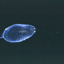
Cranchia scabra is a species of glass squid. It is the only species in the genus, and is fairly small (about 150 mm). The mantle is covered by large, multi-pointed cartilagenous tubercles. When disturbed, the squid often pulls its head and arms into the mantle cavity and folds its fins tightly against the mantle to form a turgid ball. The tubercules, presumably, provide some type of protection, but it is unclear what predators are affected and how. In addition, the squid may ink into the mantle cavity, making the ball opaque. This was thought to be an aberrant behavior due to stress and confinement of shipboard aquaria until the same inking behavior was seen in cranchiids from submersibles. The function of this behavior is unknown.
The genus contains bioluminescent species. It is named for John Cranch.
- license
- cc-by-sa-3.0
- copyright
- Wikipedia authors and editors
Distribution
provided by World Register of Marine Species
circum-(sub)tropical
van der Land, J. (ed). (2008). UNESCO-IOC Register of Marine Organisms (URMO).
- license
- cc-by-4.0
- copyright
- WoRMS Editorial Board
Habitat
provided by World Register of Marine Species
meso-bathypelagic
van der Land, J. (ed). (2008). UNESCO-IOC Register of Marine Organisms (URMO).
- license
- cc-by-4.0
- copyright
- WoRMS Editorial Board
Habitat
provided by World Register of Marine Species
Known from seamounts and knolls
Stocks, K. 2009. Seamounts Online: an online information system for seamount biology. Version 2009-1. World Wide Web electronic publication.
- license
- cc-by-4.0
- copyright
- WoRMS Editorial Board