-
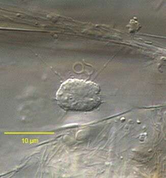
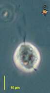
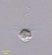
Actinomonas (act-in-o-moan-ass) is one of the pedinellid stramenopiles, a small flagellate with an apical flagellum (upper) and stalk (lower). Usually with a number of fine arms around the flagellum. These arms intercept bacteria and other suspended particles. They may also be withdrawn, as is the case here. The flagellum has not been frozen by the photography and what we see is the beat envelope. Phase contrast.
-
-
Actinomonas (ack-tin-owe-moan-ass) mirabilis is to all intents and purposes indistinguishable from Pteridomonas danica (there are small ultrastructural differences). Cells are 4 to 6 microns long and have one flagellum emerging from a small depression in the anterior end of the cell. The cells have a ring of arms around the flagellum and below the equator of the cell, the arms around the flagellum are evenly spaced. The anterior part of the cell is slightly broader than the posterior part. The single thickened flagellum is about 3 - 5 times the cell length and has an undulating beat. The cells usually swim rapidly, but occasionally attach to the substrate with a long posterior stalk trailing. Small particles are seen on the cell surface. Sometimes commonly observed.
-
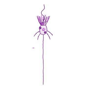
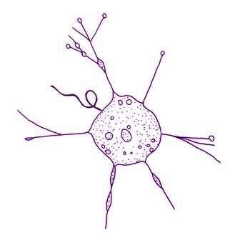
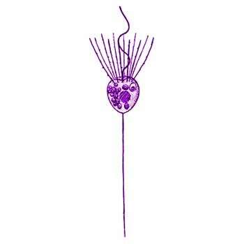
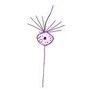
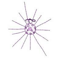
Actinomonas mirabilis Kent, 1880. Colourless pedinellid, cell body measuring 5-8 microns, and often stalked. Arms present, up to two rings of 12 arms each around the anterior flagellum and extending from other parts of the body. When disturbed may swim with arms trailing from posterior.
-
Actinomonas vernalis Stokes, 1885. Cells subspherical, the frontal border slightly emarginate, somewhat changeable in shape, free-swimming or temporarily attached by a short pedicel, flagellum active throughout its length. The flagellum equals or somewhat exceeds the diameter of the body in length, cytoplasm transparent, slightly granular, pseudopodia few in number, radiating from any part of the periphery, simple or variously branched, often capitate, sometimes curved, their length exceeding the diameter of the body, several small contractile vacuoles distributed near the periphery, nucleus spherical, subcentral. Diameter of body 16.9-21.2 microns Status of taxon dubious.
-
Pteridomonas (ter-rid-owe-moan-ass) a colourless pedinellid (stramenopile) flagellate. Like other pedinellids, it has a single apical flagellum surrounded by a wreath of fine arms, and usually attached to the substrate by a fine stalk. The stalk is contractile and may also release from the substrate. The flagellum beats with an undulating beat pattern. Phase contrast.
-
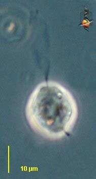
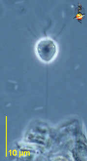
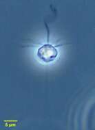
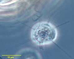
Portrait of Pteridomonas, a small silicoflagellate with a long thin stalk. Tentacles surround the single apical flagellum. Posterior contractile vacuole can be seen here. Very similar to Actinomonas, which has posterior tentacles as well. The two species also have ultrastructural differences. From freshwater pond near Boise, Idaho. Oblique illumination
-
Portrait of Pteridomonas, a small silicoflagellate with a long thin stalk. Tentacles surround the single apical flagellum. Very similar to Actinomonas, which has posterior tentacles as well. From freshwater pond near Boise, Idaho.
-
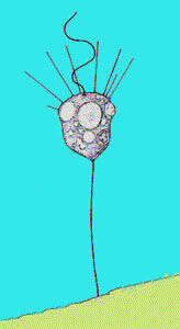
Pteridomonas pulex Penard Cells spherical, usually broader than the length, in optical profiles heart to kindney-shaped, 6-12 microns long. Stalk is long and thin. Nucleus is in central. Flagellum is 3-4 times the cell length. 12-18 pseudopodia arise at the base of the flagellum and surround the anterior end of the cell.
-
-
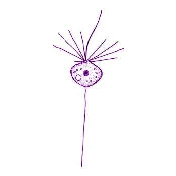
Pteridomonas danica Patterson and Fenchel, 1985. A colourless pedinellid, body 4.5-5 microns long, with one apical flagellum emerging from a slight depression at the anterior of the cell. The flagellum is surrounded by 12 stiffarms. Posterioly, the body gives rise to a stalk. The stalk and arms may be withdrawn in swimming cells.
-
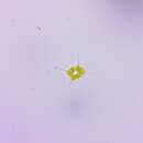
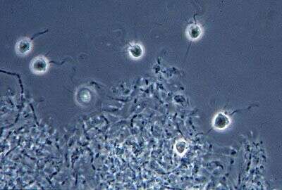
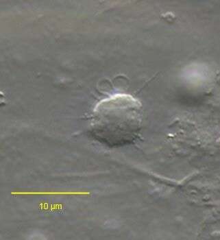
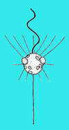
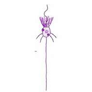
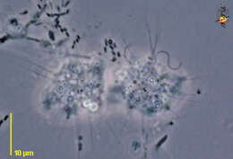
Phase contrast image of a small group of these heterotrophic flagelates. A single apical flagellum draws water twoards the thin arms that intercept food particles.
-
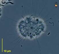
Ciliophrys (silly-off-rees), a pedinellid stramenopile, with a single flagellum. The cell may exist as a non-swimming form with radiating arms, and with the flagellum inactive or beating in a languid figure of 8 motion. The arms can be resorbed, and the cell can then swim with the flagellum pulling the cell forward. This is the heliozoon form, with a nucleus in the centre of the cell. Phase contrast.
-
-
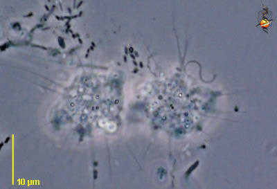
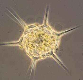
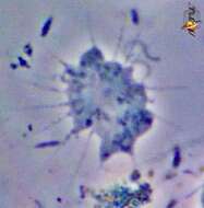
Ciliophrys (silly-off-riss) infusionum Cienkowski, 1876. Helioflagellate, in the heliozoan stage the cells are about 4 - 9 microns across, and have a central nucleus and one flagellum held in a figure of eight. The cells are spherical with delicate pseudopodia extending radially from the body and bearing extrusomes. The cells may change from the heliozoan stage with pseudopodia and a slow beating flagellum to a swimming flagellate without pseudopodia and with the flagellum beating rapidly. In swimming cells, the nucleus is located apically. Observed to consume suspended bacteria. When feeding, bacteria adhere to the pseudopodia and then are drawn to the body. The cells eat diatoms up to 18 microns long. Sometimes common.
-
Ciliophrys infusionum Cienkowski, 1876. Ciliophrys, in the heliozoan stage the cells are about 4 - 9 microns across, and have a central nucleus and one flagellum held in a figure of eight. The cells are spherical with delicate pseudopodia extending radially from the body and bearing extrusomes. The cells may change from the heliozoan stage with pseudopodia and a slow beating flagellum to a swimming flagellate without pseudopodia and with the flagellum beating rapidly. In swimming cells, the nucleus is located apically. Observed to consume suspended bacteria. When feeding, bacteria adhere to the pseudopodia and then are drawn to the body. The cells eat diatoms up to 18 microns long.
-
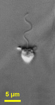
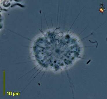
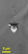
Portrait of Ciliophrys infusionum (Cienkowski,1876), one of the colourless pedinellid flagellates (also referred to as a helioflagellate). When at rest on the substrate tentacles(axopodia supported by one triplet of microtubules) of with fine granules are distributed over the entire surface and the single flagellum is held in a tight S configuration hardly moving. When swimming, tentacles withdraw and the cell surface appears smooth, the flagellum beating more rapidly in sine wave fashion. From standing freshwater near Boise, Idaho. Phase contrast.
-
Portrait of Ciliophrys infusionum (Cienkowski,1876), one of the colourless pedinellid flagellates (also referred to as a helioflagellates). When at rest on the substrate, tentacles (axopodia supported by one triplet of microtubules) with fine granules are distributed over the entire cell surface and the single flagellum is held in a tight S configuration hardly moving. When swimming, tentacles withdraw and the cell surface appears smooth, the flagellum beating more rapidly in sine wave fashion. From a commercial marine aquarium in Boise, Idaho. DIC.
-
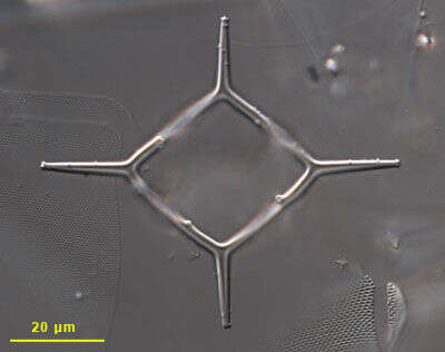
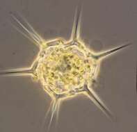
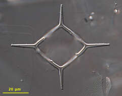
Dictyocha (dick-tee-oke-a) is a silicoflagellate, a type of flagellate which is quite common in marine waters, although there are very few species. The cells contain an open polygonal skeleton of silica with radiating spines. The skeleton survives when the organism dies, and so is often seen on its own. This cell is distressed . Phase contrast image by Dave Caron.
-
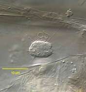
This skeleton of the flagellate Dictyocha was prepared by bleaching and cleaning from a marine water column sample taken off Martha's Vineyard in Vineyard Sound, Massachusetts. Differential interference contrast optics, image by Charley O'Kelly.
-
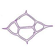
Dictyocha fibula Ehrenberg, 1839. Silicoflagellate with siliceous skeleton (basal ring) with four sides, but without the apical ring. Spines project from the corners of the outer hexagon, internal projections form irregular connections. Bars also arise inwards and may fuse to form a central arch. The cytoplasm extends over the skeleton, fine pseudopodia usually project from the tops of the spines. Many small plastids, single emergent flagellum. Mostly 30-80microns Marine