-
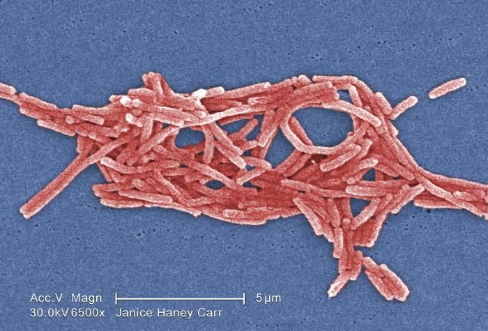
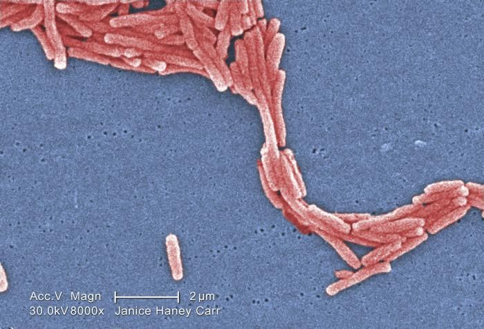
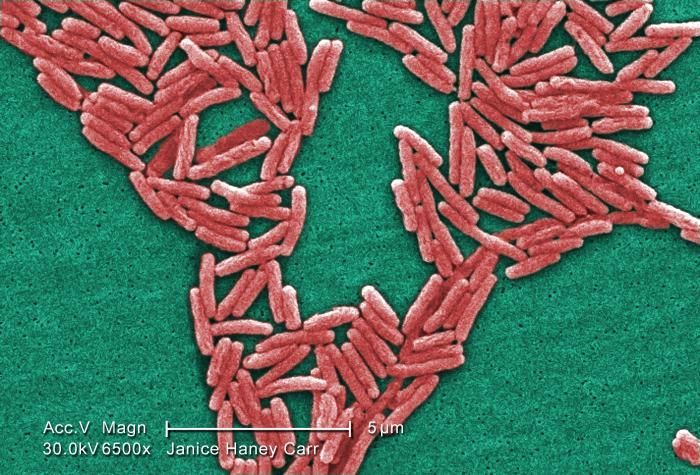
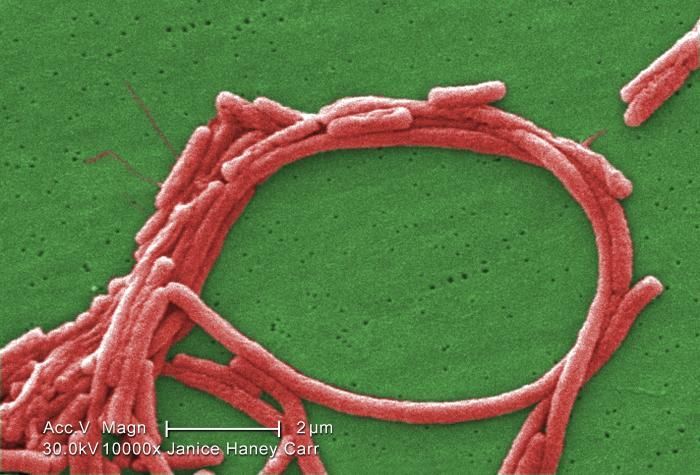
Under a moderately-high magnification of 6500X, this colorized scanning electron micrograph (SEM) depicted a grouping of Gram-negative Legionella pneumophila bacteria. Please see PHIL 11092 through 11160 for additional SEMs of these organisms, specifically PHIL 11121 for a black and white version of this image. In some of these views you'll note the presence of flagellar appendages.Youll note that a number of these bacteria seem to display an elongated-rod morphology. L. pneumophila are known to most frequently exhibit this configuration when grown in broth, however, they can also elongate when plate-grown cells age, as it was in this case, especially when theyve been refrigerated. The usual L. pneumophila morphology consists of stout, fat bacilli, which is the case for the vast majority of the organisms depicted here.Created: 2009
-
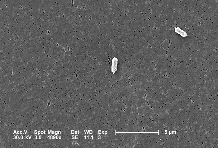
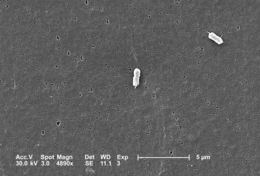
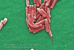
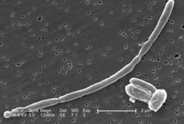
This scanning electron micrograph (SEM) depicts two Escherichia coli bacteria, clearly displaying one or more peritrichous flagella, i.e., flagella that may originate from anywhere on the bacteriums cell wall surface.; Magnification 4890x.Created: 2005
-
Under a moderately-high magnification of 8000X, this colorized scanning electron micrograph (SEM) depicted a grouping of Gram-negative Legionella pneumophila bacteria. Please see PHIL 11092 through 11152 for additional SEMs of these organisms, specifically PHIL 11119 for a black and white version of this image.Youll note that a number of these bacteria seem to display an elongated-rod morphology. L. pneumophila are known to most frequently exhibit this configuration when grown in broth, however, they can also elongate when plate-grown cells age, as it was in this case, especially when theyve been refrigerated. The usual L. pneumophila morphology consists of stout, fat bacilli, which is the case for the vast majority of the organisms depicted here.Created: 2009
-
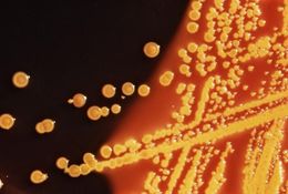
These are colonies of Escherichia coli bacteria grown on a Hektoen enteric (HE) agar plate medium; colonies of E. coli grown on HE agar display a raised morphology, and are yellow, to orange-yellow in coloration.Created: 1976
-
Under a moderately-high magnification of 6500X, this colorized scanning electron micrograph (SEM) depicted a scattered grouping of Gram-negative Legionella pneumophila bacteria. Please see PHIL 11092 through 11152 for additional SEMs of these organisms, specifically PHIL 11117 for a black and white version of this image. In some of these views you'll note the presence of flagellar appendages.Youll note that a number of these bacteria seem to display an elongated-rod morphology. L. pneumophila are known to most frequently exhibit this configuration when grown in broth, however, they can also elongate when plate-grown cells age, as it was in this case, especially when theyve been refrigerated. The usual L. pneumophila morphology consists of stout, fat bacilli, which is the case for the vast majority of the organisms depicted here.Created: 2009
-
At an extremely high magnification of 44, 818X, twice that of PHIL 10574 and 10575, this scanning electron micrograph (SEM) revealed some of the morphologic details displayed by a single Gram-negative Escherichia coli bacterium. This bacterium was a member of the strain, 0:169 H41 ETEC (Enterotoxigenic E. coli). See PHIL 10577 for a colorized version of this image.Enterotoxigenic E. coli, a common cause of bacterial diarrhea
Enterotoxigenic Escherichia coli, or ETEC, is an important cause of bacterial diarrheal illness. Infection with ETEC is the leading cause of travelers' diarrhea and a major cause of diarrheal disease in underdeveloped nations, especially among children. ETEC is transmitted by food or water contaminated with animal or human feces. Although ETEC causes a significant amount of illness worldwide, the infection will end on its own and is rarely life-threatening.Created: 2008
-
Under a moderately-high magnification of 6500X, this scanning electron micrograph (SEM) depicted a scattered grouping of Gram-negative Legionella pneumophila bacteria. Please see PHIL 11092 through 11152 for additional SEMs of these organisms, specifically PHIL 11118 for a colorized version of this image. In some of these views you'll note the presence of flagellar appendages.Youll note that a number of these bacteria seem to display an elongated-rod morphology. L. pneumophila are known to most frequently exhibit this configuration when grown in broth, however, they can also elongate when plate-grown cells age, as it was in this case, especially when theyve been refrigerated. The usual L. pneumophila morphology consists of stout, fat bacilli, which is the case for the vast majority of the organisms depicted here.Created: 2009
-
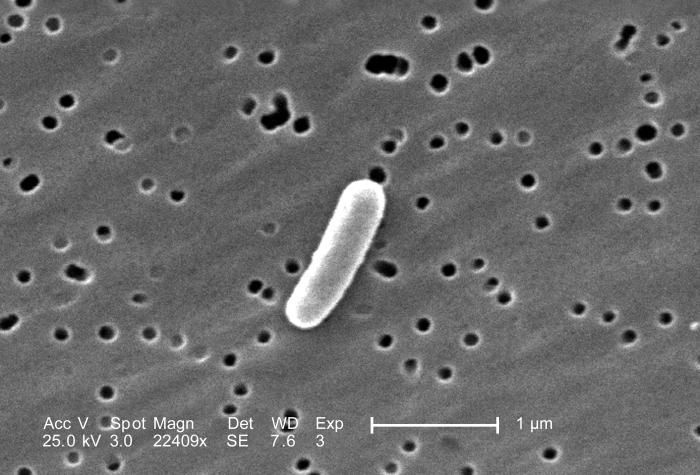
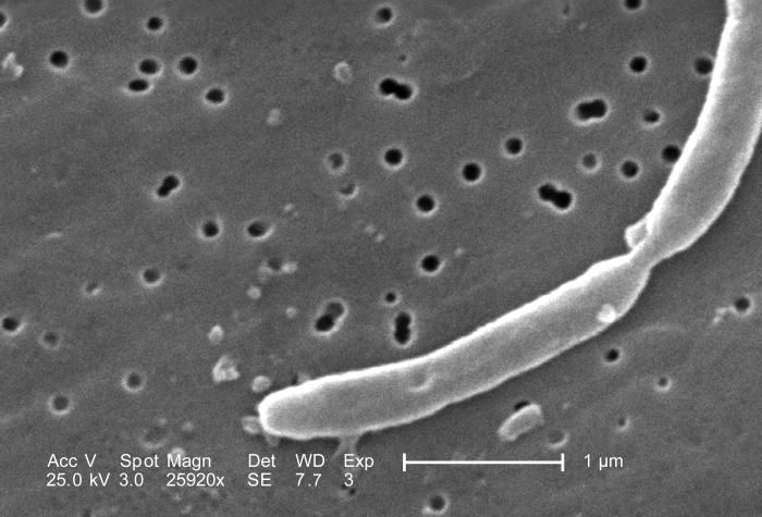
At a very high magnification of 22,409X, this scanning electron micrograph (SEM) revealed some of the morphologic details displayed by a single Gram-negative Escherichia coli bacterium. This bacterium was a member of the strain, 0:169 H41 ETEC (Enterotoxigenic E. coli).Enterotoxigenic E. coli, a common cause of bacterial diarrhea
Enterotoxigenic Escherichia coli, or ETEC, is an important cause of bacterial diarrheal illness. Infection with ETEC is the leading cause of travelers' diarrhea and a major cause of diarrheal disease in underdeveloped nations, especially among children. ETEC is transmitted by food or water contaminated with animal or human feces. Although ETEC causes a significant amount of illness worldwide, the infection will end on its own and is rarely life-threatening.Created: 2008
-
Under a moderately-high magnification of 6500X, this colorized scanning electron micrograph (SEM) depicted a number of Gram-negative Legionella pneumophila bacteria. Please see PHIL 11092 through 11152 for additional SEMs of these organisms, specifically PHIL 11115 for a black and white version of this image. Some views in this series reveals the presence of flagellar appendages.Youll note that a number of these bacteria seem to display an elongated-rod morphology. L. pneumophila are known to most frequently exhibit this configuration when grown in broth, however, they can also elongate when plate-grown cells age, as it was in this case, especially when theyve been refrigerated. The usual L. pneumophila morphology consists of stout, fat bacilli, which is the case for the vast majority of the organisms depicted here.Created: 2009
-
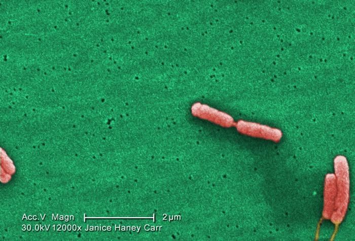
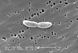
At a very high magnification of 22,409X, this scanning electron micrograph (SEM) revealed some of the morphologic details displayed by two joined Gram-negative Escherichia coli bacteria. These bacteriia were members of the strain, 0:169 H41 ETEC (Enterotoxigenic E. coli).Enterotoxigenic E. coli, a common cause of bacterial diarrhea
Enterotoxigenic Escherichia coli, or ETEC, is an important cause of bacterial diarrheal illness. Infection with ETEC is the leading cause of travelers' diarrhea and a major cause of diarrheal disease in underdeveloped nations, especially among children. ETEC is transmitted by food or water contaminated with animal or human feces. Although ETEC causes a significant amount of illness worldwide, the infection will end on its own and is rarely life-threatening.Created: 2008
-
Under a high magnification of 12000X, this colorized scanning electron micrograph (SEM) depicted a number of Gram-negative Legionella pneumophila bacteria. Please see PHIL 11092 through 11152 for additional SEMs of these organisms, specifically PHIL 11114 for a black and white version of this image. Note that a few of these bacteria are sporting their flagella.Youll note that a number of these bacteria seem to display an elongated-rod morphology. L. pneumophila are known to most frequently exhibit this configuration when grown in broth, however, they can also elongate when plate-grown cells age, as it was in this case, especially when theyve been refrigerated. The usual L. pneumophila morphology consists of stout, fat bacilli, which is the case for the vast majority of the organisms depicted here.Created: 2009
-
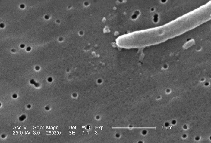
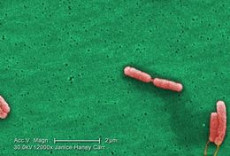
At a very high magnification of 25,920X, twice that of PHIL 10570 and 10571, this scanning electron micrograph (SEM) revealed some of the morphologic details displayed a single Gram-negative Escherichia coli bacterium. This bacterium was a member of the strain, 0:169 H41 ETEC (Enterotoxigenic E. coli).What is ETEC?ETEC was first recognized as a cause of human diarrheal illness in the 1960s. It have since emerged as a major bacterial cause of diarrhea among travelers and children in the developing world. ETEC is increasingly recognized as an important cause of foodborne illness in developed nations, such as the United States.ETEC produces two toxins, a heat-stable toxin (known as ST) and a heat-labile toxin (LT). Although different strains of ETEC can secrete either one or both of these toxins, the illness caused by each toxin is similar.Created: 2008
-
Under a moderately-high magnification of 6500X, this colorized scanning electron micrograph (SEM) depicted a large number of Gram-negative Legionella pneumophila bacteria. Please see PHIL 11092 through 11152 for additional SEMs of these organisms, specifically PHIL 11111 for a black and white version of this image.Youll note that a number of these bacteria seem to display an elongated-rod morphology. L. pneumophila are known to most frequently exhibit this configuration when grown in broth, however, they can also elongate when plate-grown cells age, as it was in this case, especially when theyve been refrigerated. The usual L. pneumophila morphology consists of stout, fat bacilli, which is the case for the vast majority of the organisms depicted here.Created: 2009
-
At a very high magnification of 25,920X, twice that of PHIL 10570 and 10571, this scanning electron micrograph (SEM) revealed some of the morphologic details displayed by a two joined Gram-negative Escherichia coli bacteria. These bacteria were members of the strain, 0:169 H41 ETEC (Enterotoxigenic E. coli).Enterotoxigenic E. coli, a common cause of bacterial diarrhea
Enterotoxigenic Escherichia coli, or ETEC, is an important cause of bacterial diarrheal illness. Infection with ETEC is the leading cause of travelers' diarrhea and a major cause of diarrheal disease in underdeveloped nations, especially among children. ETEC is transmitted by food or water contaminated with animal or human feces. Although ETEC causes a significant amount of illness worldwide, the infection will end on its own and is rarely life-threatening.Created: 2008
-
Highly magnified 12000X, this colorized scanning electron micrograph (SEM) depicted six Gram-negative Legionella pneumophila bacteria. Note that youre able to see a flagella emanating from the lower right organisms. Also note that one bacteria is about to become two, separate entities, as they were finishing the process of cell division. Please see PHIL 11092 through 11152 for additional SEMs of these organisms, specifically PHIL 11109 for a black and white version of this image.Youll note that a number of these bacteria seem to display an elongated-rod morphology. L. pneumophila are known to most frequently exhibit this configuration when grown in broth, however, they can also elongate when plate-grown cells age, as it was in this case, especially when theyve been refrigerated. The usual L. pneumophila morphology consists of stout, fat bacilli, which is the case for the vast majority of the organisms depicted here.Created: 2009
-
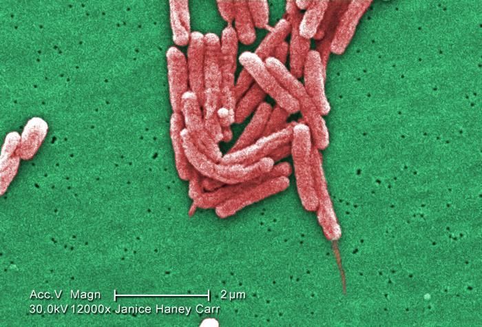
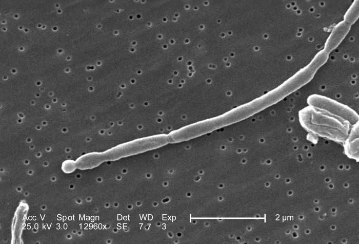
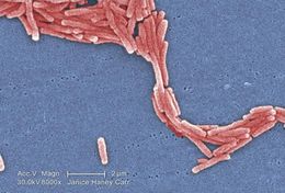
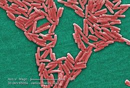
At a high magnification of 12,960X, this scanning electron micrograph (SEM) revealed some of the morphologic details displayed by a number of joined Gram-negative Escherichia coli bacteria. These bacteria were members of the strain, 0:169 H41 ETEC (Enterotoxigenic E. coli).Enterotoxigenic E. coli, a common cause of bacterial diarrhea
Enterotoxigenic Escherichia coli, or ETEC, is an important cause of bacterial diarrheal illness. Infection with ETEC is the leading cause of travelers' diarrhea and a major cause of diarrheal disease in underdeveloped nations, especially among children. ETEC is transmitted by food or water contaminated with animal or human feces. Although ETEC causes a significant amount of illness worldwide, the infection will end on its own and is rarely life-threatening.Created: 2008
-
Under a high magnification of 10000X, this colorized scanning electron micrograph (SEM) depicted a grouping of Gram-negative Legionella pneumophila bacteria. Note that youre able to see a number of the flagella emanating from these organisms. Please see PHIL 11092 through 11152 for additional SEMs of these organisms, specifically PHIL 11107 for a black and white version of this image.Youll note that a number of these bacteria seem to display an elongated-rod morphology. L. pneumophila are known to most frequently exhibit this configuration when grown in broth, however, they can also elongate when plate-grown cells age, as it was in this case, especially when theyve been refrigerated. The usual L. pneumophila morphology consists of stout, fat bacilli, which is the case for the vast majority of the organisms depicted here.Created: 2009
-
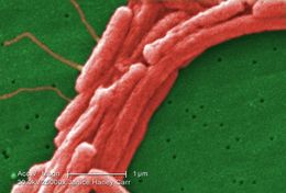
At a high magnification of 12,960X, this scanning electron micrograph (SEM) revealed some of the morphologic details displayed by a number of joined Gram-negative Escherichia coli bacteria. These bacteria were members of the strain, 0:169 H41 ETEC (Enterotoxigenic E. coli). See PHIL 11379 for a colorized version of this image.What is ETEC?Escherichia colii is a bacterium that normally lives in the intestines of humans and other animals. Most types of E. coli are harmless, but some can cause disease. Disease-causing E. coli are grouped according to the different ways by which they cause illness. Enterotoxigenic Escherichia coli, or ETEC, is the name given to a group of E. coli that produce special toxins which stimulate the lining of the intestines causing them to secrete excessive fluid, thus producing diarrhea. The toxins and the diseases that ETEC causes are not related to E. coli O157:H7.Created: 2008
-
Under a very high magnification of 25000X, this colorized scanning electron micrograph (SEM) depicted a grouping of Gram-negative Legionella pneumophila bacteria. Note that in this view youre able to see a number of the flagella emanating from these organisms. Please see PHIL 11092 through 11152 for additional SEMs of these organisms, specifically PHIL 11105 for a black and white version of this image.Youll note that a number of these bacteria seem to display an elongated-rod morphology. L. pneumophila are known to most frequently exhibit this configuration when grown in broth, however, they can also elongate when plate-grown cells age, as it was in this case, especially when theyve been refrigerated. The usual L. pneumophila morphology consists of stout, fat bacilli, which is the case for the vast majority of the organisms depicted here.Created: 2009
-
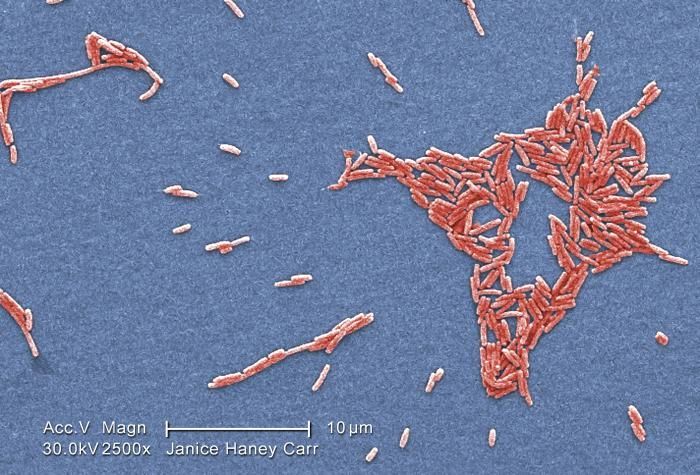
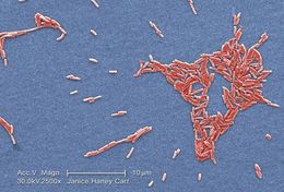
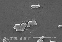
Under a magnification of 7075x, this scanning electron micrograph (SEM) depicted a number of Gram-negative Escherichia coli bacteria of the strain O157:H7, which is one of hundreds of strains of this bacterium. Although most strains are harmless, and live in the intestines of healthy humans and animals, this strain produces a powerful toxin, which can cause severe illness.E. coli O157:H7 was first recognized as a cause of illness in 1982 during an outbreak of severe bloody diarrhea; the outbreak was traced to contaminated hamburgers. Since then, most infections have come from eating undercooked ground beef.The combination of letters and numbers in the name of the bacterium refers to the specific markers found on its surface, which distinguishes it from other types of E. coli. See PHIL 10066 for a colorized version of this image.Created: 2006
-
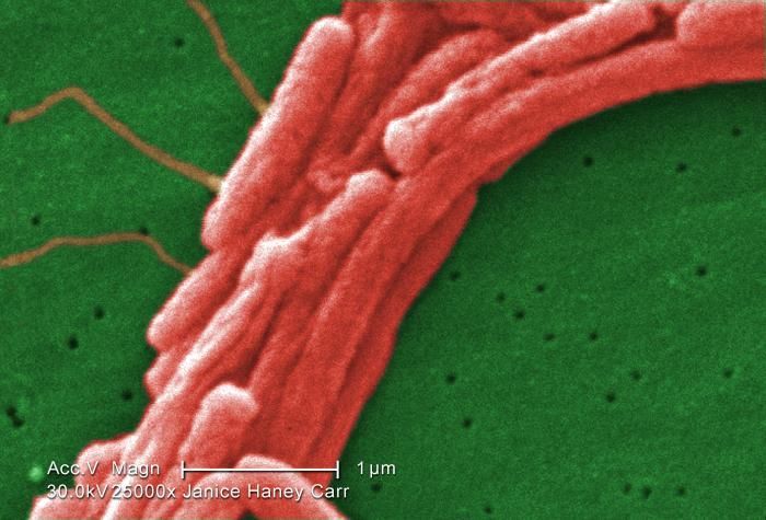
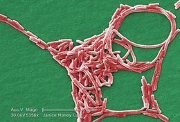
Under a moderately-high magnification of 5356X, this colorized scanning electron micrograph (SEM) depicted a grouping of Gram-negative Legionella pneumophila bacteria. Note that in some of these views youre able to see a number of the flagella emanating from these organisms. Please see PHIL 11092 through 11160 for additional SEMs of these organisms, specifically PHIL 11103 for a black and white version of this image.Youll note that a number of these bacteria seem to display an elongated-rod morphology. L. pneumophila are known to most frequently exhibit this configuration when grown in broth, however, they can also elongate when plate-grown cells age, as it was in this case, especially when theyve been refrigerated. The usual L. pneumophila morphology consists of stout, fat bacilli, which is the case for the vast majority of the organisms depicted here.Created: 2009
-
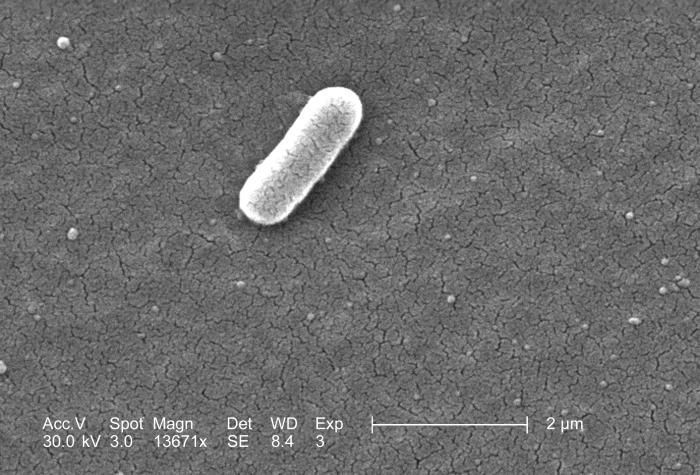
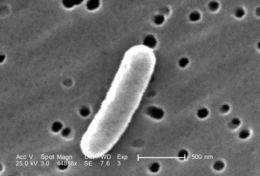
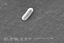
Under a high magnification of 13671x, this scanning electron micrograph (SEM) depicted a single Gram-negative Escherichia coli bacterium of the strain O157:H7, which is one of hundreds of strains of this bacterium. Although most strains are harmless, and live in the intestines of healthy humans and animals, this strain produces a powerful toxin, which can cause severe illness.E. coli O157:H7 was first recognized as a cause of illness in 1982 during an outbreak of severe bloody diarrhea; the outbreak was traced to contaminated hamburgers. Since then, most infections have come from eating undercooked ground beef.The combination of letters and numbers in the name of the bacterium refers to the specific markers found on its surface, which distinguishes it from other types of E. coli. See PHIL 10067 for a colorized version of this image.Created: 2006
-
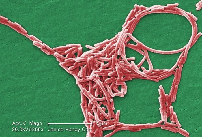
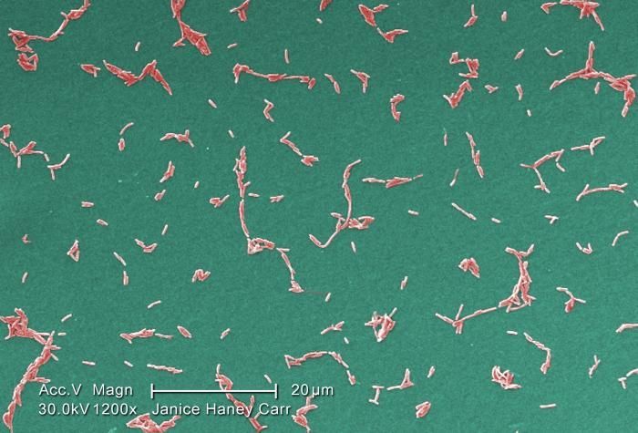
Under a moderate magnification of 1200X, this colorized scanning electron micrograph (SEM) depicted a diffuse group of Gram-negative Legionella pneumophila bacteria. Please see PHIL 11092 through 11152 for additional SEMs of these organisms, specifically PHIL 11101 for a black and white version of this image.Youll note that a number of these bacteria seem to display an elongated-rod morphology. L. pneumophila are known to most frequently exhibit this configuration when grown in broth, however, they can also elongate when plate-grown cells age, as it was in this case, especially when theyve been refrigerated. The usual L. pneumophila morphology consists of stout, fat bacilli, which is the case for the vast majority of the organisms depicted here. These bacteria originated on a 1 week-old culture plate (+/- 1 day), which had incubated a single colony, at 37oC upon a buffered charcoal yeast extract (BCYE) medium with no antibiotics.Created: 2009
-
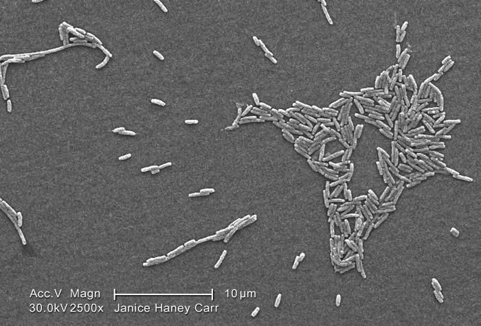
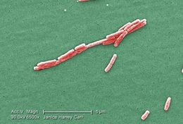
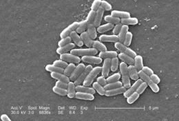
Under a magnification of 6836x, this scanning electron micrograph (SEM) depicted a number of Gram-negative Escherichia coli bacteria of the strain O157:H7, which is one of hundreds of strains of this bacterium. Although most strains are harmless, and live in the intestines of healthy humans and animals, this strain produces a powerful toxin, which can cause severe illness.E. coli O157:H7 was first recognized as a cause of illness in 1982 during an outbreak of severe bloody diarrhea; the outbreak was traced to contaminated hamburgers. Since then, most infections have come from eating undercooked ground beef.The combination of letters and numbers in the name of the bacterium refers to the specific markers found on its surface, which distinguishes it from other types of E. coli.Created: 2006