Image of Legionella pneumophila
Description:
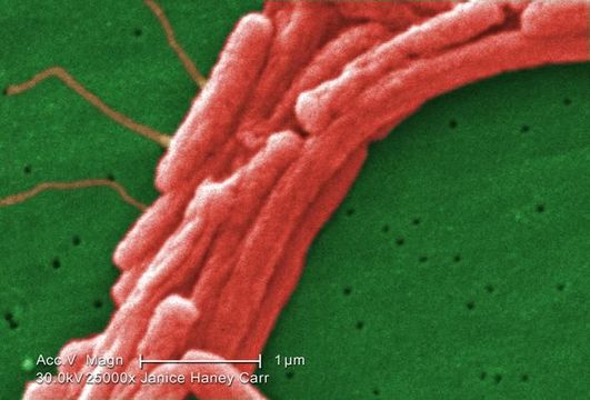
Under a very high magnification of 25000X, this colorized scanning electron micrograph (SEM) depicted a grouping of Gram-negative Legionella pneumophila bacteria. Note that in this view youre able to see a number of the flagella emanating from these organisms. Please see PHIL 11092 through 11152 for additional SEMs of these organisms, specifically PHIL 11105 for a black and white version of this image.
Youll note that a number of these bacteria seem to display an elongated-rod morphology. L. pneumophila are known to most frequently exhibit this configuration when grown in broth, however, they can also elongate when plate-grown cells age, as it was in this case, especially when theyve been refrigerated. The usual L. pneumophila morphology consists of stout, fat bacilli, which is the case for the vast majority of the organisms depicted here.
Created: 2009
Included On The Following Pages:
- Life (creatures)
- Cellular (cellular organisms)
- Bacteria
- Proteobacteria (Purple Bacteria & relatives)
- Gammaproteobacteria
- Legionellales
- Legionellaceae
- Legionella
- Legionella pneumophila
This image is not featured in any collections.
Source Information
- license
- cc-publicdomain
- photographer
- Janice Haney Carr
- provider
- Public Health Image Library
- original
- original media file
- visit source
- partner site
- Public Health Image Library
- ID