-
Michael G. Rix, Mark S. Harvey
Zookeys
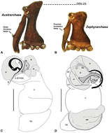
Figure 4.Diagnostic characters of Zephyrarchaea gen. n. and Austrarchaea Forster & Platnick. A–B, Cephalothorax, lateral view, showing differences in carapace height and the position of accessory setae on male chelicerae: A, male Austrarchaea harmsi Rix & Harvey; B, male Zephyrarchaea marki sp. n. C–D, Expanded male pedipalps, retro-ventral view, showing differences in the articulation and fusion of the conductor sclerites: C, Austrarchaea helenae Rix & Harvey; D, Zephyrarchaea marae sp. n. bH = basal haematodocha; C = conductor; C1–2 = conductor sclerites 1–2; Cy = cymbium; E = embolus; Es = embolic sclerite; H = distal haematodocha; T = tegulum; (TS)1–3 = tegular sclerites 1–3. Scale bars: C–D = 0.2 mm.
-
Michael G. Rix, Mark S. Harvey
Zookeys
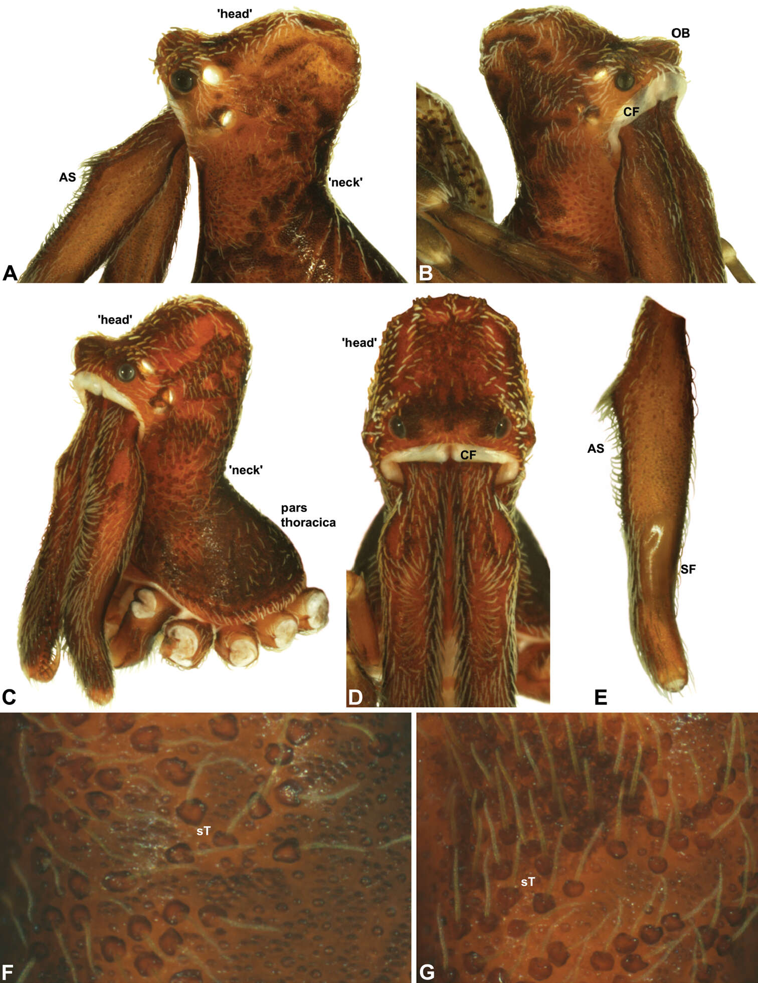
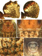
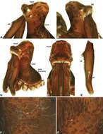
Figure 5.Carapace morphology of Zephyrarchaea species. A–B, Zephyrarchaea mainae (Platnick): A, male pars cephalica, dorso-lateral view, showing accessory setae (AS) on and adjacent to proximal cheliceral bulge; B, female pars cephalica, antero-lateral view, showing cheliceral foramen (CF) and ocular bulge (OB). C–D, Zephyrarchaea marae sp. n.: C, male cephalothorax, antero-lateral view; D, male pars cephalica and chelicerae, frontal view, showing dorsal ‘head’ region and cheliceral foramen (CF). E, Cheliceraeof male Zephyrarchaea mainae, lateral view, showing proximal accessory setae (AS) and ectal stridulatory file (SF). F–G, Detail of carapace of male Zephyrarchaea marae, lateral view, showing granulate cuticle and setose tubercles (sT).
-
Michael G. Rix, Mark S. Harvey
Zookeys
Figure 6.Abdominal morphology of Zephyrarchaea species. A–B, Male abdomens, dorso-lateral view, showing dorsal scutes (S) and additional dorsal sclerites (ds) on hump-like tubercles: A, Zephyrarchaea mainae (Platnick); B, Zephyrarchaea marae sp. n. C, Female epigastric region of Zephyrarchaea mainae, ventral view, showing setose book lung covers (BL) and median genital plate (GP). D, Male epigastric region of Zephyrarchaea mainae, ventral view, showing fusion of epigastric sclerites. E, Abdominal cuticle of male Zephyrarchaea marae, lateral view, showing sclerotic spots (ss) surrounded by short setae. F, Spinnerets of female Zephyrarchaea mainae, posterior view (ventral side uppermost), showing anterior lateral (ALS) and posterior lateral (PLS) spinnerets anterior to anal tubercle (AT), and the absence of a colulus.
-
Michael G. Rix, Mark S. Harvey
Zookeys
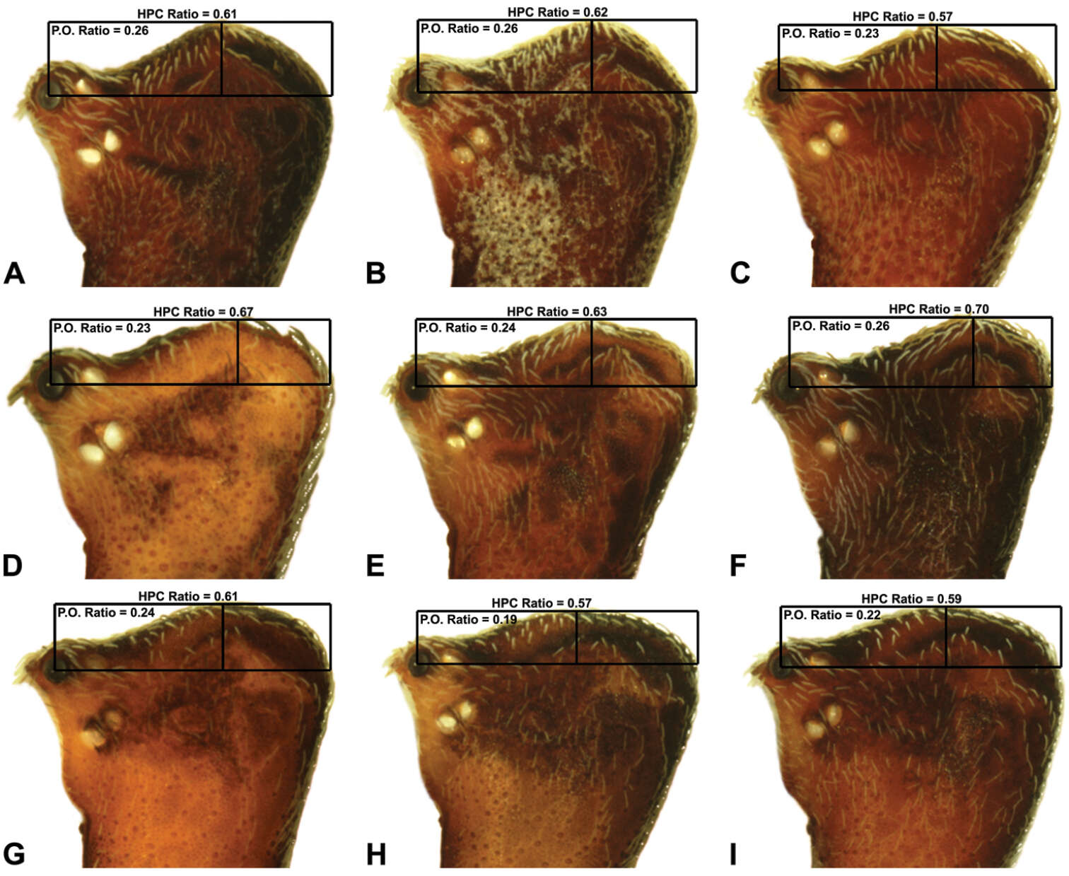
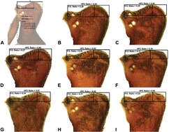
Figure 8.Lateral ‘head’ profiles of males of species of Zephyrarchaea, showing variation in carapace shape as quantified by the post-ocular ratio (P.O. Ratio) and ratio of highest point of carapace relative to post-ocular length (HPC Ratio): A, holotype Zephyrarchaea marae sp. n., showing the derivation of morphometric ratios; B, holotype Zephyrarchaea vichickmani sp. n.; C, holotype Zephyrarchaea marae sp. n.; D, holotype Zephyrarchaea porchi sp. n.; E, holotype Zephyrarchaea melindae sp. n.; F, holotype Zephyrarchaea barrettae sp. n.; G, holotype Zephyrarchaea mainae (Platnick, 1991b); H, holotype Zephyrarchaea marki sp. n.; I, holotype Zephyrarchaea janineae sp. n.
-
Michael G. Rix, Mark S. Harvey
Zookeys
Figure 9.Lateral ‘head’ profiles of females of species of Zephyrarchaea, showing variation in carapace shape as quantified by the post-ocular ratio (P.O. Ratio) and ratio of highest point of carapace relative to post-ocular length (HPC Ratio) (see Fig. 8): A, allotype Zephyrarchaea vichickmani sp. n.; B, allotype Zephyrarchaea marae sp. n.; C, holotype Zephyrarchaea grayi sp. n.; D, holotype Zephyrarchaea austini sp. n.; E, Zephyrarchaea mainae (Platnick, 1991b); F, allotype Zephyrarchaea janineae sp. n.; G, holotype Zephyrarchaea robinsi (Harvey, 2002a); H, allotype Zephyrarchaea melindae sp. n.; I, allotype Zephyrarchaea barrettae sp. n.
-
Michael G. Rix, Mark S. Harvey
Zookeys
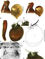
Figure 17.Zephyrarchaea marae sp. n. A–B, Cephalothorax and abdomen, lateral view: A, allotype female (MV K5921) from Gunyah Rainforest State Reserve, Victoria; B, holotype male (MV K11580) from Tarra-Bulga National Park, Victoria. C, Holotype male chelicerae, lateral view, showing accessory setae. D–F, Holotype male pedipalp: D–E, bulb, retrolateral view, with inset showing twisted apex of tegular sclerite 1 in retroventral view; F, detail of distal tegular sclerites, prolateral view. G, Allotype female internal genitalia, dorsal view. C1–2 = conductor sclerites 1–2; E = embolus; GP = genital plate; T = tegulum; (TS)1–3 = tegular sclerites 1–3. Scale bars: A–B = 1.0 mm; E = 0.2 mm.
-
Michael G. Rix, Mark S. Harvey
Zookeys
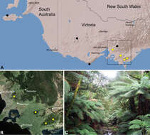
Figure 27.Zephyrarchaea marae sp. n., distribution and habitat: A, topographic map showing the known distribution of Archaeidae in Victoria and South Australia, with collection localities for Zephyrarchaea marae highlighted in yellow; B, satellite image showing detail of inset (A); C, cool-temperate Nothofagus rainforest at the type locality – Tarra Valley, Tarra-Bulga National Park, Victoria (April 2010). Image (C) by M. Rix.
-

Description: English: Zephyrarchaea marae. A–B, Cephalothorax and abdomen, lateral view: A, allotype female (MV K5921) from Gunyah Rainforest State Reserve, Victoria; B, holotype male (MV K11580) from Tarra-Bulga National Park, Victoria. C, Holotype male chelicerae, lateral view, showing accessory setae. D–F, Holotype male pedipalp: D–E, bulb, retrolateral view, with inset showing twisted apex of tegular sclerite 1 in retroventral view; F, detail of distal tegular sclerites, prolateral view. G, Allotype female internal genitalia, dorsal view. C1–2 = conductor sclerites 1–2; E = embolus; GP = genital plate; T = tegulum; (TS)1–3 = tegular sclerites 1–3. Scale bars: A–B = 1.0 mm; E = 0.2 mm. Date: 7 May 2012. Source: Rix M, Harvey M (2012) Australian Assassins, Part II: A review of the new assassin spider genus Zephyrarchaea (Araneae, Archaeidae) from southern Australia. ZooKeys 191: 1-62.
doi:10.3897/zookeys.191.3070. Author:
Michael G. Rix and Mark S. Harvey, Western Australia Museum.
: This image is uploaded as part of a
collaboration between
Wikispecies and
ZooKeys বাংলা |
català |
čeština |
Deutsch |
English |
français |
italiano |
македонски |
polski |
português do Brasil |
русский |
+/−. : This file is licensed under the
Creative Commons Attribution 3.0 Unported license. :. You are free: to share – to copy, distribute and transmit the work to remix – to adapt the work Under the following conditions: attribution – You must give appropriate credit, provide a link to the license, and indicate if changes were made. You may do so in any reasonable manner, but not in any way that suggests the licensor endorses you or your use. https://creativecommons.org/licenses/by/3.0 CC BY 3.0 Creative Commons Attribution 3.0 truetrue.
-

Description: English: Abdominal morphology of Zephyrarchaea species. A–B, Male abdomens, dorso-lateral view, showing dorsal scutes (S) and additional dorsal sclerites (ds) on hump-like tubercles: A, Zephyrarchaea mainae (Platnick); B, Zephyrarchaea marae. C, Female epigastric region of Zephyrarchaea mainae, ventral view, showing setose book lung covers (BL) and median genital plate (GP). D, Male epigastric region of Zephyrarchaea mainae, ventral view, showing fusion of epigastric sclerites. E, Abdominal cuticle of male Zephyrarchaea marae, lateral view, showing sclerotic spots (ss) surrounded by short setae. F, Spinnerets of female Zephyrarchaea mainae, posterior view (ventral side uppermost), showing anterior lateral (ALS) and posterior lateral (PLS) spinnerets anterior to anal tubercle (AT), and the absence of a colulus. Date: 7 May 2012. Source: Rix M, Harvey M (2012) Australian Assassins, Part II: A review of the new assassin spider genus Zephyrarchaea (Araneae, Archaeidae) from southern Australia. ZooKeys 191: 1-62.
doi:10.3897/zookeys.191.3070. Author:
Michael G. Rix and Mark S. Harvey, Western Australia Museum.
: This image is uploaded as part of a
collaboration between
Wikispecies and
ZooKeys বাংলা |
català |
čeština |
Deutsch |
English |
français |
italiano |
македонски |
polski |
português do Brasil |
русский |
+/−.mw-parser-output.responsive-license-cc{clear:both;text-align:center;box-sizing:border-box;width:100%;justify-content:space-around;align-items:center;margin:0.5em auto;background-color:#f9f9f9;border:2px solid #e0e0e0;border-spacing:8px;display:flex}.mw-parser-output.responsive-license-cc div{margin:4px}.mw-parser-output.rlicense-text div{margin:0.5em auto}@media screen and (max-width:640px){.mw-parser-output.responsive-license-cc{flex-flow:column}.mw-parser-output.rlicense-text{order:1}} This file is licensed under the
Creative Commons Attribution 3.0 Unported license. You are free: to share – to copy, distribute and transmit the work to remix – to adapt the work Under the following conditions: attribution – You must give appropriate credit, provide a link to the license, and indicate if changes were made. You may do so in any reasonable manner, but not in any way that suggests the licensor endorses you or your use. https://creativecommons.org/licenses/by/3.0CC BY 3.0 Creative Commons Attribution 3.0 truetrue.
-

Description: English: Carapace morphology of Zephyrarchaea species. A–B, Zephyrarchaea mainae (Platnick): A, male pars cephalica, dorso-lateral view, showing accessory setae (AS) on and adjacent to proximal cheliceral bulge; B, female pars cephalica, antero-lateral view, showing cheliceral foramen (CF) and ocular bulge (OB). C–D, Zephyrarchaea marae. C, male cephalothorax, antero-lateral view; D, male pars cephalica and chelicerae, frontal view, showing dorsal ‘head’ region and cheliceral foramen (CF). E, Chelicerae of male Zephyrarchaea mainae, lateral view, showing proximal accessory setae (AS) and ectal stridulatory file (SF). F–G, Detail of carapace of male Zephyrarchaea marae, lateral view, showing granulate cuticle and setose tubercles (sT). Date: 7 May 2012. Source: Rix M, Harvey M (2012) Australian Assassins, Part II: A review of the new assassin spider genus Zephyrarchaea (Araneae, Archaeidae) from southern Australia. ZooKeys 191: 1-62.
doi:10.3897/zookeys.191.3070. Author:
Michael G. Rix and Mark S. Harvey, Western Australia Museum.
: This image is uploaded as part of a
collaboration between
Wikispecies and
ZooKeys বাংলা |
català |
čeština |
Deutsch |
English |
français |
italiano |
македонски |
polski |
português do Brasil |
русский |
+/−.mw-parser-output.responsive-license-cc{clear:both;text-align:center;box-sizing:border-box;width:100%;justify-content:space-around;align-items:center;margin:0.5em auto;background-color:#f9f9f9;border:2px solid #e0e0e0;border-spacing:8px;display:flex}.mw-parser-output.responsive-license-cc div{margin:4px}.mw-parser-output.rlicense-text div{margin:0.5em auto}@media screen and (max-width:640px){.mw-parser-output.responsive-license-cc{flex-flow:column}.mw-parser-output.rlicense-text{order:1}} This file is licensed under the
Creative Commons Attribution 3.0 Unported license. You are free: to share – to copy, distribute and transmit the work to remix – to adapt the work Under the following conditions: attribution – You must give appropriate credit, provide a link to the license, and indicate if changes were made. You may do so in any reasonable manner, but not in any way that suggests the licensor endorses you or your use. https://creativecommons.org/licenses/by/3.0CC BY 3.0 Creative Commons Attribution 3.0 truetrue.


