Nematodes within the Secernentea have phasmids, which are unicellular glands. Phasmids likely function as chemoreceptors. Females may produce pheromones to attract males.
Nematodes in general have papillae, setae and amphids as the main sense organs. Setae detect motion (mechanoreceptors), while amphids detect chemicals (chemoreceptors).
Communication Channels: tactile ; chemical
Other Communication Modes: pheromones
Perception Channels: tactile ; chemical
The life cycle of S. vulgaris involves five juvenile stages and its equine host. Eggs are found in its host feces, where they hatch and the juveniles feed on the feces through the third larval stage. At this point, they crawl onto vegetation the equines feed on, where they are ingested by the host. Once in the small intestine, the third-stage juvenile penetrates through the intestinal wall and molts into its fourth larval stage. The larvae penetrate the surrounding arteries and make their way to the mesenteric arteries, where they develop into the fifth larval stage, immature adults. The immature adults make their way back to the intestines, where they encapsulate themselves and develop into adults. The adults hatch from the capsules and mate, producing eggs which are passed out in the feces.
Strongylus vulgaris has at times been a very common parasite in horses; it was estimated that it was present in 90% to 100% of horses in the U.S. The inflammation caused by the strongyle travelling throughout the arteries and intestines can cause blood clots to form. The clots can block oxygen passage to the intestines, causing parts of them to die. Ultimately, besides causing abdominal pain (colic) in equines, the complications from S. vulgaris infestation can lead to death.
S. vulgaris is no longer considered as much of a threat to horses, because it can be controlled by drugs such as benzimidazoles, ivermectin, and moxidectin, which kill both larvae and adult stages of the worm.
Negative Impacts: causes or carries domestic animal disease
Strongylus vulgaris is parasitic on horses.
Ecosystem Impact: parasite
Species Used as Host:
Through the first three larval stages of development S. vulgaris feeds upon the feces of its parent's host. The later larval stages are found in the circulatory system of equines, feeding on blood and tissue as it makes its way through different areas of the body. The adults are primarily found in the intestine and associated organs, where their feeding can cause major damage to the gastrointestinal organs.
Animal Foods: blood; body fluids
Other Foods: dung; microbes
Primary Diet: carnivore (Eats body fluids); coprophage
This parasite can be found worldwide; its dispersal is dependent on where its hosts (equines) are found.
Biogeographic Regions: nearctic ; palearctic ; oriental ; ethiopian ; neotropical ; australian
Other Geographic Terms: cosmopolitan
Strongylus vulgaris is found primarily in grasslands and pastures. They are essentially located where equine populations are concentrated.
Habitat Regions: temperate ; tropical ; terrestrial
Terrestrial Biomes: tundra ; taiga ; desert or dune ; savanna or grassland ; chaparral ; forest ; rainforest ; scrub forest ; mountains
Wetlands: marsh ; swamp
Other Habitat Features: urban ; suburban ; agricultural
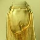
These worms are cylindrical, dark red in color, with male adults approximately 2.5 to 4 cm in length, while females range from 4 to 6 cm. There are five larval stages before adulthood is reached. The buccal capsule, which adults use to feed, is relatively large and heavily sclerotized, with two lobed teeth. The anterior region is dorsally curved, giving the appearance characteristic of hookworms.
The cuticle has three or more main outer layers made of collagen and other compounds. The outer layers are non-cellular and are secreted by the epidermis. The cuticle layer protects the nematodes so they can invade the digestive tracts of animals.
Nematodes have longitudinal muscles along the body wall. The muscles are obliquely arranged in bands. Dorsal, ventral and longitudinal nerve cords are connected to the main body of the muscle.
Range length: 2.5 to 6 cm.
Other Physical Features: ectothermic ; heterothermic ; bilateral symmetry
Sexual Dimorphism: female larger; sexes shaped differently
These parasites are probably not preyed on directly, but are likely ingested by various animals. Larval mortality is high as most of the parasites do not reach appropriate hosts.
Females may produce a phermomone to attract males. The male coils around a female with his curved area over the female genital pore. The gubernaculum, made of cuticle tissue, guides spicules which extend through the cloaca and anus. Males use spicules to hold the female during copulation. Nematode sperm are amoeboid-like and lack flagella.
Key Reproductive Features: sexual ; fertilization (Internal ); oviparous
Parental Investment: pre-fertilization (Provisioning)
Palisadeorm (latin: Strongylus vulgaris) er en rundorm, der som udvokset er en blodsugende tarmsnylter hos hesten.
Der Pferdepalisadenwurm (Strongylus vulgaris) ist ein Endoparasit, der im Dickdarm von Pferd, Esel, Maultier und Zebra lebt. Er zählt zu den Rundwürmern und zur Parasitengruppe der großen Strongyliden. Von den Palisadenwürmern sind etwa 50 Arten beim Pferd bekannt.[1][2] Er entwickelt sich teilweise im Freien und wird ohne Zwischenwirt direkt vom Wirt oral aufgenommen.
Das Männchen erreicht eine Größe von 1,9 cm, das Weibchen eine Größe von bis zu 2,5 cm.
Die infektiöse Larve wird mit der Nahrung aufgenommen. In Blind- und Dickdarm bohren sich die Larven dann in die untere Darmschleimhaut ein und entwickeln sich dort in den Darmarterien zum adulten Tier. Nach dieser Entwicklung wandern sie zurück in den Darm. Dort werden erneut Eier gebildet, die dann mit dem Kot ausgeschieden werden. Dort entwickelt sich die Larve bis zu ihrem dritten Juvenilstadium.
Durch einen Massenbefall der Larven in den Darmarterien kommt es zu Gefäßerweiterungen. Es kann dort zu Blutgerinnseln führen. Da der Pferdepalisadenwurm Blut saugt, kann es auch dadurch zu Schädigungen kommen. Allerdings treten meist nur geringe Entzündungen auf.
Die Larven der kleinen Strongyliden (Cyathostominae) verkapseln sich während der Wintermonate, danach kann es zum Massenbefall (simultanes Auswandern, sog. larvale Cyathostominose) kommen mit Koliken, Durchfall und Abmagerung – es treten schwerwiegende Schleimhauterosionen im Dick- und Blinddarm auf.[1][2]
Oral zu verabreichende Anthelminthika (Wurmmittel) zeigen unterschiedliche Wirksamkeit. Manche Substanzen können das verkapselte Stadium nicht bekämpfen, gegen andere (Benzimidazole) haben die Parasiten bereits Resistenzen entwickelt[3].
Man versucht, die Übertragung durch Hygiene (Kot entfernen, Weide wechseln) zu begrenzen, jedoch sind auch Wildtiere Träger. Pferde haben sehr unterschiedliche Befallsintensitäten, die meisten[1] sind Träger, bestimmte und besonders ältere Pferde scheiden jedoch wesentlich mehr Eier aus als andere. Man versucht, Resistenzentwicklung zu vermindern, indem man nur den Tieren Wurmmittel verabreicht, die eine bestimmte Eierkonzentration im Kot überschreiten (z. B. 200 pro Gramm[4][3]).

Der Pferdepalisadenwurm (Strongylus vulgaris) ist ein Endoparasit, der im Dickdarm von Pferd, Esel, Maultier und Zebra lebt. Er zählt zu den Rundwürmern und zur Parasitengruppe der großen Strongyliden. Von den Palisadenwürmern sind etwa 50 Arten beim Pferd bekannt. Er entwickelt sich teilweise im Freien und wird ohne Zwischenwirt direkt vom Wirt oral aufgenommen.
Strongylus vulgaris (large strongyles),[1] commonly known as the blood worm,[2] is a common horse parasite in the phylum Nematoda. It looks like a long worm with a large biting mouth. They are usually reddish in color because of all the blood they take from the equine host.[1] This nematode is considered to be one of the "most pathogenic" of the large strongyles subphylum and is distributed worldwide, wherever there are grassland and temperate environments.[3]
During the infective stage of the S. vulgaris life cycle, the larvae that have matured in the intestinal tract of the horse will migrate into the surrounding blood vessels. Once in the blood vessels, the larvae will continue their migration throughout the body to various organs causing damage to the blood vessels along the way. This can cause anemia or blockage of blood flow caused by the detachment of blood clots from the vessel wall resulting in tissue death.[4] S. vulgaris are commonly found in pastures and stalls where feces are present. The larvae crawl up on the grass which is where they are then eaten by the horse. Harsh environments, such as freezing, do not kill S. vulgaris.[5]
Symptoms can range depending on how severe the infection is. In mild cases the most common clinical signs are weightloss, compromised performance, and a dull hair coat.[6] Other clinical signs can range from diarrhea, weakness, anorexia, anemia if there are significant blood loss and abdominal discomfort. Severe cases can show signs of having severe colic, rupture of the intestines, and death.[7] These signs may also be observed with other equine-related infections or illnesses and their presence alone should not be used to diagnose an infection of S. vulgaris. A veterinarian is required for further diagnosis when infection with S. vulgaris is suspected.
Large strongyle infections are commonly diagnosed through the observations of fecal samples. These fecal samples can be viewed under a microscope to identify the eggs of S. vulgaris. Diagnosis via egg identification is not enough to determine if the horse has a S. vulgaris infection as the fecal samples often contain a mixture of large and small strongyle eggs that are very similar in appearance. The best diagnostic method to determine if a horse has a S. vulgaris infection is through a fecal culture. Here the eggs grow and develop into the infective larval stage that is specific to the large strongyle parasite.[8]
The adults of S. vulgaris are primarily found in the cecum and colon of infected equids and it is here that the females will lay their eggs. These eggs will pass out of the horse's body in the feces. Once outside the body the eggs will hatch and develop into the infective larval stage L3. The larvae enter the host when the horse grazes a patch of grass containing the larval stage. Back inside the host the infective larval stage migrates to the small intestine where it can enter the intestinal mucosa and further develop into the L4 stage. It is this stage that can enter the blood vessels and migrate throughout the body for a period of up to six months. During this time the L4 stage matures into the L5 or immature adult stage before returning to the intestinal wall. The L5 stage resides primarily in the cecum and colon where the males and females copulate thus starting the cycle over again.[9]
Dewormers that have ivermectin or moxidectin are the most effective against these parasites.[10] Treating a horse with regular dewormer when they have the worms inside of them may cause the worms to migrate all at once, causing rupture of the intestines and lead to death.[11] Recommendations for deworming horses include performing fecal egg counts to determine what kind of protection each individual horse requires and alternating products in the fall and spring. An example would be using ivermectin based products (Equell or Zimectrin) in the spring and moxidectin based products in combination with praziquantel (Quest Plus) in the fall or vice versa.[12] A deworming schedule should be discussed with a veterinarian.
Horses are able to pick up this parasite from food and water contaminated by feces. This can be avoided by cleaning water tanks and picking out pastures often, avoidance of feeding on the ground, and avoidance of overstocking pastures.[13] As foals can be extremely susceptible to this parasite, it is best to treat broodmares before they give birth and move them to uninfected pastures to avoid transfer.
{{cite web}}: CS1 maint: archived copy as title (link) {{cite web}}: Missing or empty |title= (help) Strongylus vulgaris (large strongyles), commonly known as the blood worm, is a common horse parasite in the phylum Nematoda. It looks like a long worm with a large biting mouth. They are usually reddish in color because of all the blood they take from the equine host. This nematode is considered to be one of the "most pathogenic" of the large strongyles subphylum and is distributed worldwide, wherever there are grassland and temperate environments.
Strongylus vulgaris es un parásito nematodo de la familia strongylidae que afecta a los equinos, se localiza en el intestino grueso pero durante su ciclo biológico realiza migraciones por el organismo de su hospedador. La contaminación ocurre por vía oral por la ingestión de la larva III, inicialmente esta llega al intestino delgado, lo perforan y se dirigen a los vasos sanguíneos; para completar su ciclo biológico deben ingresar específicamente a las arterias, si llegan a las venas o a los vasos linfáticos no son viables. No tienen un tropismo específico por las arterias, por lo tanto los parásitos que logran llegar a ellas migran contra corriente y se van adhiriendo al endotelio vascular, alimentándose de este.
Las principales arterias en las que se localiza son las arterias mesentéricas, las ilíacas o ileocólicas. Los parásitos más agresivos migran hasta la arteria aorta y al corazón, pero es poco probable.
En las arterias mencionadas anteriormente el parásito se transforma en larva IV, posteriormente se suelta del endotelio y se dirige al intestino grueso por el flujo sanguíneo en donde se convierte en larva V. Luego el parásito adulto se reproduce y los huevos son eliminados por materia fecal; en el medio ambiente los huevos eclosionan y se desarrolla la larva I y la larva II, las cuales no producen infección en el animal en caso de ser ingeridas, al desarrollarse el parásito como larva III es cuando tiene facultad infectante.
El ciclo de vida del parásito dentro del equino tiene una duración de aproximadamente 6 meses.
El curso de la enfermedad en el animal se divide en dos etapas: las lesiones que causa la larva y las que produce el parásito adulto. Lesiones que causa la larva, el endotelio vascular es lesionado cuando la larva se adhiere a éste y se alimenta, por lo tanto sobre la lesión se genera acumulación plaquetaria, que se desprende y queda libre dentro del vaso sanguíneo formando un émbolo que generará isquemia por obstrucción de los vasos sanguíneos. Dependiendo en que arteria este localizado el parásito se producirán cólicos de tipo infartante, o claudicaciones en caliente si la larva se localiza en iliacas y compromete la circulación del tren posterior, la causa de la cojera es debido a la hipoxia que sufren los músculos y por lo tanto el metabolismo anaerobio produce ácido láctico que estimula receptores nociceptivos.
En algunos casos se puede producir aneurismas ya que al lesionar al endotelio éste se hace más delgado y los glóbulos rojos hacen presión sobre la pared. Daños que causa el adulto, se produce colitis, diarreas, hematoquesia, secreción mucosa, posible contaminación bacteriana secundaria.
Strongylus vulgaris es un parásito nematodo de la familia strongylidae que afecta a los equinos, se localiza en el intestino grueso pero durante su ciclo biológico realiza migraciones por el organismo de su hospedador. La contaminación ocurre por vía oral por la ingestión de la larva III, inicialmente esta llega al intestino delgado, lo perforan y se dirigen a los vasos sanguíneos; para completar su ciclo biológico deben ingresar específicamente a las arterias, si llegan a las venas o a los vasos linfáticos no son viables. No tienen un tropismo específico por las arterias, por lo tanto los parásitos que logran llegar a ellas migran contra corriente y se van adhiriendo al endotelio vascular, alimentándose de este.
Las principales arterias en las que se localiza son las arterias mesentéricas, las ilíacas o ileocólicas. Los parásitos más agresivos migran hasta la arteria aorta y al corazón, pero es poco probable.
En las arterias mencionadas anteriormente el parásito se transforma en larva IV, posteriormente se suelta del endotelio y se dirige al intestino grueso por el flujo sanguíneo en donde se convierte en larva V. Luego el parásito adulto se reproduce y los huevos son eliminados por materia fecal; en el medio ambiente los huevos eclosionan y se desarrolla la larva I y la larva II, las cuales no producen infección en el animal en caso de ser ingeridas, al desarrollarse el parásito como larva III es cuando tiene facultad infectante.
El ciclo de vida del parásito dentro del equino tiene una duración de aproximadamente 6 meses.
De grote bloedworm (Strongylus vulgaris) is de naam van een parasitaire maagdarmworm uit de familie van de Strongylidae uit de orde Strongylida van de Nematoda. Het zijn rondwormen die leven als parasiet in het spijsverteringskanaal en de slagaderen van paarden. Deze bloedworm behoort tot de zogenaamde grote strongyliden waartoe ook rondwormen met een vergelijkbare leefwijze behoren zoals Strongylus edentatus, S. equinus en soorten uit de geslachten Triodontophorus en Craterostomum.
De besmetting van gezonde paarden met deze worm begint als paarden grazen in een weiland waarin besmette paarden hebben gelopen en hun ontlasting hebben achtergelaten. Hierin zitten volwassen wormen en eitjes die door de mest tegen uitdroging worden beschermd. Deze eitjes worden na één tot twee weken besmettelijke larven in het stadium L3. Deze larven in het milieu kunnen het twee jaar uithouden. Bij het grazen komen deze larven in de dikke darm van het paard en dringen door de darmwand en verplaatsen zich langs de wanden van de slagaders, tegen de stroom in, van de dikke darm naar de grote lichaamsslagader. In de bloedbaan doorlopen de larven een volgend stadium. Daarna dringen ze weer via de darmwand de darmen binnen en worden daar binnen zes tot acht weken volwassen.
De larven in de bloedbaan kunnen leiden tot ontstekingen in de darmslagader waardoor bloedpropjes ontstaan die elders in de slagaders die naar de darmen lopen verstoppingen (infarcten) veroorzaken. Stukken darmwand kunnen daardoor afsterven en in het ergste geval leiden tot een koliek die fataal wordt voor het paard.
Door het gebruik van goede ontwormingsmiddelen is de grote bloedworm de laatste decennia afgenomen. Op veel paardenbedrijven komen grote strongyliden niet meer voor.
Het woord bloedworm wordt ook wel gebruikt voor de larven van dansmuggen (geslacht Chironomus) die gebruikt worden als aas en voedsel voor aquariumvissen.
Bronnen, noten en/of referentiesDe grote bloedworm (Strongylus vulgaris) is de naam van een parasitaire maagdarmworm uit de familie van de Strongylidae uit de orde Strongylida van de Nematoda. Het zijn rondwormen die leven als parasiet in het spijsverteringskanaal en de slagaderen van paarden. Deze bloedworm behoort tot de zogenaamde grote strongyliden waartoe ook rondwormen met een vergelijkbare leefwijze behoren zoals Strongylus edentatus, S. equinus en soorten uit de geslachten Triodontophorus en Craterostomum.
Strongylus vulgaris[1] är en rundmaskart som först beskrevs av Looss 1900. Strongylus vulgaris ingår i släktet Strongylus och familjen Strongylidae.[1][2][3] Arten är reproducerande i Sverige.[3] Inga underarter finns listade i Catalogue of Life.[1]
Strongylus vulgaris är den farligaste inälvsparasiten som förekommer hos hästar och den kallas även för stora blodmasken[4]. Strongylus vulgaris är den vanligaste av tre arter och den förekommer över hela världen. Förekomsten är dock varierande beroende på geografiskt läge, då klimatförhållanden skapar olika spridningsmönster, men även betestrycket verkar spela in. I en studie utförd av Eva Osterman Lind var det högre förekomst av ägg i träcken i södra Sverige (50 %) än i de mellersta (37 %) och norra (13 %) delarna av landet. I Sverige rapporterades det under 1949 att cirka 90 % av hästarna som obducerades hade artärbråck som orsakats av Strongylus vulgaris[5]. Under 1970-talet påvisades ägg från Strongylus vulgaris i 65-70 % av de svenska hästarna men på 2000-talet har andelen positiva minskat till 5 %, då det nu finns effektivare avmaskningsmedel[6]. För att inte parasiterna ska utveckla resistens mot avmaskningsmedlet är det viktigt att inte avmaska i onödan. Istället kan parasitproblemet förebyggas med betesplanering och minskning av parasitsmitta på betet[7]. Under våren sker den största ökningen av ägg i träcken. Dessa ägg är producerade av larver som intogs föregående säsong och har levt i hästen under vintern. Den högsta halten av ägg kan ses under juli-september då förhållandena är optimala för utveckling av de frilevande larvstadierna på betesmarkerna[5].
Alla rundmaskar har en cylindrisk form och kroppen är täckt med en färglös kutikula. Det som skiljer Strongylus vulgaris från övriga arter är framförallt huvudet med sina väl utvecklade mundelar med karakteristiska tänder[5]. De vuxna maskarna är 2-3 cm långa och har en rödbrun färg[4].
När en häst blir infekterad av Strongylus vulgaris sker en lång utvecklingsperiod på 6 månader innan man ser spår av ägg i träcken. Äggen som kommer ut med träcken utvecklas under goda förhållanden till första larvstadiet (L1) inom 2-4 dagar[5]. Äggen tål temperaturer under 5°C men vid denna temperatur utvecklas de inte, utan befinner sig i ett vilostadium[8]. I motsats till äggen är larverna i första larvstadiet (L1) känsliga för att torka ut och även för låga temperaturer[9]. Larver som befinner sig i detta stadium lever i träcken och livnär sig på bakterier. De utvecklas vidare till larvstadium två (L2) som klarar av torka bättre men inte låga temperaturer[5]. Vid höga temperaturer, upp till 35°C, kan ägg utvecklas till larver i tredje stadiet (L3) på tre dagar, medan det tar längre tid vid lägre temperaturer[8]. L3-larver klarar av extrem kyla och de klarar sig ännu bättre om ett snötäcke skyddar mot klimatets variationer[5]. De larver som är farliga är de som utvecklas från ägg i den begynnande betessäsongen. Under sensommarens nederbörd ser man ofta en kraftig ökning av larver som befinner sig i det tredje stadiet[6]. Det är dessa larver som hästen får i sig när den betar. I detta stadium har larverna fortfarande en porös kutikula runt sig från föregående stadium. Väl inuti hästen släpper kutikulan när larven passerar genom mag- och tarmkanalen. Larven penetrerar slemhinnorna (mucosa och submucosa) i tunntarmen, blindtarmen och en del av tjocktarmen (ventrala kolon). I submucosan utvecklas larven till det fjärde stadiet (L4) och tar sig sedan vidare in i artärerna och börjar vandra mot blodflödet[10] till krösroten som är ett stort blodkärl i tarmpaketet[6]. I dessa blodkärl finner man majoriteten av L4. Det tar 14-21 dagar för larven att ta sig dit och här tillväxer larven betydligt under 3-4 månader innan den övergår till det femte och sista stadiet (L5). I detta stadium släpper larven taget och följer med blodflödet i artärerna till väggarna på blindtarmen och ventrala kolon där de innesluter sig i subserosa som noduler, 5-8 mm i diameter. När dessa brister blir larverna fria i tarmen[5] och fäster vid tarmslemhinnan där de suger blod[6]. Här utvecklas de vidare och blir könsmogna, det tar ungefär 6-8 veckor innan de börjar producera ägg som följer med träcken ut[5].
Hästar som är infekterade med Strongylus vulgaris visar oftast inga symtom alls. Det vanligaste symptomet som uppstår vid kraftiga parasitangrepp är kolik. Då förflyttar sig larverna mellan tarm och blodkärl och orsakar inflammation och blodproppar, vilket skapar syrebristskador i tarmen. Angreppen kan även ge minskat blodflöde till tarmarna, nervskador och förändrad tarmmotorik som också kan orsaka kolik[6]. Hälta kan uppstå då små blodproppar spridit sig från tarmkärlen till benen[4]. Larverna kan även orsaka artärbråck som försvagar artärväggarna, ett resultat av deras migration mot krösroten. Föl som har utsatts för experimentella infektioner med Strongylus vulgaris har visat på feber, matvägran, depression och buksmärtor efter några veckors infektion[5]. Febern uppstår av vävnadsskadorna som Strongylus vulgaris orsakar eller på grund av toxiska substanser som larverna utsöndrar[10]. Under normala förhållanden ser man inte dessa svåra symtom. Däremot är föl och unghästar de som drabbas hårdast av parasitangrepp och lider större risk att drabbas av kolik. Kolik som uppstår av Strongylus vulgaris sker vanligtvis på vintern då den högsta förekomsten av larver uppträder i artärerna[5] som förser tarmen med syrerikt blod[6]. Hästar som tidigare i livet har utsatts för infektion av Strongylus vulgaris har ofta immunitet när de blir infekterade senare i livet och de uppvisar även färre symtom[5].
Det är svårt att fastställa en tidig infektion av Strongylus vulgaris[10]. Det vanligaste är att påvisa ägg som kommer ut med träcken. Med denna metod är det svårt att fastställa exakt vilken art äggen tillhör och för artbestämning måste man odla fram larverna under 10-14 dagar i 20-25°C. Larverna utvecklas då till det tredje stadiet (L3) och kan därmed artbestämmas[5]. Med hjälp av mikroskop kan man särskilja den farliga Strongylus vulgaris från den lilla blodmasken, som är betydligt ofarligare[11]. Det är även möjligt att använda sig av molekylära tekniker för att identifiera vilken art äggen tillhör[12].
Om det har bekräftats att hästen lider av en Strongylus vulgaris-infektion bör hästen behandlas med avmaskningsmedel. Det är dock viktigt att inte avmaska i onödan då resistens kan utvecklas hos inälvsmaskarna. Det är bra att försöka identifiera de hästar i en grupp, vanligtvis 20 %, som är bärare av inälvsparasiter. Denna minoritet av individer brukar stå för 80-90 % av produktionen av maskägg, det bästa är att enbart avmaska dessa bärare och inte hela flocken[13].
För att minska användningen av avmaskningsmedel är det bra att förebygga parasitsmitta i hagarna och därmed minska spridningen av resistens mot avmaskningsmedel hos parasiterna. Betesplanering är ett alternativ för att förebygga parasitsmitta, man skiljer då på vinter- och sommarhagar. Ston med föl och unghästar bör släppas på parasitfria beten. Man bör byta beteshagar innan växtligheten är nedbetad för att inte tvinga hästarna att beta nära gödselhögar som leder till att de får i sig parasitlarver[7]. Det har visat sig vara bra att ta bort träcken från hagarna minst en gång i veckan, för att vara säker på att L3-larver inte utvecklas och sprider sig till omkringliggande gräsmarker. Detta är minst lika effektivt eller bättre än att använda avmaskningsmedel mot parasitsmitta på betet. Det har länge varit känt att man ska sprida ut gödselhögarna för att minska förekomsten av smitta, vilket stämmer i torrt och soligt väder då larverna dör, det har däremot en omvänd effekt om det är fuktigt. Larverna sprids då över stora områden vilket leder till ökad risk för parasitsmitta[5]. För att minska parasitsmittan på betet kan man låta hästar växelbeta med nötkreatur och/eller får, då Strongylus vulgaris är artspecifik. Betet kan också vila ett helt år från betande djur för att nästkommande år ha en mycket låg smitta. Eventuellt kan det vara ett alternativ att låta betet vila från hästar under första halvan av säsongen och släppa djuren på bete först i juli[7].
Strongylus vulgaris är en rundmaskart som först beskrevs av Looss 1900. Strongylus vulgaris ingår i släktet Strongylus och familjen Strongylidae. Arten är reproducerande i Sverige. Inga underarter finns listade i Catalogue of Life.
