-
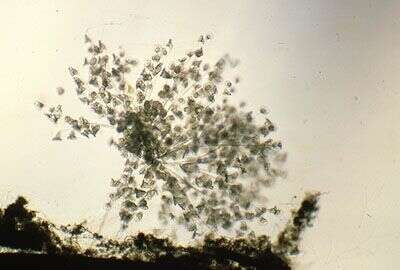
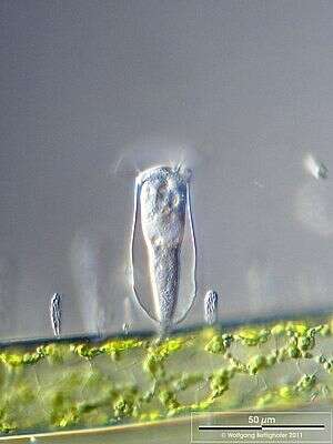
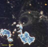
This peritrich is one of a number of genera that occurs in colonies. It is distinguishable from Zoothamnium in that the whole colony does not contract together when a single cell is stimulated. feeds on bacteria. Bright field illumination.
-
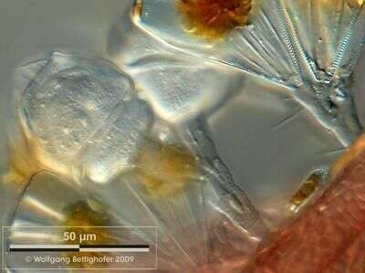
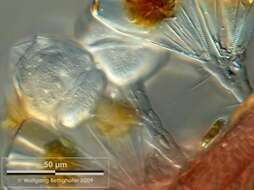
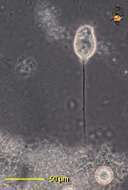
Scale bar indicates 50 µm. Collected from Bodden, the brackish waters lying between the isles of Hiddensee and Ruegen (German Baltic Sea). The image was built up using several photomicrographic frames with manual stacking technique. This image was taken using Zeiss Universal with Olympus C7070 CCD camera.
-
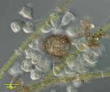
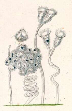
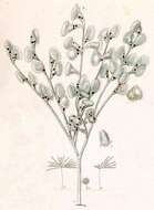
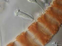
Colony of Carchesium spec. sitting on red alga Polysiphonia fibrillosa. The cells are redy to transform into free swimmung swarmers by developing the telotroch, a marginally ciliary fringe. Collected from Bodden, the brackish waters lying between the isles of Hiddensee and Ruegen (German Baltic Sea). This image was taken using Zeiss Universal with Olympus C7070 CCD camera.
-
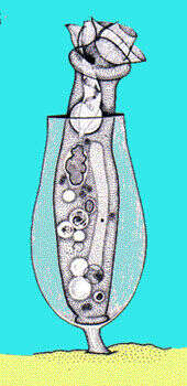
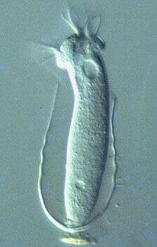
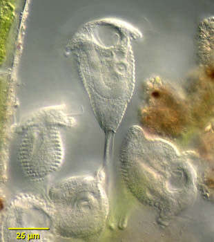
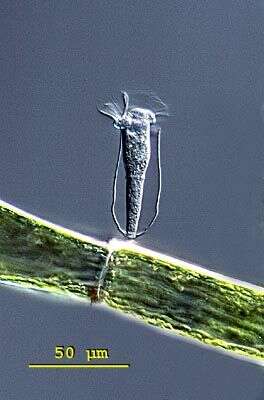
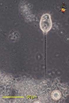
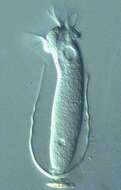
Cothurnia (co-thur-knee-a) is a sessile peritrich ciliate. The cells live within a lorica which is itself stalked. Cells attach to the base of the lorica by the posterior ends of the cell. Cells can contract into the lorica. The oral cilia form a wreath around the anterior end of the cell. One of the peritrich ciliates, distinguished by having a wreath of cilia around the anterior of the cell. Normally feeding on bacteria. No cilia on the body. Phase contrast.
-
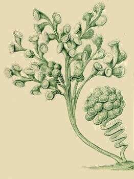
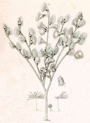
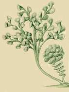
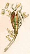
Carchesium polypinum.
-
-
-
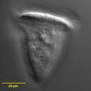
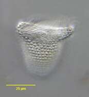
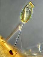
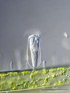
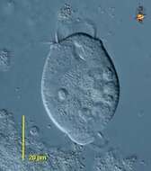
Differential interference contrast image showing living cell and lorica with a short stalk.
-
-
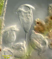
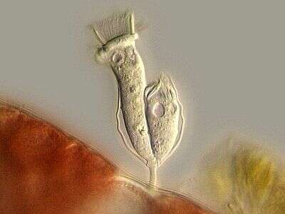
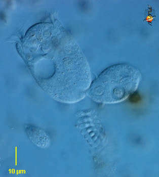
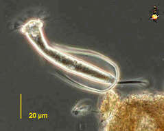
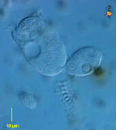
Cothurnia spec living as Aufwuchs on Ceramium diaphanum, shortly after binary fission. Now the swarmer (right cell) has to leave the lorica and build his own. Multi layer image using 8 frames generating depth of focus, stacked manually using Corel Photopaint. Collected from Bodden, the brackish waters lying between the isles of Hiddensee and Ruegen (German Baltic Sea). This image was taken using Zeiss Universal with Olympus C7070 CCD camera.
-
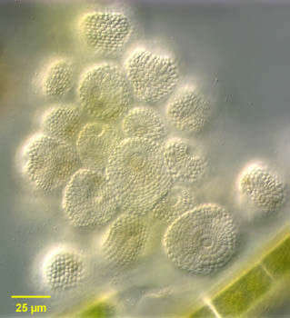
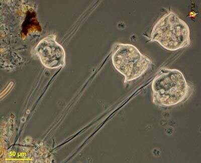
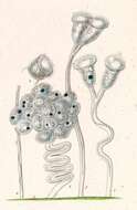
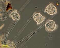
Group portrait of the gregarious but non-colonial peritrich ciliate, Pseudovorticella monilata (Tatem, 1870) Foissner and Sciffmann, 1975, all in contracted state (anterior apical view). Stalks are not visible here. The prominent pellicular blebs consist of paraglycogen, a carbohydrate storage product. From freshwater pond near Boise, Idaho. Differential interference contrast.
-
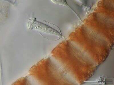
Cothurnia spec. on a young branch of the red alga Ceramium diaphanum. The optical longitudinal section of the axial cells oft the red alga thallus shows their central strand of cytoplasma. Collected from Bodden, the brackish waters lying between the isles of Hiddensee and Ruegen (German Baltic Sea). This image was taken using Zeiss Universal with Olympus C7070 CCD camera.
-
Group portrait of the gregarious but non-colonial peritrich ciliate, Pseudovorticella monilata (Tatem, 1870) Foissner and Sciffmann, 1975. Some individuals have separated from their contractile stalks. From freshwater pond near Boise, Idaho. Differential interference contrast.(Tatem, 1870) Foissner and Sciffmann, 1975
-
Cothurnia spec. on a branch of the green alga Cladophora accompanied by colonies of Zoogloea (Eubacteria). Scale bar indicates 50 µm. Sample from Lake Constance (Bodensee, Southern Germany) near Bodman. This image was taken using Zeiss Universal with Olympus C7070 CCD camera.
-
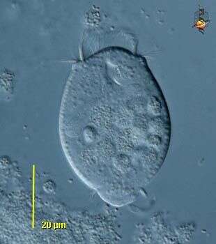
Portrait of the peritrich ciliate, Pseudovorticella monilata (Tatem, 1870) Foissner and Sciffmann, 1975. Designated as Vorticella monilata in many compendia, Foissner assigns this organism as the type species for the genus Pseudovorticella. Pseudovorticella is distinguished from Vorticella by silver staining which reveals a lattice-like silver line system in the former and circumferential lines without vertical connections in the latter. Pseudovorticella also has two contractile vacuoles (only one of which is seen here). The species is distinguished by circumferential rows of prominent pellicular blebs consisting of paraglycogen, a carbohydrate storage product. The macronucleus is J-shaped, lying in the long axis of the cell. The stalk consisting of a sheathed myonemes is seen here. Feeds on bacteria. From freshwater pond near Boise, Idaho. Differential interference contrast.
-
-
Pellicular detail of Pseudovorticella monilata (Tatem, 1870) Foissner and Sciffmann, 1975. The species is distinguished by circumferential rows of prominent pellicular blebs seen well in this image. They consist of paraglycogen, a carbohydrate storage product. DIC.
-
Cothurnia (coe-thurr-knee-ah) annulata built up a colourless transparent lorica. The lorica is fixed by a stalk on the ground. Cothurnia annulata has a distinctive ringshape bulge in the middle of the cell. Differential interference contrast.
-
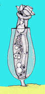
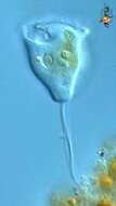
Vorticella (vort-ee-sell-a) the iconic peritrich ciliate. The feeding cells in sessile peritrich ciliates have lost all of the somatic cilia and only have feeding cilia. The feeding cilia form a wreath which extends around the front of the cell and descends into a narrowing buccal cavity. This cavity ends at the cytostome where food is packaged into food vacuoles . If the cells become unhappy, they produce a temporary wreath of basal cilia (trochal cilia), break away from their stalk and use these to swim. Daughter cells are produced near the base of the parental cells and have the trochal cilia so that they can swim around when they break from at the end of division. The stalk, contains a contractile filament, and when this contracts it coils up a bit like a spring. Phase contrast.
-
Vorticella (vort-ee-sell-a) the iconic peritrich ciliate. The feeding cells in sessile peritrich ciliates have lost all of the somatic cilia and only have the feeding cilia. The feeding cilia form a wreath which extends around the front of the cell and descends into a narrowing buccal cavity. This cavity ends at the cytostome where food is packaged into food vacuoles and can be seen at the top of this cell, as can many food vacuoles. If the cells become unhappy, they produce a temporary wreath of basal cilia (trochal cilia), break away from their stalk and use these to swim. This cell has the trochal cilia. Daughter cells are produced near the base of the parental cells and have the trochal cilia so that they can swim around when they break from at the end of division. Most peritrichs have a large curving macronucleus, and this is also evident towards the top of the cell. The stalk, not evident here, contains a contractile filament, and when this contracts it coils up a bit like a spring. Differential interference contrast.
-
Vorticella (vort-ee-sell-a) the iconic peritrich ciliate. The feeding cells in sessile peritrich ciliates have lost all of the somatic cilia and only have the feeding cilia. The feeding cilia form a wreath which extends around the front of the cell and descends into a narrowing buccal cavity. This image was taken with a long exposure and illustrates the currents created by the action of the feeding cilia of a single cell. Modified dark ground, long exposure.
-
Vorticella (vort-ee-sell-a) the iconic peritrich ciliate. The feeding cells in sessile peritrich ciliates have lost all of the somatic cilia and only have the feeding cilia. The feeding cilia form a wreath which extends around the front of the cell and descends into a narrowing buccal cavity. This cavity ends at the cytostome where food is packaged into food vacuoles. If the cells become unhappy, they produce a temporary wreath of basal cilia (trochal cilia), break away from their stalk and use these to swim. Daughter cells, like the one shown here, are produced near the base of the parental cells and have the trochal cilia so that they can swim around when they break from at the end of division. The stalk contains a contractile filament, and when this contracts it coils up a bit like a spring. Differential interference contrast.
-
Vorticella (vort-ee-sell-a) the iconic peritrich ciliate. The feeding cells in sessile peritrich ciliates have lost all of the somatic cilia and only have the feeding cilia. The feeding cilia form a wreath which extends around the front of the cell and descends into a narrowing buccal cavity. This cavity ends at the cytostome where food is packaged into food vacuoles. If the cells become unhappy, they produce a temporary wreath of basal cilia (trochal cilia), break away from their stalk and use these to swim. Daughter cells are produced near the base of the parental cells and have the trochal cilia so that they can swim around when they break from at the end of division. The stalk contains a contractile filament, and when this contracts it coils up a bit like a spring. Differential interference contrast.
-
Vorticella (vort-ee-sell-a) the iconic peritrich ciliate. The feeding cells in sessile peritrich ciliates have lost all of the somatic cilia and only have the feeding cilia. The feeding cilia form a wreath which extends around the front of the cell and descends into a narrowing buccal cavity. The stalk contains a contractile filament, and when this contracts it coils up a bit like a spring. Phase contrast.