portrait
Description:
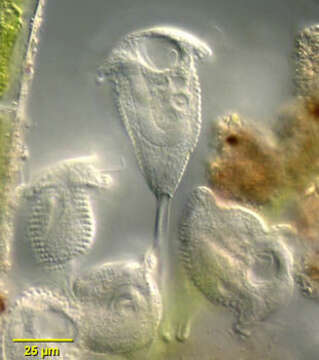
Portrait of the peritrich ciliate, Pseudovorticella monilata (Tatem, 1870) Foissner and Sciffmann, 1975. Designated as Vorticella monilata in many compendia, Foissner assigns this organism as the type species for the genus Pseudovorticella. Pseudovorticella is distinguished from Vorticella by silver staining which reveals a lattice-like silver line system in the former and circumferential lines without vertical connections in the latter. Pseudovorticella also has two contractile vacuoles (only one of which is seen here). The species is distinguished by circumferential rows of prominent pellicular blebs consisting of paraglycogen, a carbohydrate storage product. The macronucleus is J-shaped, lying in the long axis of the cell. The stalk consisting of a sheathed myonemes is seen here. Feeds on bacteria. From freshwater pond near Boise, Idaho. Differential interference contrast.
Included On The Following Pages:
- Life (creatures)
- Cellular (cellular organisms)
- Eukaryota (eukaryotes)
- SAR (Stramenopiles, Alveolates, Rhizaria)
- Alveolata (alveolates)
- Ciliophora (ciliates)
- Intramacronucleata
- Oligohymenophorea
- Peritrichia
- Sessilida
- Vorticellidae
- Pseudovorticella
- Pseudovorticella monilata
This image is not featured in any collections.
Source Information
- license
- cc-by-nc
- author
- William Bourland
- provider
- micro*scope
- original
- original media file
- visit source
- partner site
- micro*scope
- ID