-
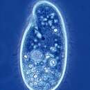
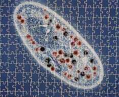
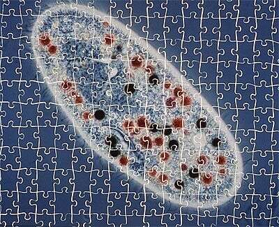
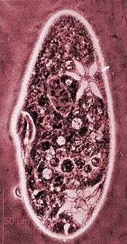
This cell has been fed on stained bacteria and indian ink to monitor the rate of food vacuole formation. Oh, and we made a jigsaw puzzle out of the picture.
-
Ventrolateral view of the infraciliature of the hymenostome ciliate, Dexiotricha granulosa (Kent, 1881) Foissner, 1994. Synonym of Loxocephulus granulosa. The cell is ovoid, broadly rounded posteriorly and truncate anteriorly. Regular longitudinal kineties terminate at a subapical band of circumferential kineties demarcating a cilia-free truncate apical area or frontal plate. Fibrils radiate anteriorly from the kinetids of the anteriormost paratene (seen here). There is a single long caudal cilium. The oral aperture is small and difficult to visualize in vivo. It is located in the anterior quarter with an undulating membrane on the right (seen here) and 3 membranelles (the posterior most seen here). The macronucleus is spheroid and located in the mid-cell. Single contractile vacuole. From freshwater pond near Boise, Idaho. Silver carbonate stain (see Foissner, W. Europ. J. Protistol., 27:313-330;1991).Brightfield. Black and white.
-
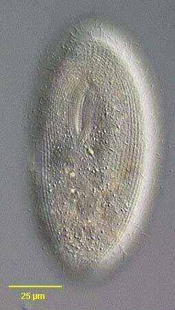
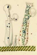
Portrait of Clathrostoma viminale (Penard, 1922) a frontoniine ciliate. Similar in overall appearance to but smaller than Frontonia leucas. The cytostome is located in a slight depression in the anterior half of the body. There is a cytopharyngeal basket (seen well here) composed of fibrils (as found in Loxodes) rather than true trichites. The somatic ciliation is uniform with a postoral suture. The sausage-shaped macronucleus is seen here with an adjacent cluster of several (1-4) micronuclei at its posterior end. A single contractile vacuole is located in the posterior 1/3. Collected from a freshwater agricultural irrigation ditch near McCall, Idaho in September 2003. DIC optics.
-
Surface view of the endocommensal astomatid ciliate, Anoplophrya maupasi (Cépède,1910) from the digestive tract of the oligochaete, Aelosoma hemprichi. The metachronal ciliary wave patterns can be seen in this image. See description of characteristics in: Lom, J., Arch. Protistenk.101:p.286,1956. Collected from a freshwater pond near Boise, Idaho. June 2005.Phase contrast.
-
-
These three individuals of this small species of Paramecium have been allowed to dry in a suspension of nigrosin. The stain dries around the cell, showing off the dimpling of the surface, and the ingestion area (in the lower cell we can see the arc-shaped buccal cavity. Crystals inside the cell appear light blue. Dark lines outside the cell are trichocysts (a type of extrusome) that have been expelled from the cells.
-
Dorsal view of the infraciliature of the hymenostome ciliate, Dexiotricha granulosa (Kent, 1881) Foissner, 1994. Synonym of Loxocephulus granulosa. The cell is ovoid, broadly rounded posteriorly and truncate anteriorly. Regular longitudinal kineties terminate at a subapical band of circumferential kineties demarcating a cilia-free truncate apical area or frontal plate. Fibrils radiate anteriorly from the kinetids of the anteriormost paratene (seen here). There is a single long caudal cilium. The oral aperture is small and difficult to visualize in vivo. It is located in the anterior quarter with an undulating membrane on the right (seen here) and 3 membranelles (the posterior most seen here). The macronucleus is spheroid and located in the mid-cell. Single contractile vacuole. From freshwater pond near Boise, Idaho. Silver carbonate stain (see Foissner, W. Europ. J. Protistol., 27:313-330;1991).Brightfield. Black and white.
-
Portrait of Clathrostoma viminale (Penard, 1922) a frontoniine ciliate (surface view). Similar in overall appearance to but smaller than Frontonia leucas. The cytostome is located in a slight depression in the anterior half of the body (seen well in this image). There is a cytopharyngeal basket composed of fibrils (as found in Loxodes) rather than true trichites. The somatic ciliation is uniform with a postoral suture (seen in this image). The sausage-shaped macronucleus (not seen in this image) has an adjacent cluster of several (1-4) micronuclei at its posterior end. A single contractile vacuole is located in the posterior 1/3. Collected from a freshwater agricultural irrigation ditch near McCall, Idaho in September 2003. DIC optics.
-
Anterolateral view of the endocommensal astomatid ciliate, Anoplophrya maupasi (Cépède,1910) from the digestive tract of the oligochaete, Aelosoma hemprichi. The longitudinal kineties meet at a small anterior transverse suture (seen here). Skeletal elemnts and specialized attachment area are absent. See description of characteristics in: Lom, J., Arch. Protistenk.101:p.286,1956. Collected from a freshwater pond near Boise, Idaho.Stained by the silver carbonate technique (see Foissner, W. Europ. J. Protistol., 27:313-330;1991) June 2005.Brightfield.
-
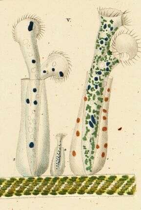
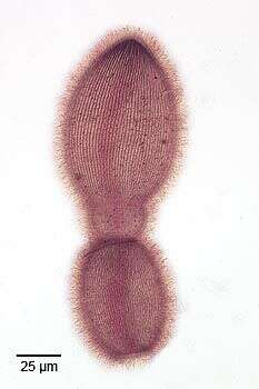
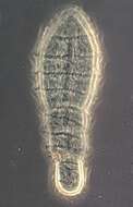
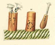
Vaginicola (vadge-in-ick-cola) tincta, two individuals in their lorica which has a flat bottom and no stalk. This specimen was collected in a pond near Konstanz, Germany. Differential interference contrast.
-
-
Ventral view of the infraciliature of late division of the hymenostome ciliate, Dexiotricha granulosa (Kent, 1881) Foissner, 1994. Synonym of Loxocephulus granulosa. The cell is ovoid, broadly rounded posteriorly and truncate anteriorly. Regular longitudinal kineties terminate at a subapical band of circumferential kineties demarcating a cilia-free truncate apical area or frontal plate. Fibrils radiate anteriorly from the kinetids of the anteriormost paratene (seen here). There is a single long caudal cilium. The oral aperture is small and difficult to visualize in vivo. It is located in the anterior quarter with an undulating membrane on the right (seen here) and 3 membranelles ( seen most clearly in the proter). The macronucleus is spheroid and located in the mid-cell. Single contractile vacuole. From freshwater pond near Boise, Idaho. Silver carbonate stain (see Foissner, W. Europ. J. Protistol., 27:313-330;1991).Brightfield. Black and white.
-
Portrait of Clathrostoma viminale (Penard, 1922) a frontoniine ciliate. Similar in overall appearance to but smaller than Frontonia leucas. The cytostome is located in a slight depression in the anterior half of the body. There is a cytopharyngeal basket composed of fibrils (as found in Loxodes) rather than true trichites. The somatic ciliation is uniform with a postoral suture. The sausage-shaped macronucleus is seen here with an adjacent cluster of several (1-4) micronuclei at its posterior end. A single contractile vacuole is located in the posterior 1/3. Collected from a freshwater agricultural irrigation ditch near McCall, Idaho in September 2003. DIC optics.
-
Anterolateral view of the endocommensal astomatid ciliate, Anoplophrya maupasi(Cépède,1910), from the digestive tract of the oligochaete,Aelosoma hemprichi. The densely packed longitudinal kineties meet at a small apical transverse suture (seen here).Catenation or repeated incomplete fissions resulting in chains of individuals is seen here (catenoid colonies). See description of characteristics in: Lom, J., Arch. Protistenk.,101:p.286,195. Collected from a freshwater pond near Boise, Idaho.Stained by the silver carbonate technique (see Foissner, W. Europ. J. Protistol., 27:313-330;1991) June 2005.Brightfield.
-
Originally described by Ehrenberg under the name Vaginicola tinctus.
-
-
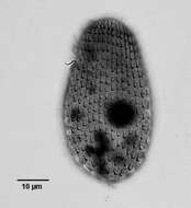
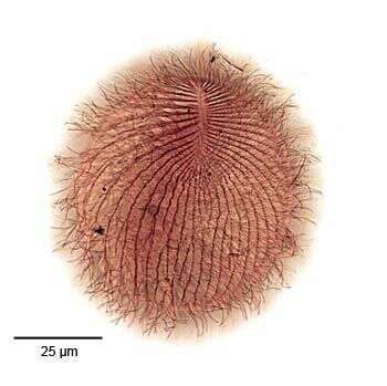
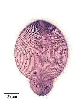
Portrait of the hymenostome ciliate, Dexiotricha granulosa (KENT,1881) FOISSNER, 1994, synonymous with Loxocephulus granulosus. The cell is ovoid, broadly rounded posteriorly and truncate anteriorly. Regular longitudinal kineties terminate at a subapical band of circumferential kineties demarcating a cilia-free truncate apical area or frontal plate. There is a single long caudal cilium. The oral aperture is small and difficult to visualize in vivo. It is located in the anterior quarter with an undulating membrane on the right (seen faintly here) and 3 membranelles (not seen here). The macronucleus is spheroid and located in the mid-cell. The contractile vacuole is seen here to the left of the macronucleus. The spherical micronucleus is not seen here. The cytoplasm contains many small refractile ring-shaped glycogen granules, which are diagnostic for the species (see detail images). Dexitricha is bactiverous. From freshwater pond near Boise, Idaho. Differential interference contrast.
-
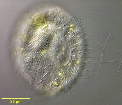
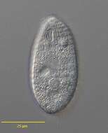
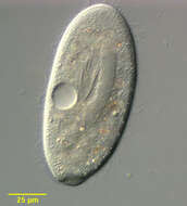
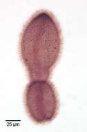
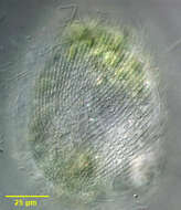
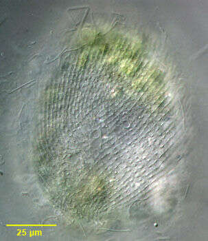
.Portrait of the frontoniid ciliate, Disematostoma buetschlii LAUTERBORN, 1894. D. buetschlii contains endosymbiotic algae but may lose (or digest) them during fall and winter (Ulrike-G.Berninger, Bland J.Finlay, and Hilda M.Canter; The Spatial Distribution and Ecology of Zoochlorellae-Bearing Ciliates in a Productive Pond. J.Protozool. 33(4):557-563, 1986). Although this specimen is slightly smaller (80 microns) than what is commonly reported for D. buetschlii (110 microns) it is otherwise indistinguishable. Kahl describes a smaller species without endosymbiotic algae (D. minor) (A.Kahl; [Urtiere oder Protozoa I: Wimpertiere oder Ciliata (Infusoria) 2. Holotricha]. Die Tierwelt Deutschlands und der angrenzenden Meeresteile. Germany:Verlag von Gustav Fischer. (2)-398). However it is unclear whether this is simply a small variety of D. buetschlii with algal endosymbionts. The cell shape is obovoid tapering to a blunt slightly curved point posteriorly. Dorsal surface convex with a flattened ventral surface. The cytostome (seen well in this image) is located in the anterior 1/3 with 3 left adoral membranelles and an inconspicuous undulating membrane on the right. 4-5 dense vestibular ciliary rows are found on the right of the cytostome. The reniform macronucleus is seen well here. The contractile vacuole is in the posterior half. The longitudinal somatic kineties terminate on prominent ladder-like preoral and postoral suture (the polar band). The preoral suture is seen well in this image. D. buetschlii is primarily algivorous and some of the green algae seen in the cytoplasm in this image may be in food vacuoles. Collected from freshwater pond near Boise, Idaho September 2003. DIC.
-
Ventral view of the endocommensal astomatid ciliate, Anoplophrya maupasi (Cé°¨de,1910), from the digestive tract of the oligochaete,Aelosoma hemprichi. The densely packed longitudinal kineties meet at a small apical transverse suture.Catenation or repeated incomplete fissions resulting in chains of individuals is seen here (catenoid colonies).Skeletal elements are absent (see description of characteristics in: Lom, J., Arch. Protistenk.,101:p.286,1956). Collected from a freshwater pond near Boise, Idaho.Stained by the silver carbonate technique (see Foissner, W. Europ. J. Protistol., 27:313-330;1991) June 2005.Brightfield.
-
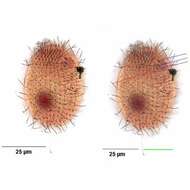
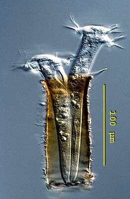
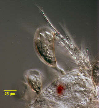
Portrait of the peritrich ciliate, Cyclodonta bipartita (Stokes, 1885) Matthes, 1958. Usually found as an epibiont of freshwater copepods. The cells are contained in a vase-shaped transparent lorica that has fine longitudinal striations. The lorica has a short, stout noncontractile stalk. The cell is attached to the posterior portion of the lorica by a series of membranes and does not protrude from the lorica. The cell body is cylindrical in cross section, rounded posteriorly and transversely truncate anteriorly. The cell surface has fine transverse striations. The macronucleus is ellipsoid. There is a single contractile vacuole. Found on the surface of a cyclopoid copepod collected from a freshwater pond near Boise, Idaho March 2005. DIC
-
-

Ventrolateral view of the infraciliature of the hymenostome ciliate, Dexiotricha granulosa (Kent, 1881) Foissner, 1994. Synonym of Loxocephulus granulosa. The cell is ovoid, broadly rounded posteriorly and truncate anteriorly. Regular longitudinal kineties terminate at a subapical band of circumferential kineties demarcating a cilia-free truncate apical area or frontal plate. Fibrils radiate anteriorly from the kinetids of the anteriormost paratene. There is a single long caudal cilium (green line). The oral aperture is small and difficult to visualize in vivo. It is located in the anterior quarter with a paraoral membrane on the right (red line) and 3 adoral membranelles (dark blue lines). Closely spaced basal bodies of the somatic kinaty to the right of the oral aperture form a "pseudomembrane" (light blue line). The macronucleus is spheroid and located in the mid-cell. Single contractile vacuole. From freshwater pond near Boise, Idaho. Silver carbonate stain (see Foissner, W. Europ. J. Protistol., 27:313-330;1991).Brightfield.
-
Dorsal surface of the frontoniid ciliate, Disematostoma buetschlii LAUTERBORN, 1894. D. buetschlii contains endosymbiotic algae but may lose (or digest) them during fall and winter (Ulrike-G.Berninger, Bland J.Finlay, and Hilda M.Canter; The Spatial Distribution and Ecology of Zoochlorellae-Bearing Ciliates in a Productive Pond. J.Protozool. 33(4):557-563, 1986). Although this specimen is slightly smaller (80 microns) than what is commonly reported for D. buetschlii (about 110 microns) it is otherwise indistinguishable. Kahl describes a smaller species without endosymbiotic algae (D. minor) (A.Kahl; [Urtiere oder Protozoa I: Wimpertiere oder Ciliata (Infusoria) 2. Holotricha]. Die Tierwelt Deutschlands und der angrenzenden Meeresteile. Germany:Verlag von Gustav Fischer. (2)-398). However it is unclear whether this is simply a small variety of D. buetschlii without algal endosymbionts. The cell shape is obovoid tapering to a blunt slightly curved point posteriorly. Dorsal surface convex with a flattened ventral surface. The cytostome is located in the anterior 1/3 with 3 left adoral membranelles and an inconspicuous undulating membrane on the right. 4-5 dense vestibular ciliary rows are found on the right of the cytostome. The reniform macronucleus is seen well here. The longitudinal somatic kineties terminate on prominent ladder-like preoral and postoral suture (the polar band). The polar band can be seen curving to the viewer's right in this image. The solitary dorsal excretory pore of the contractile vacuole is seen here to the viewer's right of the anterior part of the polar band.D. buetschlii is primarily algivorous and some of the green algae seen in the cytoplasm in this image may be in food vacuoles. Collected from freshwater pond near Boise, Idaho September 2003. DIC.
-
Phase contrast micrograph showing clearly one contractile vacuole with five filled channels.