in vivo
Description:
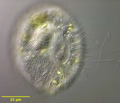
.Portrait of the frontoniid ciliate, Disematostoma buetschlii LAUTERBORN, 1894. D. buetschlii contains endosymbiotic algae but may lose (or digest) them during fall and winter (Ulrike-G.Berninger, Bland J.Finlay, and Hilda M.Canter; The Spatial Distribution and Ecology of Zoochlorellae-Bearing Ciliates in a Productive Pond. J.Protozool. 33(4):557-563, 1986). Although this specimen is slightly smaller (80 microns) than what is commonly reported for D. buetschlii (110 microns) it is otherwise indistinguishable. Kahl describes a smaller species without endosymbiotic algae (D. minor) (A.Kahl; [Urtiere oder Protozoa I: Wimpertiere oder Ciliata (Infusoria) 2. Holotricha]. Die Tierwelt Deutschlands und der angrenzenden Meeresteile. Germany:Verlag von Gustav Fischer. (2)-398). However it is unclear whether this is simply a small variety of D. buetschlii with algal endosymbionts. The cell shape is obovoid tapering to a blunt slightly curved point posteriorly. Dorsal surface convex with a flattened ventral surface. The cytostome (seen well in this image) is located in the anterior 1/3 with 3 left adoral membranelles and an inconspicuous undulating membrane on the right. 4-5 dense vestibular ciliary rows are found on the right of the cytostome. The reniform macronucleus is seen well here. The contractile vacuole is in the posterior half. The longitudinal somatic kineties terminate on prominent ladder-like preoral and postoral suture (the polar band). The preoral suture is seen well in this image. D. buetschlii is primarily algivorous and some of the green algae seen in the cytoplasm in this image may be in food vacuoles. Collected from freshwater pond near Boise, Idaho September 2003. DIC.
Included On The Following Pages:
- Life (creatures)
- Cellular (cellular organisms)
- Eukaryota (eukaryotes)
- SAR (Stramenopiles, Alveolates, Rhizaria)
- Alveolata (alveolates)
- Ciliophora (ciliates)
- Intramacronucleata
- Oligohymenophorea
- Peniculida (Peniculid)
- Frontoniidae
- Disematostoma
- Disematostoma buetschlii
This image is not featured in any collections.
Source Information
- license
- cc-by-nc
- author
- William Bourland
- provider
- micro*scope
- original
- original media file
- visit source
- partner site
- micro*scope
- ID