-

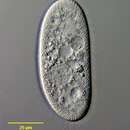
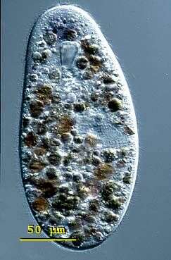
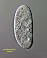
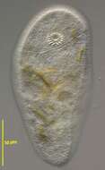
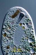
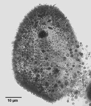
Ventral view of the nassulid ciliate, Furgasonia trichocystis (Stokes, 1894) Jankowski, 1964. Synonym: Cyclogramma. The cell shape is a slightly dorsoventrally flattened ellipsoid. The left side is flattened and the right side slightly convex. The cytostome is in the anterior 1/5 of the cell in a shallow depression. It is supported by a prominent basket of obliquely oriented cytopharyngeal trichites. The somatic ciliature consists of about 32 to 26 longitudinal kineties. On the ventral surface the right kineties arch to the left anterior to the cytostome to terminate on a short but wide preoral suture. The straight left kineties terminate on this suture to the left of the cytostome. There is a short curved right paraoral membrane. There are three approximately rectangular paroral polykineties. The most anterior (M1) is also least conspicuous. It is obliquely oriented in the preoral suture. The middle membrane (M2) is to the left of the cytostome and almost perpendicular to the long axis of the cell. The most posterior membranelle (M3) is posterior to the cytostome (often obscured by the trichites in silver carbonate preparations) almost parallel to the long axis of the cell. These three distinctive small polykineties distinguish Furgasonia from other nassulid genera. The spherical macronucleus and adjacent micronucleus are slightly posterior to the equator. The single contractile vacuole (visible here posterior to the cytopharyngeal basket) is located in the cell center with an excretory pore on its ventral aspect. There is a prominent layer of fusiform subpellicular extrusomes (mucocysts). The cytoplasm is colorless in these bactivorous individuals. It is unclear whether this species is synonymous with F. rubens which is orange to blue colored due to ingested cyanobacteria. Morphologically the two species are quite similar aside from this coloration (see Faur�-Fremiet, E. Le Genre Cyclogramma, Perty, 1852. J. Protozool. 14: 456-464, 1967.) Collected from a temporary rainwater pool with abundant decaying grass near Boise, Idaho. March, 2005. DIC.
-
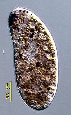
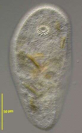
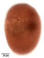
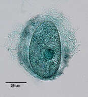
Nassulopsis (nass-you-lop-sis). Nassulopsis elegans is a conspicuous pink- or blue coloured nassulid found in pond and lake plankton. This cell was collected from floating colonies of Oscillatoria in a freshwater pond near Konstanz, Germany. The body is cylindrical in shape and measures 150 - 300 X 50 - 100 microns. Nassulopsis can be distinguished from Nassula and Obertrumia by 5-7 contractile vacuoles arranged in a ventral row. The macronucleus is cylindrical with rounded ends. The cytopharyngeal basket is located in the anterior third of the cell. The cells fill up with food vacuoles of different colour (green, orange), depending on stage of digestion. Free-swimming cell, 250 microns long. Differential interference contrast.
-

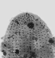
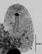
Right dorsolateral view of the infraciliature of the large nassulid ciliate Obertrumia aurea (Ehrenberg, 1833; Foissner 1987). Synonym of Nassula aurea. Obertrumia is distinguished by it's bipartite "hypostomial frange", linear arrays of ciliary tufts. A sigmoid ventrolateral frange runs anteriorly and to the left. The dorsal termination of this part is seen as a vertical arrangement of rectangular collections of kinetids on the viewer's left in this image. It terminates at the left end of the horizontal line of ciliary tufts on the dorsal surface.The uniform longitudinal somatic kineties are well seen here. In vivo the cell appears brightly colored (orange, green or violet) due to multiple food vacuoles containing ingested cyanobacteria. Numerous small mucocysts give the cortex a roughly granular appearance. O. aurea feeds mainly on cyanobacteria. Silver carbonate stain (see Foissner, W.Europ. J. Protistol.27,313-330;1991). Collected from a freshwater aquaculture pond near Boise,Idaho November 2004. Brightfield.
-
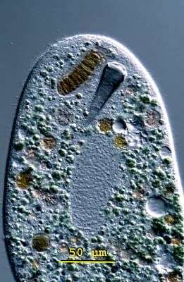
Zosterodasys transversa (Kahl,1928) Foissner,1994. (previously known as Chilodontopsis vorax), a large nassulid ciliate. Z. transversa is elongate and dorsoventrally flattened. Both dorsal and ventral surfaces bear uniform longitudinal rows of somatic cilia. A distinctive synhymenion or hypostomial frange runs obliquely posterior to the cytostome around half the cell circumference. Unlike the similar Synhymenia there is no preoral suture. The cytoplasm is highly vacuolated. There are multiple contractile vacuoles. The macronucleus is ovoid. There is a very prominent anterior cytopharyngeal basket composed of stout nematodesmata. From freshwater pond near Boise, Idaho. DIC.
-
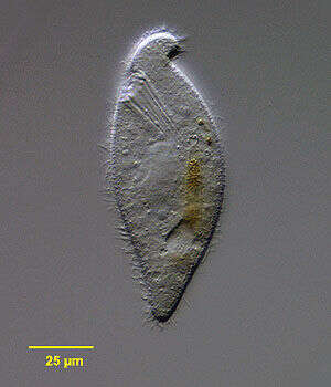
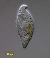
Portrait of the nassophorean marine ciliate, Orthodonella hamatus (Bhatia, 1936) . The elongate cell is dorsoventrally flattened with the anterior end bent to the left in a blunt hook shape. The posterior is bluntly tapered. The oral aperture is right anterior. There is a cytopharyngeal apparatus composed of stout nematodesmata. The longitudinal somatic kineties are uniform. There is a sigmoid post-oral frange extending obliquely from the anterior end just posterior to the cytostome almost to the right edge (not seen in this image). The ovoid macronucleus and adjacent micronucleus are located in the mid-cell. A posterior peripheral contractile vacuole is seen in this image. O. hamatus is also found in freshwater habitats. Orthodonella is a monotypic genus. Collected from a commercial saltwater aquarium in Boise, Idaho February 2004. DIC optics.
-
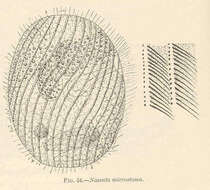
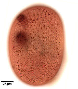
Nassula microstoma.
-
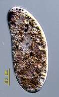
Dorsal view of the nassulid ciliate, Furgasonia trichocystis (Stokes, 1894) Jankowski, 1964. Synonym: Cyclogramma. The cell shape is a slightly dorsoventrally flattened ellipsoid. The left side is flattened and the right side slightly convex. There is a prominent layer of fusiform subpellicular extrusomes (mucocysts). The cytoplasm is colorless in these bactivorous individuals. It is unclear whether this species is synonymous with F. rubens which is orange to blue colored due to ingested cyanobacteria. Morphologically the two species are quite similar aside from this coloration (see Faur�-Fremiet, E. Le Genre Cyclogramma, Perty, 1852. J. Protozool. 14: 456-464, 1967.) Collected from a temporary rainwater pool with abundant decaying grass near Boise, Idaho. March, 2005. DIC.
-
Nassulopsis (nass-you-lop-sis). Nassulopsis elegans is a conspicuous pink- or blue coloured nassulid found in pond and lake plankton. This cell was collected from floating colonies of Oscillatoria in a freshwater pond near Konstanz, Germany. The body is cylindrical in shape and measures 150 - 300 X 50 - 100 microns. Nassulopsis can be distinguished from Nassula and Obertrumia by 5-7 contractile vacuoles arranged in a ventral row. The macronucleus is cylindrical with rounded ends. The cytopharyngeal basket is located in the anterior third of the cell. The cells fill up with food vacuoles of different colour (green, orange), depending on stage of digestion. Slightly flattened cell, 260 microns long. Differential interference contrast.
-
Ventral infraciliature of the large nassulid ciliate Obertrumia aurea (Ehrenberg, 1833; Foissner 1987). Synonym of Nassula aurea. Obertrumia is distinguished by it's bipartite "hypostomial frange",linear arrays of ciliary tufts. A sigmoid ventrolateral frange begins posterior to the cytostome running anteriorly and to the left (viewer's right) around to the dorsal surface. The dorsal termination is not seen in this image. The ventrolateral part of the frange terminates at the left end of the horizontal line of ciliary tufts on the dorsal surface. The uniform longitudinal somatic kineties, interrupted by the hypostomial frange, are well seen here. The multiple darkly stained micronuclei are seen overlying the spherical macronucleus in this image.The small circular structure to the viewer's left just posterior to the macronucleus is the excretory pore of the contractile vacuole. The thin dark longitudinal line posterior to this is the cytopyge (cell anus).In vivo the cell appears brightly colored (orange, green or violet) due to multiple food vacuoles containing ingested cyanobacteria. Numerous small mucocysts give the cortex a roughly granular appearance. O. aurea feeds mainly on cyanobacteria. Silver carbonate stain (see Foissner, W.Europ. J. Protistol.27,313-330;1991). Collected from a freshwater aquaculture pond near Boise, Idaho,Idaho November 2004. Brightfield optics.
-
Zosterodasys transversa (Kahl,1928) Foissner,1994. (previously known as Chilodontopsis vorax), a large nassulid ciliate. Z. transversa is elongate and dorsoventrally flattened. Both dorsal and ventral surfaces bear uniform longitudinal rows of somatic cilia. A distinctive synhymenion or hypostomial frange runs obliquely posterior to the cytostome around half the cell circumference (seen well in accompanying images). Unlike the similar Synhymenia there is no preoral suture. The cytoplasm is highly vacuolated. There are multiple contractile vacuoles. The macronucleus is ovoid. There is a very prominent anterior cytopharyngeal basket composed of stout nematodesmata. From freshwater pond near Boise, Idaho. Differential interference contrast.
-
Portrait of the nassophorean marine ciliate, Orthodonella hamatus (Bhatia, 1936). The elongate cell is dorsoventrally flattened with the anterior end bent to the left in a blunt hook shape. The posterior is bluntly tapered. The oral aperture is right anterior. There is a cytopharyngeal apparatus composed of stout nematodesmata (seen clearly in this image). The longitudinal somatic kineties are uniform. There is a sigmoid post-oral frange extending obliquely from the anterior end just posterior to the cytostome almost to the right edge (not seen in this image). The ovoid macronucleus and adjacent micronucleus are located in the mid-cell. A posterior peripheral contractile vacuole is seen in this image. O. hamatus is also found in freshwater habitats. Orthodonella is a monotypic genus. Collected from a commercial saltwater aquarium in Boise, Idaho February 2004. DIC optics.
-
Ventral anterior view of the nassulid ciliate, Furgasonia trichocystis (Stokes, 1894) Jankowski, 1964. Synonym: Cyclogramma. The cell shape is a slightly dorsoventrally flattened ellipsoid. The left side is flattened and the right side slightly convex. The cytostome is in the anterior 1/5 of the cell in a shallow depression. It is supported by a prominent basket of obliquely oriented cytopharyngeal trichites. The somatic ciliature consists of about 32 to 26 longitudinal kineties. On the ventral surface the right kineties arch to the left anterior to the cytostome to terminate on a short but wide preoral suture. The straight left kineties terminate on this suture to the left of the cytostome. There is a short curved right paraoral membrane. There are three approximately rectangular paroral polykineties. The most anterior (M1) is obliquely oriented in the preoral suture. The middle membrane (M2) is to the left of the cytostome and almost perpendicular to the long axis of the cell. The most posterior membranelle (M3) is posterior to the cytostome (often obscured by the trichites in silver carbonate preparations) almost parallel to the long axis of the cell. These three distinctive small polykineties distinguish Furgasonia from other nassulid genera. The spherical macronucleus and adjacent micronucleus are slightly posterior to the equator. The single contractile vacuole (visible here posterior to the cytopharyngeal basket) is located in the cell center with an excretory pore on its ventral aspect. There is a prominent layer of fusiform subpellicular extrusomes (mucocysts). The cytoplasm is colorless in these bactivorous individuals. It is unclear whether this species is synonymous with F. rubens which is orange to blue colored due to ingested cyanobacteria. Morphologically the two species are quite similar aside from this coloration (see Faurè-Fremiet, E. Le Genre Cyclogramma, Perty, 1852. J. Protozool. 14: 456-464, 1967). Collected from a temporary rainwater pool with abundant decaying grass near Boise, Idaho. March, 2005. Silver carbonate stain (see Foissner, W. Europ. J. Protistol., 27:313-330;1991). Brightfield.
-
Nassulopsis (nass-you-lop-sis). Nassulopsis elegans is a conspicuous pink- or blue coloured nassulid found in pond and lake plankton. This cell was collected from floating colonies of Oscillatoria in a freshwater pond near Konstanz, Germany. The body is cylindrical in shape and measures 150 - 300 X 50 - 100 microns. Nassulopsis can be distinguished from Nassula and Obertrumia by 5-7 contractile vacuoles arranged in a ventral row. The macronucleus is cylindrical with rounded ends. The cytopharyngeal basket is located in the anterior third of the cell. The cells fill up with food vacuoles of different colour (green, orange), depending on stage of digestion. The cytopharyngeal basket is anterior. To the right of the basket is an ingested cyanobacteria. Below the basket an ovoid-shaped macronucleus is visible. Differential interference contrast.
-
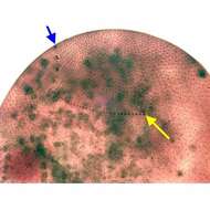
Dorsal infraciliature of the large nassulid ciliate Obertrumia aurea (Ehrenberg, 1833; Foissner 1987). Synonym of Nassula aurea. Obertrumia is distinguished by it's bipartite "hypostomial frange", linear arrays of ciliary tufts. A sigmoid ventrolateral frange runs anteriorly and to the left. The dorsal termination of this part is seen as a vertical arrangement of rectangular collections of kinetids (blue arrow). It terminates at the left end of the horizontal line of ciliary tufts on the dorsal surface (yellow arrow). The uniform longitudinal somatic kineties are well seen here. In vivo the cell appears brightly colored (orange, green or violet) due to multiple food vacuoles containing ingested cyanobacteria. Numerous small mucocysts give the cortex a roughly granular appearance. O. aurea feeds mainly on cyanobacteria. Silver carbonate stain (see Foissner, W.Europ. J. Protistol.27,313-330;1991). Collected from a freshwater pond near Idaho City, Idaho Septmeber 2004. Brightfield optics.
-
Ventrolateral surface view of the nassophorean marine ciliate, Orthodonella hamatus (Bhatia, 1936). The elongate cell is dorsoventrally flattened with the anterior end bent to the left in a blunt hook shape. The posterior is bluntly tapered. The oral aperture is right anterior. There is a cytopharyngeal apparatus composed of stout nematodesmata (not seen in this image). The longitudinal somatic kineties are uniform. There is a sigmoid post-oral frange extending obliquely from the anterior end just posterior to the cytostome almost to the right edge (seen well in this image). The ovoid macronucleus and adjacent micronucleus are located in the mid-cell. A posterior peripheral contractile vacuole is seen in this image. O. hamatus is also found in freshwater habitats. Orthodonella is a monotypic genus. Collected from a commercial saltwater aquarium in Boise, Idaho February 2004. DIC optics.
-
Ventral infraciliature of the nassulid ciliate, Furgasonia trichocystis (Stokes, 1894) Jankowski, 1964. Synonym: Cyclogramma. The cell shape is a slightly dorsoventrally flattened ellipsoid. The left side is flattened and the right side slightly convex. The cytostome is in the anterior 1/5 of the cell in a shallow depression. It is supported by a prominent basket of obliquely oriented cytopharyngeal trichites. The somatic ciliature consists of about 32 to 26 longitudinal kineties. On the ventral surface the right kineties arch to the left anterior to the cytostome to terminate on a short but wide preoral suture. The straight left kineties terminate on this suture to the left of the cytostome. There is a short curved right paraoral membrane. There are three approximately rectangular paroral polykineties. The most anterior (M1) is obliquely oriented in the preoral suture. The middle membrane (M2) is to the left of the cytostome and almost perpendicular to the long axis of the cell. The most posterior membranelle (M3) is posterior to the cytostome (often obscured by the trichites in silver carbonate preparations) almost parallel to the long axis of the cell. These three distinctive small polykineties distinguish Furgasonia from other nassulid genera. The spherical macronucleus and adjacent micronucleus are slightly posterior to the equator. The single contractile vacuole (visible here posterior to the cytopharyngeal basket) is located in the cell center with an excretory pore on its ventral aspect. There is a prominent layer of fusiform subpellicular extrusomes (mucocysts). The cytoplasm is colorless in these bactivorous individuals. It is unclear whether this species is synonymous with F. rubens which is orange to blue colored due to ingested cyanobacteria. Morphologically the two species are quite similar aside from this coloration (see Faurè-Fremiet, E. Le Genre Cyclogramma, Perty, 1852. J. Protozool. 14: 456-464, 1967). Collected from a temporary rainwater pool with abundant decaying grass near Boise, Idaho. March, 2005. Silver carbonate stain (see Foissner, W. Europ. J. Protistol., 27:313-330;1991). Brightfield.
-
Nassulopsis (nass-you-lop-sis). Nassulopsis elegans is a conspicuous pink- or blue coloured nassulid found in pond and lake plankton. This cell was collected from floating colonies of Oscillatoria in a freshwater pond near Konstanz, Germany. The body is cylindrical in shape and measures 150 - 300 X 50 - 100 microns. Nassulopsis can be distinguished from Nassula and Obertrumia by 5-7 contractile vacuoles arranged in a ventral row. The macronucleus is cylindrical with rounded ends. The cytopharyngeal basket is located in the anterior third of the cell. The cells fill up with food vacuoles of different colour (green, orange), depending on stage of digestion. This picture shows the pellicle of Nassulopsis elegans with its net like ornamentation. A mucocyst lies at the centre of each square. Differential interference contrast.
-
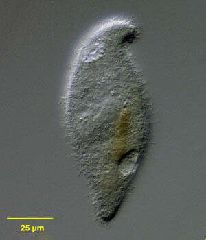
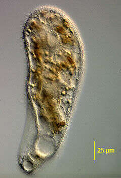
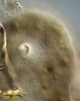
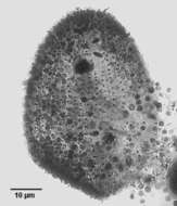
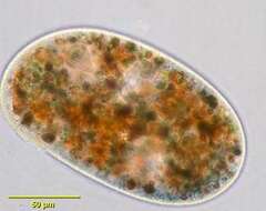
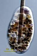
Portrait of Obertrumia aurea(Ehrenberg, 1833; Foissner 1987). Widely distributed nassulid ciliate with well developed cytopharyngeal basket or cyrtos (viewer's upper left). Fed organisms may contain brightly colored orange, blue and purple food vacuoles. O. aurea has a bipartite hypostomial frange (not seen here). From a freshwater pond near Boise, Idaho. Oblique illumination This image was taken by William Bourland. He now uses a Zeiss Axioskop 2 with a Spot Insight CCD camera (Diagnostic Instruments).
-

Ventral infraciliature of the nassulid ciliate, Furgasonia trichocystis (Stokes, 1894) Jankowski, 1964. Synonym: Cyclogramma. The cell shape is a slightly dorsoventrally flattened ellipsoid. The left side is flattened and the right side slightly convex. The cytostome is in the anterior 1/5 of the cell in a shallow depression. It is supported by a prominent basket of obliquely oriented cytopharyngeal trichites. The somatic ciliature consists of about 32 to 26 longitudinal kineties. On the ventral surface the right kineties arch to the left anterior to the cytostome to terminate on a short but wide preoral suture. The straight left kineties terminate on this suture to the left of the cytostome. There is a short curved right paraoral membrane. There are three approximately rectangular paroral polykineties. The most anterior (M1) is obliquely oriented in the preoral suture. The middle membrane (M2) is to the left of the cytostome and almost perpendicular to the long axis of the cell. The most posterior membranelle (M3) is posterior to the cytostome (often obscured by the trichites in silver carbonate preparations) almost parallel to the long axis of the cell. These three distinctive small polykineties distinguish Furgasonia from other nassulid genera. The spherical macronucleus and adjacent micronucleus are slightly posterior to the equator. The single contractile vacuole (visible here posterior to the cytopharyngeal basket) is located in the cell center with an excretory pore on its ventral aspect. There is a prominent layer of fusiform subpellicular extrusomes (mucocysts). The cytoplasm is colorless in these bactivorous individuals. It is unclear whether this species is synonymous with F. rubens which is orange to blue colored due to ingested cyanobacteria. Morphologically the two species are quite similar aside from this coloration (see Faurè-Fremiet, E. Le Genre Cyclogramma, Perty, 1852. J. Protozool. 14: 456-464, 1967). Collected from a temporary rainwater pool with abundant decaying grass near Boise, Idaho. March, 2005. Silver carbonate stain (see Foissner, W. Europ. J. Protistol., 27:313-330;1991). Brightfield.
-
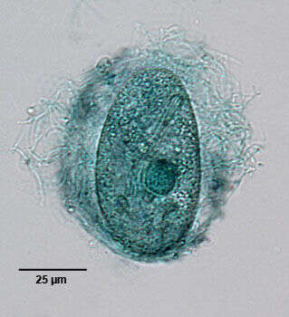
Nassulopsis (nass-you-lop-sis). Nassulopsis elegans is a conspicuous pink- or blue coloured nassulid found in pond and lake plankton. This cell was collected from floating colonies of Oscillatoria in a freshwater pond near Konstanz, Germany. The body is cylindrical in shape and measures 150 - 300 X 50 - 100 microns. Nassulopsis can be distinguished from Nassula and Obertrumia by 5-7 contractile vacuoles arranged in a ventral row. The macronucleus is cylindrical with rounded ends. The cytopharyngeal basket is located in the anterior third of the cell. The cells fill up with food vacuoles of different colour (green, orange), depending on stage of digestion. This picture shows the mouth and an anterior pigment spot consisting of 20 - 50 vacuoles filled with strongly coloured blue or violet oil. Differential interference contrast.
-
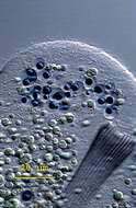
Detail of the large Nnassulid ciliate Obertrumia aurea (Ehrenberg, 1833; Foissner 1987). Synonym of Nassula aurea. Obertrumia is distinguished by it's bipartite "hypostomial frange", linear arrays of ciliary tufts. A sigmoid ventrolateral frange (seen most clearly posterior to the cytostome in this image) runs anteriorly and to the left and is separated from a horizontal line of ciliary tufts on the dorsal surface. The cell appears brightly colored (orange, green or violet) due to multiple food vacuoles containing ingested cyanobacteria. Numerous small mucocysts give the cortex a roughly granular appearance. O. aurea feeds mainly on cyanobacteria. Collected from a freshwater pond near Boise, Idaho November 2003. DIC optics.
-
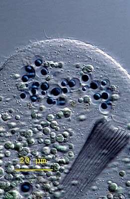
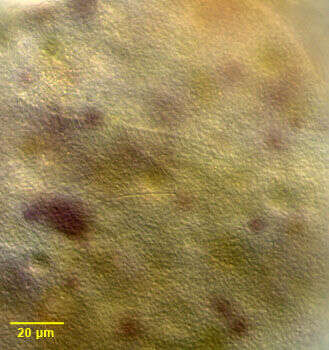
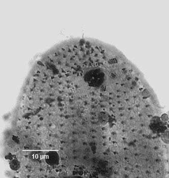
Discharged extrusomes (mucocysts) of the nassulid ciliate, Furgasonia trichocystis (Stokes, 1894) Jankowski, 1964. Synonym: Cyclogramma. The spherical macronucleus (stained dark green here)and adjacent micronucleus are slightly posterior to the equator. There is a prominent layer of fusiform subpelicular extrusomes (mucocysts). This preparation demonstartes the discharged extrusomes and mucus surrounding the cell. Collected from a temporary rainwater pool with abundant decaying grass near Boise, Idaho. March, 2005. Methyl green Pyronin-Y stain (see Foissner, W. Europ. J. Protistol., 27:313-330;1991). Brightfield.
-
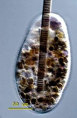
Nassulopsis (nass-you-lop-sis). Nassulopsis elegans is a conspicuous pink- or blue coloured nassulid found in pond and lake plankton. This cell was collected from floating colonies of Oscillatoria in a freshwater pond near Konstanz, Germany. The body is cylindrical in shape and measures 150 - 300 X 50 - 100 microns. Nassulopsis can be distinguished from Nassula and Obertrumia by 5-7 contractile vacuoles arranged in a ventral row. The macronucleus is cylindrical with rounded ends. The cytopharyngeal basket is located in the anterior third of the cell. The cells fill up with food vacuoles of different colour (green, orange), depending on stage of digestion. This image shows a specimen of Nassulopsis elegans beginning to ingest Oscillatoria. The cyanobacteria measures 18 (m in diameter. Differential interference contrast.
-
Detail of the large nassulid ciliate Obertrumia aurea (Ehrenberg, 1833; Foissner 1987). Synonym of Nassula aurea. Obertrumia is distinguished by it's bipartite "hypostomial frange", linear arrays of ciliary tufts. A sigmoid ventrolateral frange runs anteriorly and to the left (not seen in this image) and is separated from a horizontal line of ciliary tufts on the dorsal surface (seen here). The cell appears brightly colored (orange, green or violet) due to multiple food vacuoles containing ingested cyanobacteria. Numerous small mucocysts give the cortex a roughly granular appearance. O. aurea feeds mainly on cyanobacteria. Collected from a freshwater pond near Boise, Idaho November 2003. DIC optics.