-
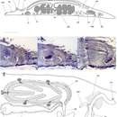
Polyclad worm.
-
This 1973 image depicts two Taenia solium cysticerci, which represent the larval, or intermediate, immature developmental stages of this pork tapeworm.Life cycle of Taenia saginata and T. solium (See PHIL 3420 for a illustrative diagram depicting the following life cycle):Taeniasis is the infection of humans with the adult tapeworm of T. saginata or T. solium. Humans are the only definitive hosts for T. saginata and T. solium. Eggs or gravid proglottids are passed with feces; the eggs can survive for days to months in the environment. Cattle (T. saginata) and pigs (T. solium) become infected by ingesting vegetation contaminated with eggs or gravid proglottids. In the animal's intestine, the oncospheres hatch, invade the intestinal wall, and migrate to the striated muscles, where they develop into cysticerci. A cysticercus can survive for several years in the animal.Created: 1973
-
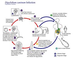
This is an illustration of the life cycle of Dipylidium caninum, which mainly infects dogs and cats, seldom in humans.Created: 2002
-
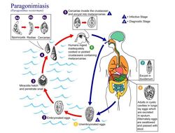
This is an illustration of the life cycle of Paragonimus westermani, one of the causal agents of Paragonimiasis.Created: 2002
-
Magnified 128X, this photomicrograph revealed some of the ultrastructural morphology of a single trematode Paragonimus westermani egg. P. westermani eggs range from 80µm to 120µm long X 45µm to 70µm wide. They are yellow-brown, ovoid or elongate, with a thick shell, and often asymmetrical with one end slightly flattened. At the large end, the operculum is clearly visible. The opposite (abopercular) end is thickened. The eggs are unembryonated when passed in sputum or feces. See PHIL 1534 for an even closer view of this egg.Created: 1979
-
Magnified 500X, this photomicrograph of an unstained, formalin-preserved stool specimen mount, revealed the presence of a Paragonimus westermani termatode egg. See PHIL 3415 for the depiction of the following life cycle.The eggs are excreted unembryonated in the sputum, or alternately they are swallowed and passed with stool (1). In the external environment, the eggs become embryonated (2), and miracidia hatch and seek the first intermediate host, a snail, and penetrate its soft tissues (3). Miracidia go through several developmental stages inside the snail (4): sporocysts (4a), rediae (4b), with the latter giving rise to many cercariae (4c), which emerge from the snail. The cercariae invade the second intermediate host, a crustacean such as a crab or crayfish, where they encyst and become metacercariae. This is the infective stage for the mammalian host (5).Created: 1973
-
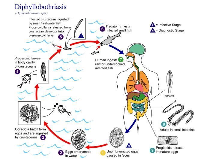
This is an illustration of the life cycle of Diphyllobothrium spp., the causal agents of Diphyllobothriasis.Created: 2002
-
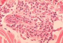
Histopathology of delayed hypersensitivity reaction to Schistosoma mansoni antigen.Created: 1972
-
Histopathology of delayed hypersensitivity reaction to Schistosoma mansoni antigen.Created: 1972
-
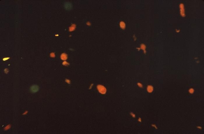
Under a low magnification of 78X, and stained using an indirect fluorescent antibody (IFA) test, this photomicrograph confirmed the presence of Schistosoma mansoni trematodes.Laboratory Diagnosis for Schistosomiasis:Microscopic identification of eggs in stool or urine is the most practical method for diagnosis. Stool examination should be performed when infection with S. mansoni or S. japonicum is suspected, and urine examination should be performed if S. haematobium is suspected. Eggs can be present in the stool in infections with all Schistosoma species. The examination can be performed on a simple smear (1 to 2 mg of fecal material).Created: 1972
-
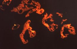
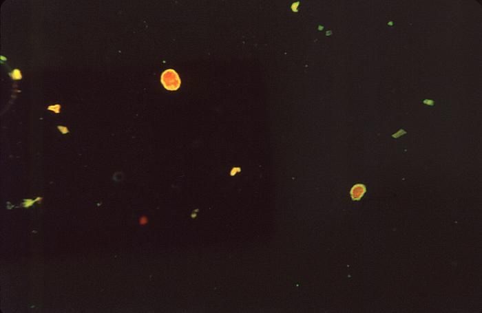
Under a low magnification of 78X, and stained using an indirect fluorescent antibody (IFA) test, this photomicrograph revealed some of the ultrastructural morphology exhibited by a number of Schistosoma mansoni trematodes.Laboratory Diagnosis for Schistosomiasis:Microscopic identification of eggs in stool or urine is the most practical method for diagnosis. Stool examination should be performed when infection with S. mansoni or S. japonicum is suspected, and urine examination should be performed if S. haematobium is suspected. Eggs can be present in the stool in infections with all Schistosoma species. The examination can be performed on a simple smear (1 to 2 mg of fecal material).Created: 1972
-
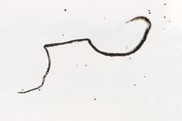
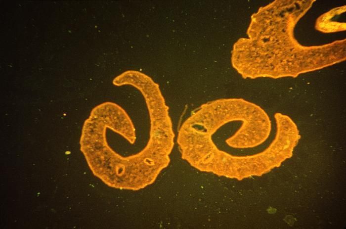
This magnified view reveals a pair of mating Schistosoma mansoni trematodes. Note that the thinner female is cradled inside the thicker male worm's gynecophoral canal.Created: 1973
-
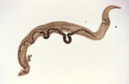
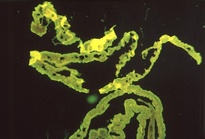
This magnified view reveals a male Schistosoma mansoni trematode. Take a look at PHIL 11193, which depicts a mating pair of worms, where the thinner female is cradled inside the thicker male worm's gynecophoral canal.Created: 1973
-
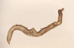
This magnified view reveals a female Schistosoma mansoni trematode. Take a look at PHIL 11193, which depicts a male S. mansoni, and PHIL 11194, which depicts two mating worms. in which case you can see the thinner female cradled inside the thicker male worm's gynecophoral canal.Created: 1973
-
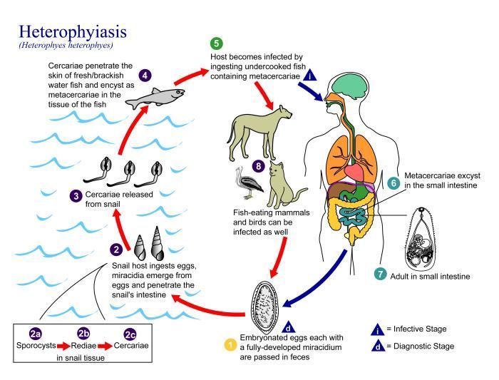
This is an illustration of the life cycle of Heterophyes heterophyes, the causal agent of Heterophyiasis.Created: 2002
-
This photograph depicted the strobilocercus, or larval stage of an unidentified tapeworm of the order Cyclophyllidea. Families that are members of this order include Dipylidiidae, i.e., cucumber tapeworm and double-pore tapeworm, Hymenolepidadae, Taeniidae, i.e., livestock parasites, and Anoplocephalidae, i.e., horse and ruminant tapeworms.Created: 1973
-
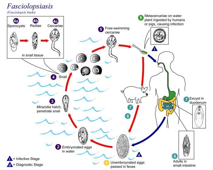
This is an illustration of the life cycle of Fasciolopsis buski, the causal agent of Fasciolopsiasis.Created: 2002
-
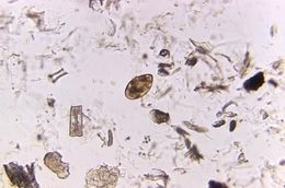
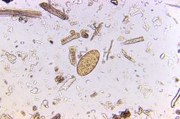
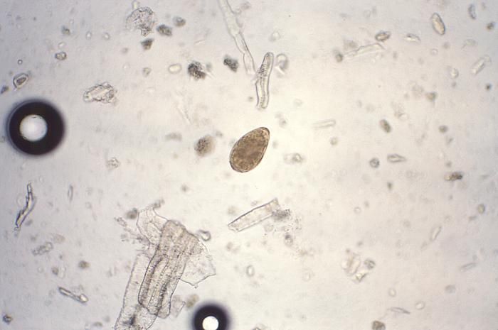
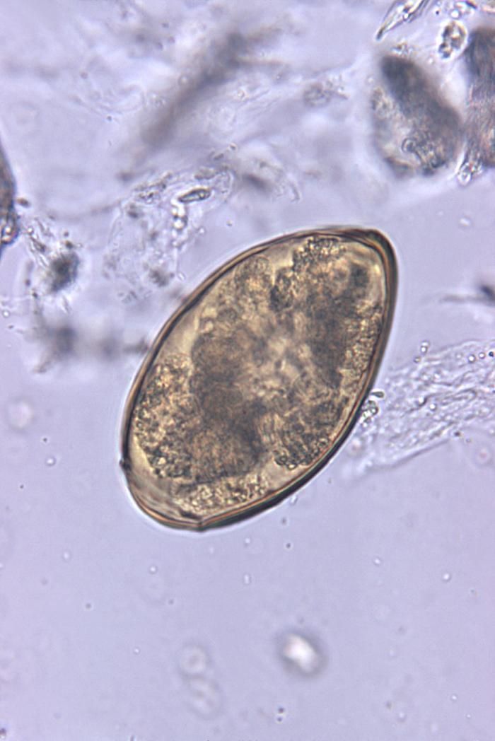
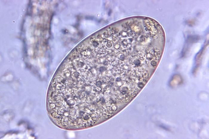
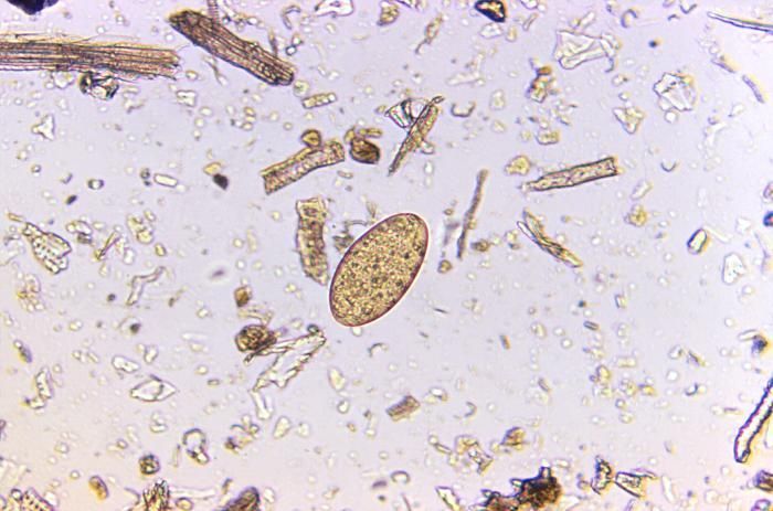
Magnified 500X, this photomicrograph revealed the presence of a Fasciolopsis buski trematode egg that was found in an unstained formalin-preserved stool sample. F. buski are the largest intestinal flukes found parasitizing human beings. These flukes inhabit Asia and the Indian subcontinent, especially in areas where humans raise pigs, and consume freshwater plants.Clinical Features:Most infections are light and asymptomatic. In heavier infections, symptoms include diarrhea, abdominal pain, fever, ascites, anasarca and intestinal obstruction.Laboratory Diagnosis:Microscopic identification of eggs, or more rarely of the adult flukes, in the stool or vomitus is the basis of specific diagnosis. The eggs are indistinguishable from those of Fasciola hepatica.Created: 1973
-
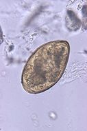
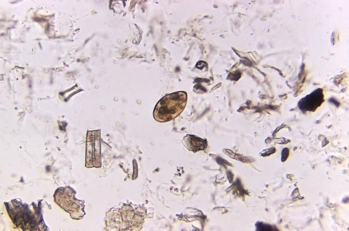
Magnified 125X, this photomicrograph revealed the presence of a Fasciolopsis buski trematode egg that was found in an unstained formalin-preserved stool sample. F. buski are the largest intestinal flukes found parasitizing human beings. These flukes inhabit Asia and the Indian subcontinent, especially in areas where humans raise pigs, and consume freshwater plants.Clinical Features:Most infections are light and asymptomatic. In heavier infections, symptoms include diarrhea, abdominal pain, fever, ascites, anasarca and intestinal obstruction.Laboratory Diagnosis:Microscopic identification of eggs, or more rarely of the adult flukes, in the stool or vomitus is the basis of specific diagnosis. The eggs are indistinguishable from those of Fasciola hepatica.Created: 1973