-
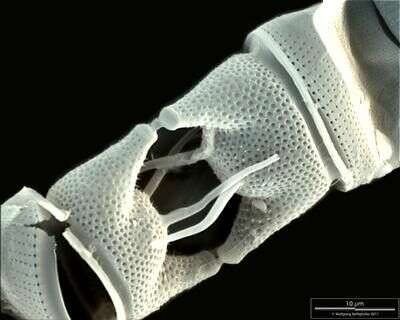
Scale bar indicates 10 µm. Sample from North Sea near Heligoland (spring diatom bloom). Use of SEM equipment courtesy of Lab Dr. Karl-Heinz Schäffner, Solingen, Germany.
-
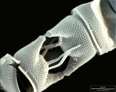
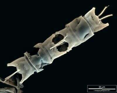
Scale bar indicates 25 µm. Sample from North Sea near Heligoland (spring diatom bloom). Use of SEM equipment courtesy of Lab Dr. Karl-Heinz Schäffner, Solingen, Germany.
-
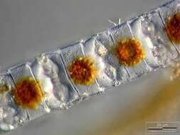
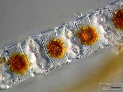
Depth-of-focus image exhibiting structure of the frustules. Chloroplasts are concentrated in cell centers due to long lasting exposition with microscope's illumination. Scale bar indicates 25 µm. The image was built up using several photomicrographic frames with manual stacking technique. Sample from North Sea near Heligoland (spring diatom bloom). Images were taken using Zeiss Universal with Olympus C7070 CCD camera.
-
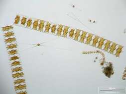
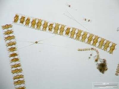
Long chains of Odontella aurita accompanied by Chaetoceros danicus and Thalassiosira nordenskjoeldii. Scale bar indicates 100 µm. The image was built up using several photomicrographic frames with manual stacking technique. Sample from North Sea near Heligoland (spring diatom bloom). Images were taken using Zeiss Universal with Olympus C7070 CCD camera.
-
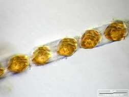
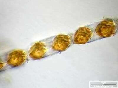
Depth-of-focus image exhibiting structure of the frustules (apical view). Oblique light. Scale bar indicates 50 µm. The image was built up using several photomicrographic frames with manual stacking technique. Sample from North Sea near Heligoland (spring diatom bloom). Images were taken using Zeiss Universal with Olympus C7070 CCD camera.
-