-
Centers for Disease Control/Division of Parasitic Diseases and Malaria
EOL staff
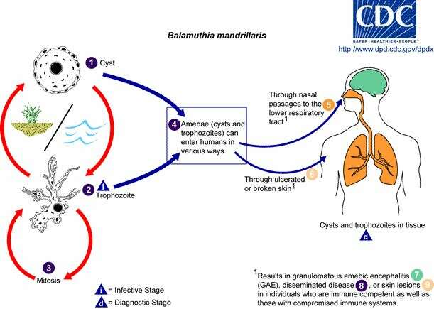
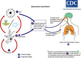
Life cycle of Balamuthia mandrillaris parasitizing humansBalamuthia mandrillaris has only recently been isolated from the environment and has also been isolated from autopsy specimens of infected humans and animals. The B. mandrillaris life cycle has only two stages, a dormant cyst stage (1) and an actively feeding and dividing trophozoite stage (2) (B. mandrillaris has no flagellated stage). The trophozoites replicate by mitosis (the nuclear membrane does not remain intact) (3). Although the trophozoites are the infective stage, both cysts and trophozoites gain entry into the body (4) through various means. Entry can occur through the nasal passages to the lower respiratory tract (5) or through ulcerated or broken skin (6). When B. mandrillaris enters the respiratory system or through the skin, it can invade the central nervous system by hematogenous dissemination causing
granulomatous amebic encephalitis (GAE) (7) or disseminated disease (8), or skin lesions (9) in individuals who are immune competent as well as those with compromised immune systems. Balamuthia mandrillaris cysts and trophozoites are found in tissue.From
Centers for Disease Control Parasites and Health website.
-
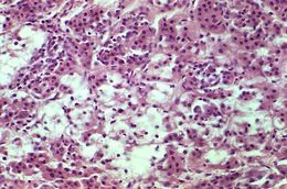
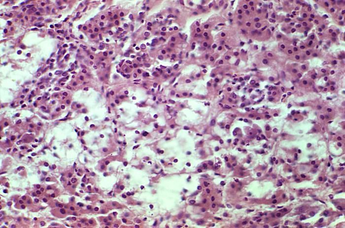
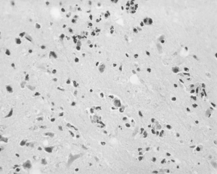
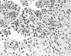
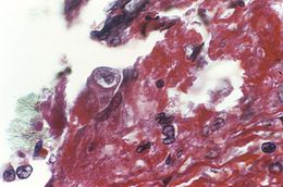
Magnified 250X, this photomicrograph revealed the presence of Acanthamoeba sp. parasitic microorganisms, which were found in a specimen of human H&E-stained adrenal gland tissue.Acanthamoeba is a microscopic, free-living ameba that is relatively common in the environment. This ameba has been isolated from water (including natural and treated water in pools or hot tubs), soil, air (in association with cooling towers, heating, ventilation and air conditioner [HVAC] systems), sewage systems, and drinking water systems (shower heads, taps). Most people will be exposed to Acanthamoeba during their lifetime and will not get sick. However, Acanthamoeba is capable of causing several infections in humans.Created: 1975
-
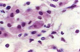
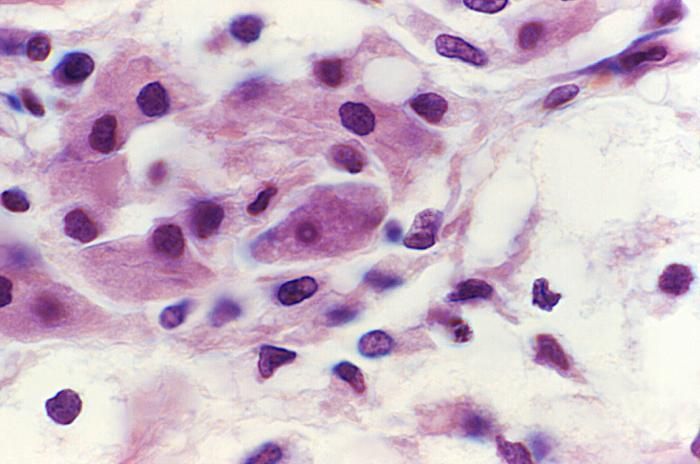
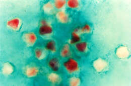
Magnified 1000X, this photomicrograph revealed the presence of Acanthamoeba sp. parasitic microorganisms, which were found in a specimen of human H&E-stained adrenal gland tissue.Acanthamoeba is a microscopic, free-living ameba that is relatively common in the environment. This ameba has been isolated from water (including natural and treated water in pools or hot tubs), soil, air (in association with cooling towers, heating, ventilation and air conditioner [HVAC] systems), sewage systems, and drinking water systems (shower heads, taps). Most people will be exposed to Acanthamoeba during their lifetime and will not get sick. However, Acanthamoeba is capable of causing several infections in humans.Created: 1975
-
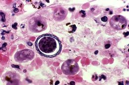
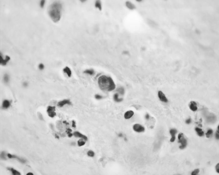
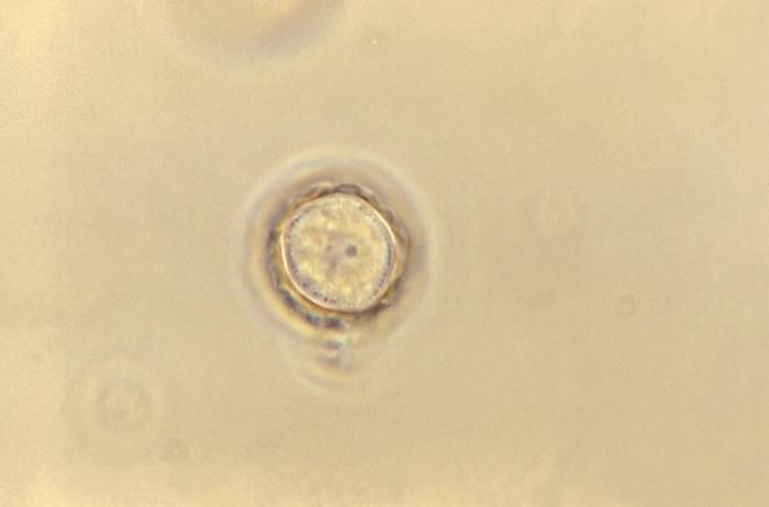
This photomicrograph depicted a magnified view of brain tissue within which was a centrally located Acanthamoeba sp. cyst. Acanthamoeba spp. are opportunistic free-living amebae, capable of causing granulomatous amebic encephalitis (GAE) in individuals with compromised immune systems.Created: 1977
-
Magnified 500X, this 1973 photomicrograph depicted a mouse brain tissue specimen stained using the hematoxylin and eosin (H&E) staining technique, and revealing the presence of Acanthamoeba polyphaga protozoa.Acanthamoeba is a microscopic, free-living ameba commonly found in the environment that can cause rare but severe illness. Acanthamoeba causes three main types of illness involving the eye (keratitis), the brain and spinal cord (Granulomatous Amebic Encephalitis), and infections that can spread from an entry point to the entire body (disseminated disease).Created: 1973
-
Magnified 1200X, this 1973 photomicrograph depicted a mouse brain tissue specimen stained using the hematoxylin and eosin (H&E) staining technique, and revealing the presence of Acanthamoeba polyphaga protozoa.Acanthamoeba is a microscopic, free-living ameba commonly found in the environment that can cause rare but severe illness. Acanthamoeba causes three main types of illness involving the eye (keratitis), the brain and spinal cord (Granulomatous Amebic Encephalitis), and infections that can spread from an entry point to the entire body (disseminated disease).Created: 1973
-
Magnified 500X, this 1973 photomicrograph depicted a mouse brain tissue specimen stained using the hematoxylin and eosin (H&E) staining technique, revealing the presence of Acanthamoeba polyphaga protozoa.Acanthamoeba is a microscopic, free-living ameba commonly found in the environment that can cause rare but severe illness. Acanthamoeba causes three main types of illness involving the eye (keratitis), the brain and spinal cord (Granulomatous Amebic Encephalitis), and infections that can spread from an entry point to the entire body (disseminated disease).Created: 1973
-
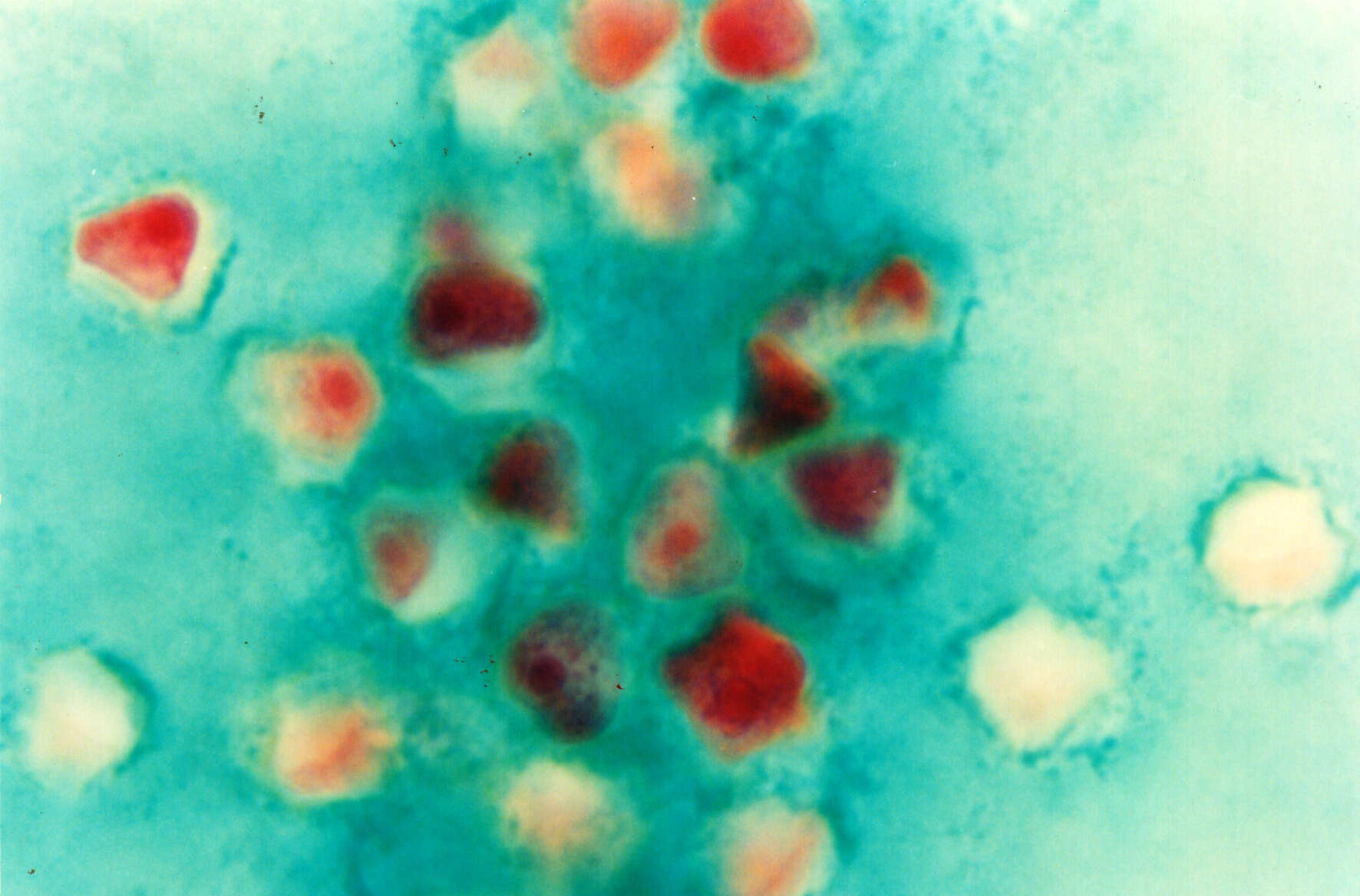
Magnified 1000X, this photomicrograph revealed the presence of Acanthamoeba polyphaga parasitic microorganisms, which were found in a specimen of human trichrome-stained corneal tissue.Acanthamoeba is a microscopic, free-living ameba that is relatively common in the environment. This ameba has been isolated from water (including natural and treated water in pools or hot tubs), soil, air (in association with cooling towers, heating, ventilation and air conditioner [HVAC] systems), sewage systems, and drinking water systems (shower heads, taps). Most people will be exposed to Acanthamoeba during their lifetime and will not get sick. However, Acanthamoeba is capable of causing several infections in humans.Created: 1975
-
Magnified 1125X, this 1973 photomicrograph depicted an Acanthamoeba polyphaga protozoan cyst.Acanthamoeba is a microscopic, free-living ameba commonly found in the environment that can cause rare, but severe illness. Acanthamoeba causes three main types of illness involving the eye (keratitis), the brain and spinal cord (Granulomatous Amebic Encephalitis), and infections that can spread from an entry point to the entire body (disseminated disease).Created: 1973
-
Description: English: Acanthamoeba spp (Cyst). Date: 1 April 2005, 15:02:47. Source: Own work. Author:
Punlop Anusonpornperm.
-
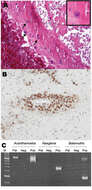
Description: A) Cysts in a vessel wall (arrows) of the patient (
hematoxylin and eosin stain, magnification ×250). Inset shows a cyst at higher magnification (hematoxylin and eosin stain, magnification ×800). B) Immunohistochemical staining with antibody to
en:Acanthamoeba cysts within vessel walls (magnification ×250). C)
Polyacrylamide gel electrophoresis of PCR products for Acanthamoeba spp.,
en:Naegleria spp., and
en:Balamuthia mandrillaris using JDP primers for a diagnostic small subunit
rDNA fragment. M, molecular mass marker; Pat, patient; Neg, negative control; Pos, positive control. Source:Meersseman W, Lagrou K, Sciot R, de Jonckheere J, Haberler C, Walochnik J, Peetermans W, van Wijngaerden E. "Rapidly Fatal Acanthamoeba Encephalitis and Treatment of Cryoglobulinemia". Emerg Infect Dis [serial on the Internet]. 2007 March [cited 2007 Feb 23]. Available from
https://www.cdc.gov/eid/content/13/3/469-G2.htm Public Domain rationale. Source: US gov. Author: US gov.
-
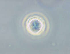
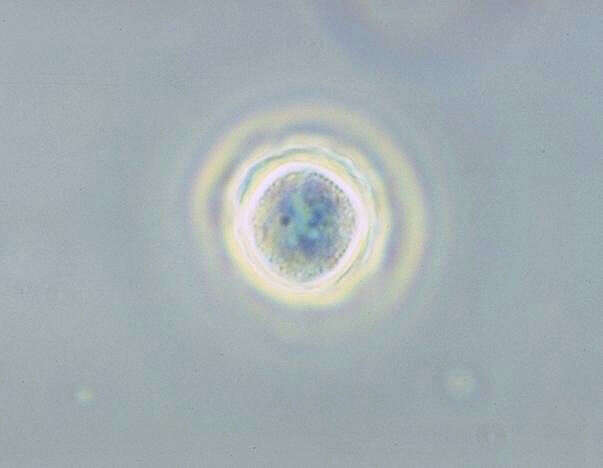
Description: English: Acanthamoeba polyphaga cyst. Phase contrast. Ameba, parasite. Date: 1973. Source: : This media comes from the
Centers for Disease Control and Prevention's
Public Health Image Library (PHIL), with identification number
#1425. Note: Not all PHIL images are public domain; be sure to check copyright status and credit authors and content providers.
English |
slovenščina |
+/−. Author: Photo Credit: Content Providers(s): CDC/ Dr. George Healy. Permission(
Reusing this file): PD-USGOV-HHS-CDC. English: None - This image is in the public domain and thus free of any copyright restrictions. As a matter of courtesy we request that the content provider be credited and notified in any public or private usage of this image.
-
-
-
-
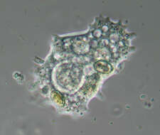
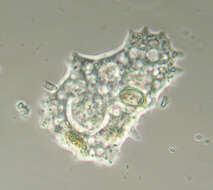
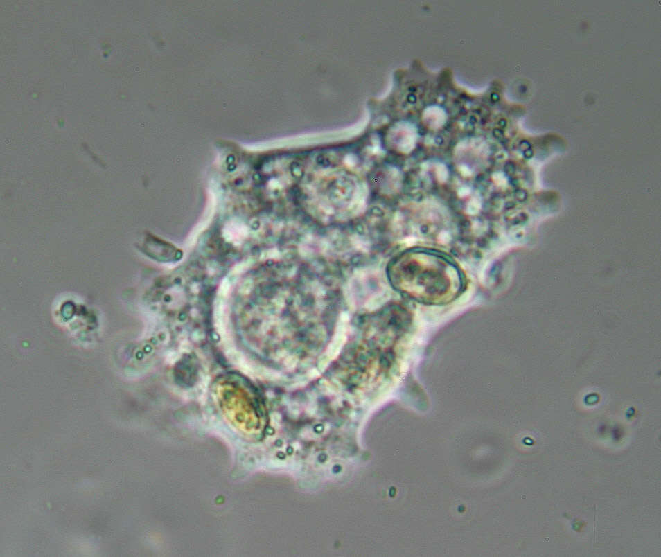
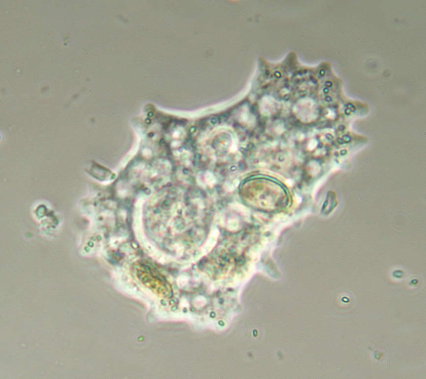
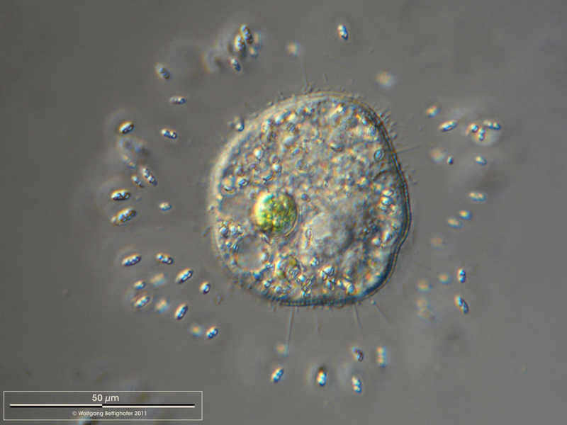
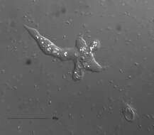
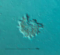
Description: Die Gattung Mayorella ist sicher, aber für eine genauere Bestimmung müsste man die genaue Form der Kristalle erkennen können. Date: 8 November 2013, 22:00. Source:
Mayorella sp. - 400x. Author:
Picturepest.
-
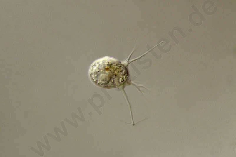
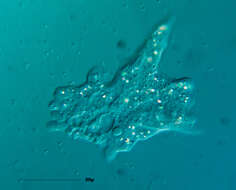
Longitude (deg): -1.2. Latitude (deg): 51.3. Longitude (deg/min): 1° 20' W. Latitude (deg/min): 51° 20' N. Vice county name: Berks. Vice county no.: 22. Country: England. Identified by: Malcolm Storey. Comment: "in alga sample, collected 18 July, cultured on shady window sill". Category: microscope photograph. Image scaling: magnified. Photographic equipment used: Canon EOS10D dSLR and Meiji microscope with x2.5 projection eye-piece. DPI defaulting to 72.
-
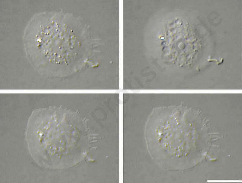
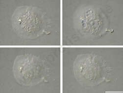
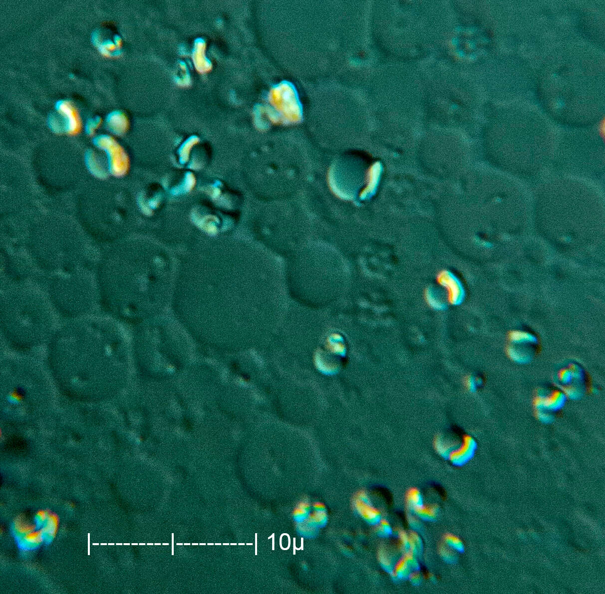
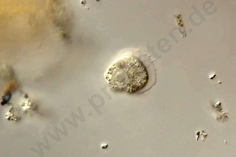
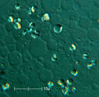
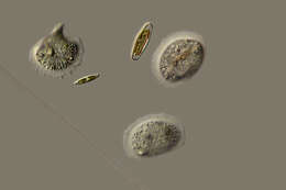
Cochliopodium spec. Several focus planes show a specimen of Cochliopodium, perhaps a member of the C. minutum group. Scale bar indicates 10 µm. The scales are very delicate and were not clearly visible. For further details see Kudryavtsev, A.: Minute species of Cochliopodium (Himatismenida): Description of three new fresh- and brackish-water species with a new diagnosis for Cochliopodium minus Page, 1976. Eu. J. Prot. 42 (2006) 7789. (doi:10.1016/j.ejop.2005.12.002). Sample from a freshwater pond on the island of Hiddensee (Baltic Sea, Germany). This image was taken using Zeiss Universal with Olympus C7070 CCD camera.Image under Creative Commons License V 3.0 (CC BY-NC-SA). Place name: Pond Suploch, Hiddensee (Germany) Latitude: 54.538638 Longitude: 13.097802 Darstellung eines Exemplars von Cochliopodium, vielleicht ein Mitglied der C. minutum-Gruppe, in mehreren Fokusebenen. Der Messbalken markiert eine Länge von 10 µm. Die Schuppen in der Pellicula sind sehr zart und waren nicht deutlich zu erkennen. Für weitere Details siehe Kudryavtsev, A.: Minute species of Cochliopodium (Himatismenida): Description of three new fresh- and brackish-water species with a new diagnosis for Cochliopodium minus Page, 1976. Eu. J. Prot. 42 (2006) 7789. (doi:10.1016/j.ejop.2005.12.002). Probe aus einem kleinen Süßwasserteich auf der Insel Hiddensee, welcher eine faszinierende Vielfalt von nackten und beschalten Amöben beherbergt. Mikrotechnik: Zeiss Universal, Kamera: Olympus C7070. Creative Commons License V 3.0 (CC BY-NC-SA). For permission to use of (high-resolution) images please contact postmaster@protisten.de.
-
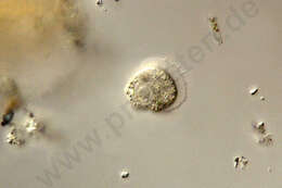
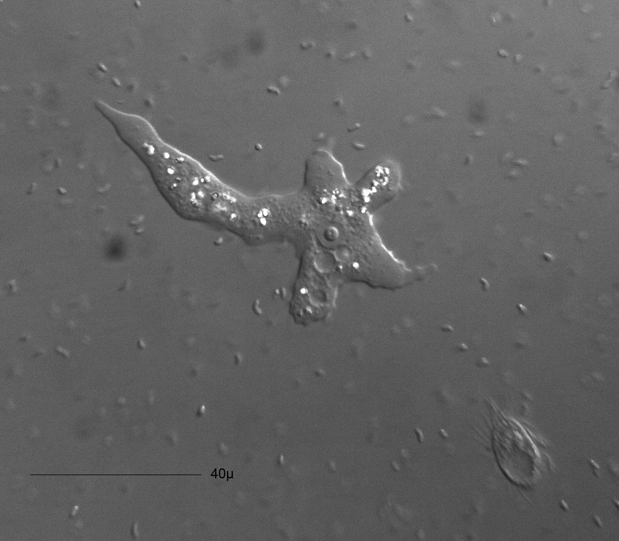
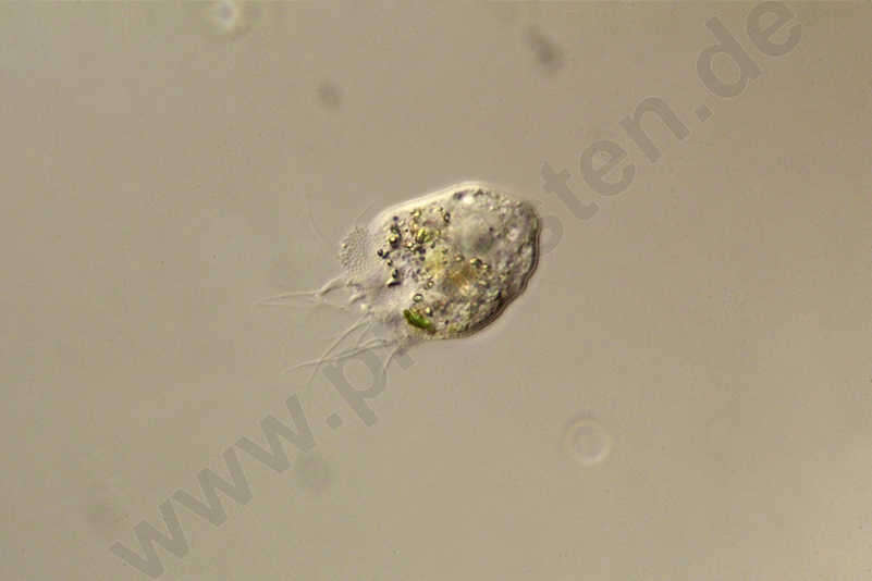
Cochliopodium spec. Sample from a pond near Großostheim, Germany. Sampling date 04/2021. Copyright Winfried Hölz, Hausen, Germany.Images were taken using Zeiss Axioskop with DSLR.Image under Creative Commons License V 3.0 (CC BY-NC-SA). Place name: Pond near Großostheim (Germany) Latitude: 49.88482168 Longitude: 9.09980822 Probe aus einem Waldteich bei Großostheim. Datum der Aufsammlung: 04/2021.Copyright Winfried Hölz, Hausen. Mikrotechnik: Zeiss Axioskop, Kamera: DSLR. Creative Commons License V 3.0 (CC BY-NC-SA). For permission to use of (high-resolution) images please contact postmaster@protisten.de.
-
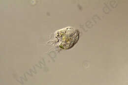
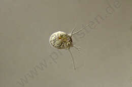
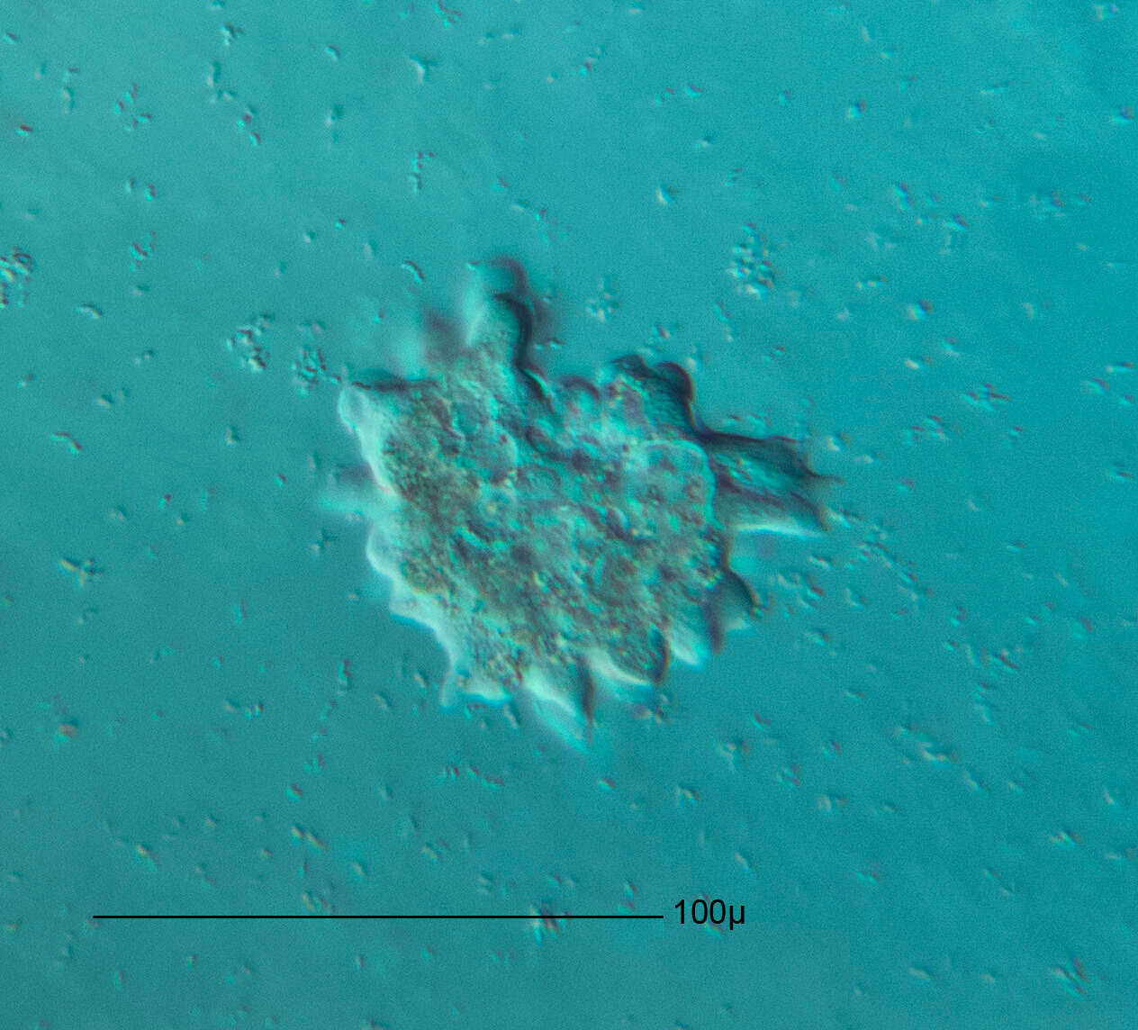
Cochliopodium vestitum Optical transversal section. Through the bacteria the mucilaginous sheeth becomes clearly visible. Scale bar indicates 50 µm. Sample from the pond Hegne Moor situated in the vicinity of Lake Constance. The image was built up using several photomicrographic frames with manual stacking technique. Images were taken using Zeiss Universal with Olympus C7070 CCD camera.Image under Creative Commons License V 3.0 (CC BY-NC-SA). Place name: Bog Hegne Moor near Lake Constance (Germany) Latitude: 47.718106 Longitude: 9.093974 Optischer Querschnitt. Durch die Bakterien wird die Schleimhülle deutlich sichtbar. Multiebenen-Abbildung, manuell gestapelt. Der Messbalken markiert eine Länge von 50 µm. Probe aus dem Simmelried nahe Konstanz. Mikrotechnik: Zeiss Universal, Kamera: Olympus C7070. Creative Commons License V 3.0 (CC BY-NC-SA). For permission to use of (high-resolution) images please contact postmaster@protisten.de.
-
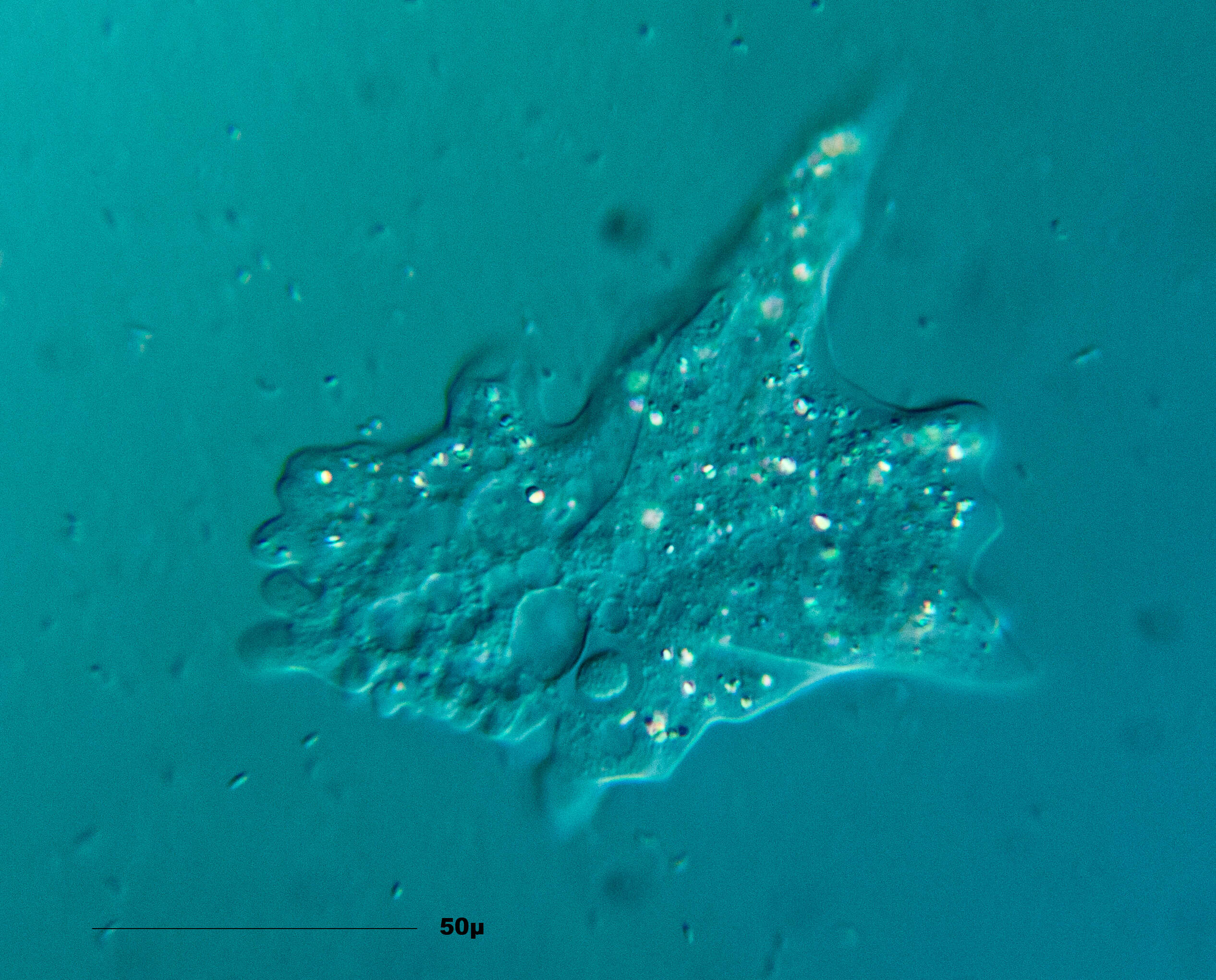
Cochliopodium vestitum The specimen was gathered in ponds near Hausen (Hessisch Lichtenau , Germany). Copyright Winfried Hölz, Hausen, Germany.Images were taken using Zeiss Axioskop with DSLR.Image under Creative Commons License V 3.0 (CC BY-NC-SA). Place name: Ponds near Hausen (Hessisch Lichtenau , Germany) Latitude: 51.21453 Longitude: 9.868894 Probe aus Gewässern nahe Hausen (Hessisch Lichtenau).Copyright Winfried Hölz, Hausen. Mikrotechnik: Zeiss Axioskop, Kamera: DSLR. Creative Commons License V 3.0 (CC BY-NC-SA). For permission to use of (high-resolution) images please contact postmaster@protisten.de.


