dcsimg

Ecology ( англиски )

добавил NMNH Marine Dinoflagellates
D. acuta is a planktonic oceanic and neritic species (Dodge 1982; Taylor et al. 1995; Steidinger & Tangen 1996). This is a bloom-forming species; blooms are often associated with shellfish toxicity (Taylor et al. 1995).
лиценца
cc-publicdomain
библиографски навод
Faust, Maria A. and Rose A. Gulledge. Identifying Harmful Marine Dinoflagellates. Smithsonian Contributions from the United States National Herbarium, volume 42: 1-144 (including 48 plates, 1 figure and 1 table).
соработничко мреж. место
NMNH Marine Dinoflagellates

Habitat and Locality ( англиски )

добавил NMNH Marine Dinoflagellates
Dinophysis acuta is widely distributed in cold and temperate waters world-wide (Larsen & Moestrup 1992; Steidinger & Tangen 1996).
лиценца
cc-publicdomain
библиографски навод
Faust, Maria A. and Rose A. Gulledge. Identifying Harmful Marine Dinoflagellates. Smithsonian Contributions from the United States National Herbarium, volume 42: 1-144 (including 48 plates, 1 figure and 1 table).
соработничко мреж. место
NMNH Marine Dinoflagellates

Morphology and Structure ( англиски )

добавил NMNH Marine Dinoflagellates
Dinophysis acuta is a photosynthetic species with yellow chloroplasts (Dodge 1982; Larsen & Moestrup 1992).
Dimorphic cells, one half resembling D. acuta and the other half resembling D. dens (the proposed gamete form), have occasionally been observed in this species (Reguera et al. 1990; Hansen 1993; Moita & Sampayo 1993). It is highly probable that these cell forms represent a stage in gametogenesis (Hansen 1993).
лиценца
cc-publicdomain
библиографски навод
Faust, Maria A. and Rose A. Gulledge. Identifying Harmful Marine Dinoflagellates. Smithsonian Contributions from the United States National Herbarium, volume 42: 1-144 (including 48 plates, 1 figure and 1 table).
соработничко мреж. место
NMNH Marine Dinoflagellates

Nomenclatural Types ( англиски )

добавил NMNH Marine Dinoflagellates
Holotype: Dinophysis acuta Ehrenberg, 1839: 124, 151, plate 4 (fide Schiller, 1933)
Type Locality: Mediterranean Sea: Gulf of Lion, France
лиценца
cc-publicdomain
библиографски навод
Faust, Maria A. and Rose A. Gulledge. Identifying Harmful Marine Dinoflagellates. Smithsonian Contributions from the United States National Herbarium, volume 42: 1-144 (including 48 plates, 1 figure and 1 table).
соработничко мреж. место
NMNH Marine Dinoflagellates

Remarks ( англиски )

добавил NMNH Marine Dinoflagellates
Many authors consider Phalacroma to be synonymous with Dinophysis (Steidinger & Tangen 1996).
лиценца
cc-publicdomain
библиографски навод
Faust, Maria A. and Rose A. Gulledge. Identifying Harmful Marine Dinoflagellates. Smithsonian Contributions from the United States National Herbarium, volume 42: 1-144 (including 48 plates, 1 figure and 1 table).
соработничко мреж. место
NMNH Marine Dinoflagellates

Reproduction ( англиски )

добавил NMNH Marine Dinoflagellates
D. acuta reproduces asexually by binary fission. Hansen (1993) speculates that sexual reproduction, with sexual dimorphism, is part of the life cycle for this species.
лиценца
cc-publicdomain
библиографски навод
Faust, Maria A. and Rose A. Gulledge. Identifying Harmful Marine Dinoflagellates. Smithsonian Contributions from the United States National Herbarium, volume 42: 1-144 (including 48 plates, 1 figure and 1 table).
соработничко мреж. место
NMNH Marine Dinoflagellates

Species Comparison ( англиски )

добавил NMNH Marine Dinoflagellates
D. acuta is very similar to D. norvegica in their general shape, and thus can easily be misidentified. D. acuta can be differentiated by its larger size and different shape: D. norvegica is widest in the middle region of the cell, whereas D. acuta is widest below the mid-section. Moreover, D. acuta has a longer left sulcal list relative to its cell length (Balech 1976; Dodge 1982; Larsen & Moestrup 1992; Taylor et al. 1995; Steidinger & Tangen 1996). D. acuta also strongly resembles a warm-water species, D. schroederi Pavillard, 1909 (Schiller 1933; Balech 1976; Burns & Mitchell 1982).
лиценца
cc-publicdomain
библиографски навод
Faust, Maria A. and Rose A. Gulledge. Identifying Harmful Marine Dinoflagellates. Smithsonian Contributions from the United States National Herbarium, volume 42: 1-144 (including 48 plates, 1 figure and 1 table).
соработничко мреж. место
NMNH Marine Dinoflagellates

Species Overview ( англиски )

добавил NMNH Marine Dinoflagellates
Dinophysis acuta is an armoured, marine, planktonic dinoflagellate species. It is a toxic species associated with DSP events and is commonly found in cold and temperate neritic waters.
лиценца
cc-publicdomain
библиографски навод
Faust, Maria A. and Rose A. Gulledge. Identifying Harmful Marine Dinoflagellates. Smithsonian Contributions from the United States National Herbarium, volume 42: 1-144 (including 48 plates, 1 figure and 1 table).
соработничко мреж. место
NMNH Marine Dinoflagellates

Taxonomic Description ( англиски )

добавил NMNH Marine Dinoflagellates
Species in this genus are laterally compressed with a small, cap-like epitheca and a much larger hypotheca (dorso-ventral depth of epitheca is 1/2 to 2/3 of hypotheca). The shape of the cell in lateral view is the most important criterion used for identification (Taylor et al. 1995).
Cells of Dinophysis acuta are large and robust, and are among the largest species in the genus Dinophysis (Fig. 1). Cells are oblong with a slightly pointed or rounded posterior end (Figs. 1-4). The left sulcal list (LSL) extends beyond the midpoint of the cell (about 2/3 of cell length) ending at or above the widest portion of the cell (Fig. 3) (Balech 1976; Dodge 1982; Larsen & Moestrup 1992; Taylor et al. 1995; Steidinger & Tangen 1996).
The thick thecal plates of the hypotheca are coarsely areolated, each areola with a central pore (Figs. 1, 2, 4). The areolation becomes very faint or disappears near the edge of the plates. Cell size ranges: 54-94 µm in length and 43-60 µm in dorso-ventral width (widest below the middle) (Fig. 3)(Balech 1976; Dodge 1982; Larsen & Moestrup 1992; Taylor et al. 1995; Steidinger & Tangen 1996).
лиценца
cc-publicdomain
библиографски навод
Faust, Maria A. and Rose A. Gulledge. Identifying Harmful Marine Dinoflagellates. Smithsonian Contributions from the United States National Herbarium, volume 42: 1-144 (including 48 plates, 1 figure and 1 table).
соработничко мреж. место
NMNH Marine Dinoflagellates

Thecal Plate Description ( англиски )

добавил NMNH Marine Dinoflagellates
The small epitheca is made up of four plates. It is low, flat or weakly convex, and is not visible in lateral view (Balech 1976; Larsen & Moestrup 1992; Taylor et al. 1995).
The cingulum is made up of four unequal plates, all with pores. Well developed cingular lists are present: an anterior cingular list (ACL), and a posterior cingular list (PCL). They are generally smooth and rarely ornamented (Fig. 3). The high ACL obscures the low epitheca (Balech 1976; Dodge 1982; Larsen & Moestrup 1992).
The sulcus is comprised of several irregularly shaped plates. The flagellar pore is housed in the sulcal area. The left sulcal list (LSL), supported by three ribs that radiate outward, is rather broad with a convex ventral margin. It is wider posteriorly and slightly areolated. The second sulcal rib is closer to the first than to the third. The third rib is the longest (Figs. 1-4) (Balech 1976; Dodge 1982; Taylor et al. 1995).
The hypotheca, with four large plates, comprises the majority of the cell. The anterior 2/3 of the hypotheca has convex margins, while the posterior third of the hypotheca forms a broad asymmetrical triangle with a straight dorsal edge and occasionally a slightly concave ventral edge (Figs. 1-4). The tapered and roughly pointed antapex is directed slightly ventrally (Figs. 1-4) (Balech 1976; Dodge 1982; Taylor et al. 1995). Balech (1976: figs. 2H, 2I) depicts two specimens with two to three small knob-like spines on the posterior end.
лиценца
cc-publicdomain
библиографски навод
Faust, Maria A. and Rose A. Gulledge. Identifying Harmful Marine Dinoflagellates. Smithsonian Contributions from the United States National Herbarium, volume 42: 1-144 (including 48 plates, 1 figure and 1 table).
соработничко мреж. место
NMNH Marine Dinoflagellates

Toxicity ( англиски )

добавил NMNH Marine Dinoflagellates
D. acuta is a toxic species that produces okadaic acid (OA), as well as Dinophysistoxin-1 (DTX1) (Lee et al. 1989; Yasumoto 1990). D. acuta has been associated with DSP outbreaks in Chile (Larsen & Moestrup 1992), Portugal (Alvito et al. 1990; Sampayo et al. 1990), Scandinavia (Dahl & Yndestad 1985; Krogh et al. 1985; Underdahl et al. 1985; Edler & Hageltorn 1990), and the USA (Freudenthal & Jijina 1985).
лиценца
cc-publicdomain
библиографски навод
Faust, Maria A. and Rose A. Gulledge. Identifying Harmful Marine Dinoflagellates. Smithsonian Contributions from the United States National Herbarium, volume 42: 1-144 (including 48 plates, 1 figure and 1 table).
соработничко мреж. место
NMNH Marine Dinoflagellates

Dinophysis acuta ( германски )

добавил wikipedia DE

Dinophysis acuta ist eine Art (Spezies) von Dinoflagellaten, die zur Gattung Dinophysis gehört. Dieser Einzeller ist einer der wenigen ungewöhnlichen photosynthetischen Protisten, die durch Endosymbiose Plastiden von Algen erwerben. Durch die Bildung massiver Blüten, insbesondere im Spätsommer und Frühjahr, verursacht er rote Tiden (englisch red tides). D. acuta produziert giftige Substanzen (Toxine), und die roten Tiden verursachen weit verbreitete Infektionen von Meeresfrüchten, insbesondere von Krabben und Muscheln. Wenn infizierte Tiere dann verzehrt werden, kommt es zu schweren Durchfällen (Diarrhoe); dieses klinische Symptom wird als diarrhöische Schalentiervergiftung bezeichnet (en. diarrhetic shellfish poisoning, DSP).[1]

 src=
Okadasäure

Die wichtigsten chemischen Toxine wurden 2006 als Okadasäure und Pectenotoxine[2][3][4][5] identifiziert.[6][7][8] Sie können in ihren Fressfeinden nicht tödliche oder auch tödliche Mengen an Toxinen produzieren, die auch beim Menschen Vergiftungen hervorrufen können.

Dinophysis acuta ist Einzeller des Jahres 2020.[9]

Beschreibung

Morphologie

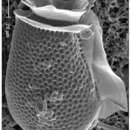
Dinophysis acuta ist ein mariner einzelliger Protist und der größte in der Gattung Dinophysis. Es handelt sich um eine gepanzerte Art mit einer ausgeprägten Körperhülle, die Theca genannt wird. Der Körper ist seitlich zusammengedrückt und die Theca besteht aus einer kleinen, kappenartigen Epitheca und einer viel größeren Hypotheca. Sie hat einen doppelten Kragen (das so genannte Cingulum) um den oberen Teil der Zelle und einen weiteren Flügel (den so genannten Sulcus), der vertikal in der Zelle verläuft. Sie hat eine längliche Form mit einem fast vollständig abgerundeten hinteren Ende, das jedoch an der Spitze leicht spitz ist. Die Größe reicht von 54 bis 94 µm in der Länge und 43 bis 60 µm in der dorso-ventralen Breite, wobei der breiteste Bereich unterhalb der Mitte liegt. Die kleine Epitheca besteht aus vier Platten. Sie ist niedrig, flach oder schwach konvex und in der Seitenansicht nicht sichtbar, was ein gutes Erkennungsmerkmal ist. Der Sulcus besteht aus mehreren unregelmäßig geformten Platten und enthält die Geißelpore. Die Hypotheca besteht aus vier großen Platten, die den größten Teil der Zelle ausmachen. Die vorderen zwei Drittel der Hypotheca haben konvexe Ränder, während das hintere Drittel ein breites asymmetrisches Dreieck mit einem geraden dorsalen Rand und gelegentlich einem leicht konkaven ventralen Rand bildet.[10][11][12]

Fortpflanzung

Die Fortpflanzung erfolgt gewöhnlich durch einfache binäre Spaltung. Lange Zeit glaubte man, dass die Dinophysis-Arten keinen Sexualzyklus (mit geschlechtlicher Fortpflanzung) hat. Inzwischen ist jedoch klar, dass sich bei D. acuminata und D. acuta Gametenzellen bilden können. Dies wurde festgestellt, als sich kleine, kugelförmige Zellen innerhalb größerer Zellen zu bilden schienen.[13]

Kleptoplastidie

Das Ungewöhnlichste an der Zellstruktur von D. acuta wie auch von D. acuminata sind zahlreiche rötlich-gelbe (phycobilinhaltige) Chloroplasten, die von ihrer Beute, Wimpertierchen der Gattung Mesodinium (M. rubrum, synonym Myrionecta rubra) stammen, die sie ihrerseits von Cryptophyceen erworben hat (Kleptoplastidie).[14][15]

Photosynthetische Arten der Gattung Dinophysis sind obligate Mixotrophe, die zum Überleben und Wachsen Licht, Nährstoffe und lebende Beute benötigen.[14]

Der bereits früher entdeckte Fressmechanismus von Phalacroma rotundatum und Dinophysis hastata, die sich von dem Wimpertierchen Tiarina fusus ernähren, ist eine Art Phagozytose (Myzozytose), bei der der Inhalt der Beute durch einen Pedunkel (englisch feeding peduncle) in die Zelle des Räuberzelle befördert wird. Eine ähnliche Struktur wird von D. acuminata und D. acuta benutzt, um sich von M. rubrum zu ernähren.[14]

Anders als früher gedacht, besitzen die photosynthetischen Arten der Gattung Dinophysis aber keine eigenen Chloroplasten. Stattdessen behalten sie die Kleptoplastiden ihrer Wimpertierchens-Beute M. rubrum vorübergehend zurück. Diese Wimpertierchen ernähren sich von Cryptophyceen (insbesondere aus dem Teleaulax/​Plagioselmis/​Geminigera-Komplex[16]), wobei sie deren Chloroplasten als Kleptoplastiden (und auch Mitochondrien) in sich aufnehmen. Man geht davon aus, dass unterschiedliche Plastiden bei Dinophysis letztlich von unterschiedlichen Cryptophyceen-Quellen stammen. Feldstudien haben bestätigt, dass Teleaulax-ähnliche Arten wie T. amphioxeia die häufigste Quelle von Plastiden in Dinophysis sind. Es kann aber die Möglichkeit nicht ausgeschlossen werden, dass auch andere Kleptoplastid-tragende Wimpertierchen wie Cyrtostrombidium, Laboea, Strombidium und/oder Tontonia neben Mesodinium als Überträger von Teleaulax-ähnlichen Plastiden auf Dinophysis in Frage kommen. Dafür spricht die Entdeckung von Plastiden aus mehreren Algenquellen in Dinophysis-Arten aus koreanischen Gewässern.[14][17][15][18]

Toxizität

Die ersten Fälle von diarrhöischen Schalentiervergiftungen (en. diarrhetic shellfish poisoning, DSP) durch D. acuta wurden 1972 in Peru festgestellt, der wissenschaftlichen Gemeinschaft jedoch erst 1991 gemeldet.[19] Es handelt sich um eine sehr milde Form von Vergiftung durch Meeresfrüchte, die sich durch schweren Durchfall bemerkbar macht.[1] Die ersten Toxine, die aus dieser Art isoliert wurden, waren im Jahr 2003 die Pectenotoxine PTX-2[4] und PTX-11[5] aus Exemplaren, die an der Westküste der Südinsel Neuseelands gesammelt wurden,[20] und PTX-12[2] unabhängig davon in Skjer im Sognefjord (auf dem Südarm Nærøyfjord) in Norwegen.[21] Im Jahr 2004 wurde das Vorhandensein von Okadasäureestern gemeldet.[22] 2006 wurden weitere dieser Verbindungen identifiziert und ihre Bedeutung als ursächliche Faktoren von DSP entdeckt.[6][7][8]

Einzelnachweise

  1. a b David Arieti, Jakob Nieva, Randolf Swiller: Prognosis Disaster: The Environment, Climate Change, Human Influences, Vectors, Disease and the Possible End of Humanity?. Au​thor House, 2011, ISBN 9781456731496, S. 404.
  2. a b Zhaoxin Li, Guo Mengmeng, Yang Shouguo, Wang Qingyin, Tan Zhijun: Investigation of Pectenotoxin Profiles in the Yellow Sea (China) Using a Passive Sampling Technique, in: MDPI Marine Drugs, Band 8, Nr. 4, S. 1263–1272, Special Issue Algal Toxins, 15. April 2010, doi:10.3390/md8041263
  3. Pectenotoxin-1, auf: Pubchem
  4. a b pectenotoxin 2, auf Chemical Book
  5. a b PTX-11 -Lösung, auf: Merck Sigma-Aldrich
  6. a b Christopher Owen Miles, Alistair L. Wilkins, Allan D. Hawkes, Dwayne J. Jensen, Janine M. Cooney, Kristofer Larsen, Dirk Petersen, Frode Rise, Veronica Beuzenberg, A. Lincoln MacKenzie: Isolation and identification of a cis-C8-diol-ester of okadaic acid from Dinophysis acuta in New Zealand. In: Toxicon. 48, Nr. 2, August 2006, S. 195–203. doi:10.1016/j.toxicon.2006.04.018. PMID 16784765.
  7. a b Christopher Owen Miles, Alistair L. Wilkins, Allan D. Hawkes, Dwayne J. Jensen, Andrew I. Selwood, Veronica Beuzenberg, A. Lincoln MacKenzie, Janine M. Cooney, Patrick T. Holland: Isolation and identification of pectenotoxins-13 and -14 from Dinophysis acuta in New Zealand. In: Toxicon. 48, Nr. 2, August 2006, S. 152–159. doi:10.1016/j.toxicon.2006.04.005. PMID 16828828.
  8. a b Toshiyuki Suzuki, John A. Walter, Patricia LeBlanc, Shawna MacKinnon, Christopher O. Miles, Alistair L. Wilkins, Rex Munday, Veronica Beuzenberg, A. Lincoln MacKenzie, Dwayne J. Jensen, Janine M. Cooney, Michael A. Quilliam: Identification of pectenotoxin-11 as 34S-hydroxypectenotoxin-2, a new pectenotoxin analogue in the toxic dinoflagellate Dinophysis acuta from New Zealand. In: Chemical Research in Toxicology. 19, Nr. 2, 2006, S. 310–318. doi:10.1021/tx050240y. PMID 16485908.
  9. Einzeller des Jahres 2020: Dinophysis acuta, Deutsche Gesellschaft für Protozoologie
  10. Identifying harmful marine dinoflagellates. Department of Botany, Smithsonian Institution. Abgerufen am 25. August 2014.
  11. P. Lassus, M. Seguel, P. Truquet: Morphological study of atypical Dinophysis acuta Ehrenberg from Chilean coastal waters by a digital pattern-recognition system. In: Botanica Marina. 41, Nr. 1–6, 1998, S. 435–441. doi:10.1515/botm.1998.41.1-6.435.
  12. D. A. Burns, J. S. Mitchell: Dinoflagellates of the genus Dinophysis Ehrenberg from New Zealand coastal waters. In: New Zealand Journal of Marine and Freshwater Research. 16, Nr. 3–4, 1982, S. 289–298. doi:10.1080/00288330.1982.9515972.
  13. Beatriz Reguera, Lourdes Velo-Suárez, Robin Raine, Myung Gil Park: Harmful Dinophysis species: A review. Harmful Algae, in: Harmful Algae, Band 14, Februar 2012, S. 87–106, doi:10.1016/j.hal.2011.10.016
  14. a b c d María García-Portela, Beatriz Reguera, Manoella Sibat, Andreas Altenburger, Francisco Rodríguez, Philipp Hess: Metabolomic Profiles of Dinophysis acuminata and Dinophysis acuta Using Non-Targeted High-Resolution Mass Spectrometry: Effect of Nutritional Status and Prey, in: MDPI Marine Drugs, Band 16, Nr. 15, 143, 26. April 2018, doi:10.3390/md16050143, PMID 29701702, PMC 5982093 (freier Volltext).
  15. a b A. Mitra: Meeresbiologie – Das Beste aus zwei Welten., Spektrum der Wissenschaft, 20. März 2019, S. 54–57.
  16. Cryptomonads. In: Tree of Life Project. 2. April 2010. Abgerufen am 7. Dezember 2021.
  17. S. Minnhagen, W. F. Carvalho, P. S. Salomon, S. Janson: Chloroplast DNA content in Dinophysis (Dinophyceae) from different cell cycle stages is consistent with kleptoplasty. In: Environ. Microbiol. Band 10, Nr. 9, September 2008, S. 2411–2417, doi:10.1111/j.1462-2920.2008.01666.x, PMID 18518896.
  18. G. Nishitani, S. Nagai, K. Baba, S. Kiyokawa, Y. Kosaka, K. Miyamura, T. Nishikawa, K. Sakurada, A. Shinada, T. Kamiyama: High-level congruence of Myrionecta rubra prey and Dinophysis species plastid identities as revealed by genetic analyses of isolates from Japanese coastal waters. In: Applied and Environmental Microbiology. Band 76, Nr. 9, 2010, S. 2791–2798, doi:10.1128/AEM.02566-09, PMID 20305031, PMC 2863437 (freier Volltext).
  19. G. Lembeye, T. Yasumoto, J. Zhao, R. Fernandez; Theodore J. Smayda, Yuzuru Shimizu (Hrsg.): DSP outbreak in Chilean Fiords. In: Toxic phytoplankton blooms in the sea. Elsevier Science Publishers, Amsterdam 1993, ISBN 9780444897190, S. 525–529.
  20. Toshiyuki Suzuki, Veronica Beuzenberg, A. Lincoln Mackenzie, Michael A. Quilliam: Liquid chromatography-mass spectrometry of spiroketal stereoisomers of pectenotoxins and the analysis of novel pectenotoxin isomers in the toxic dinoflagellate Dinophysis acuta from New Zealand. In: Journal of Chromatography A. 992, Nr. 1–2, 2003, S. 141–50. doi:10.1016/s0021-9673(03)00324-8. PMID 12735470.
  21. Christopher O. Miles, Alistair L. Wilkins, Ingunn A. Samdal, Morten Sandvik, Dirk Petersen, Michael A. Quilliam, Lars J. Naustvoll, Thomas Rundberget, Trine Torgersen, Peter Hovgaard, Dwayne J. Jensen, Janine M. Cooney: A novel pectenotoxin, PTX-12, in Dinophysis spp. and shellfish from Norway. In: Chemical Research in Toxicology. 17, Nr. 11, 2003, S. 1423–1433. doi:10.1021/tx049870a. PMID 15540940.
  22. Toshiyuki Suzuki, Veronica Beuzenberg, A. Lincoln Mackenzie, Michael A. Quilliam: Discovery of okadaic acid esters in the toxic dinoflagellate Dinophysis acuta from New Zealand using liquid chromatography/tandem mass spectrometry. In: Rapid Communications in Mass Spectrometry. 18, Nr. 10, 2004, S. 1131–1138. bibcode:2004RCMS...18.1131S. doi:10.1002/rcm.1455. PMID 15150838.
лиценца
cc-by-sa-3.0
авторски права
Autoren und Herausgeber von Wikipedia
изворно
посети извор
соработничко мреж. место
wikipedia DE

Dinophysis acuta: Brief Summary ( германски )

добавил wikipedia DE

Dinophysis acuta ist eine Art (Spezies) von Dinoflagellaten, die zur Gattung Dinophysis gehört. Dieser Einzeller ist einer der wenigen ungewöhnlichen photosynthetischen Protisten, die durch Endosymbiose Plastiden von Algen erwerben. Durch die Bildung massiver Blüten, insbesondere im Spätsommer und Frühjahr, verursacht er rote Tiden (englisch red tides). D. acuta produziert giftige Substanzen (Toxine), und die roten Tiden verursachen weit verbreitete Infektionen von Meeresfrüchten, insbesondere von Krabben und Muscheln. Wenn infizierte Tiere dann verzehrt werden, kommt es zu schweren Durchfällen (Diarrhoe); dieses klinische Symptom wird als diarrhöische Schalentiervergiftung bezeichnet (en. diarrhetic shellfish poisoning, DSP).

 src= Okadasäure

Die wichtigsten chemischen Toxine wurden 2006 als Okadasäure und Pectenotoxine identifiziert. Sie können in ihren Fressfeinden nicht tödliche oder auch tödliche Mengen an Toxinen produzieren, die auch beim Menschen Vergiftungen hervorrufen können.

Dinophysis acuta ist Einzeller des Jahres 2020.

лиценца
cc-by-sa-3.0
авторски права
Autoren und Herausgeber von Wikipedia
изворно
посети извор
соработничко мреж. место
wikipedia DE

Dinophysis acuta ( англиски )

добавил wikipedia EN

Dinophysis acuta is a species of flagellated planktons belonging to the genus Dinophysis. It is one of the few unusual photosynthetic protists that acquire plastids from algae by endosymbiosis. By forming massive blooms, particularly in late summer and spring, it causes red tides. It produces toxic substances and the red tides cause widespread infection of seafood, particularly crabs and mussels. When infected animals are consumed, severe diarrhoea occurs. The clinical symptom is called diarrhetic shellfish poisoning.[1] The main chemical toxins were identified in 2006 as okadaic acid and pectenotoxins.[2][3][4] They can produce non-fatal or fatal amounts of toxins in their predators, which can become toxic to humans.

Description

Dinophysis acuta is a marine unicellular protist, and is the largest among Dinophysis. It is an armoured species with a distinct body covering called theca or test. The body is laterally compressed with a small, cap-like epitheca and a much larger hypotheca. It has the double collars (known as cingulum) around the top of the cell, and a further wing (known as the sulcus) running vertically down the cell. It is oblong in shape with almost entirely rounded posterior end, but the tip of the end is slightly pointed. The size ranges from 54 to 94 µm in length and 43 to 60 µm in dorso-ventral width, with the widest region below the middle. The small epitheca is composed of four plates. It is low, flat or weakly convex, and is invisible in lateral view, which is a good identifying feature. The sulcus consists of several irregularly-shaped plates, and it contains the flagellar pore. The hypotheca has four large plates that constitute the majority of the cell. The anterior two-thirds of the hypotheca has convex margins, while the posterior third forms a broad asymmetrical triangle with a straight dorsal edge, and occasionally a slightly concave ventral edge. Reproduction is by simple binary fission. The most unusual cellular structure is the presence of numerous reddish-yellow chloroplasts, which are derived from its prey, which in turn had acquired from algae.[5][6][7]

Diarrhetic shellfish poisoning

The first cases of diarrhetic shellfish poisoning (DSP) due to D. acuta were recorded in 1972 in Peru, but were reported to the scientific community only in 1991.[8] It is a mildest form of seafood poisoning, indicated by severe diarrhoea.[1] The first toxins isolated from the species were pectenotoxins (PTX-2 and PTX-11) in 2003 from specimens collected from the west coast of South Island, New Zealand,[9] and PTX-12 independently at Skjer, Sognefjorden in Norway.[10] In 2004, the presence of okadaic acid esters was reported.[11] Further identification and the importance of these compounds as causal factors of DSP were discovered in 2006.[2][3][4]

References

  1. ^ a b Arieti, David; Nieva, Jakob; Swiller, Randolf (2011). Prognosis Disaster: The Environment, Climate Change, Human Influences, Vectors, Disease and the Possible End of Humanity?. Author House. p. 404. ISBN 9781456731496.
  2. ^ a b Miles, Christopher O.; Wilkins, Alistair L.; Hawkes, Allan D.; Jensen, Dwayne J.; Cooney, Janine M.; Larsen, Kristofer; Petersen, Dirk; Rise, Frode; Beuzenberg, Veronica; Lincoln MacKenzie, A. (2006). "Isolation and identification of a cis-C8-diol-ester of okadaic acid from Dinophysis acuta in New Zealand". Toxicon. 48 (2): 195–203. doi:10.1016/j.toxicon.2006.04.018. PMID 16784765.
  3. ^ a b Miles, Christopher Owen; Wilkins, Alistair L.; Hawkes, Allan D.; Jensen, Dwayne J.; Selwood, Andrew I.; Beuzenberg, Veronica; Lincoln MacKenzie, A.; Cooney, Janine M.; Holland, Patrick T. (Aug 2006). "Isolation and identification of pectenotoxins-13 and -14 from Dinophysis acuta in New Zealand". Toxicon. 48 (2): 152–159. doi:10.1016/j.toxicon.2006.04.005. PMID 16828828.
  4. ^ a b Suzuki, Toshiyuki; Walter, John A.; LeBlanc, Patricia; MacKinnon, Shawna; Miles, Christopher O.; Wilkins, Alistair L.; Munday, Rex; Beuzenberg, Veronica; MacKenzie, A. Lincoln; Jensen, Dwayne J.; Cooney, Janine M.; Quilliam, Michael A. (2006). "Identification of pectenotoxin-11 as 34S-hydroxypectenotoxin-2, a new pectenotoxin analogue in the toxic dinoflagellate Dinophysis acuta from New Zealand". Chemical Research in Toxicology. 19 (2): 310–318. doi:10.1021/tx050240y. PMID 16485908.
  5. ^ "Identifying harmful marine dinoflagellates". Department of Botany, Smithsonian Institution. Retrieved 25 August 2014.
  6. ^ Lassus, P.; Seguel, M.; Truquet, P. (1998). "Morphological study of atypical Dinophysis acuta Ehrenberg from Chilean coastal waters by a digital pattern-recognition system". Botanica Marina. 41 (1–6): 435–441. doi:10.1515/botm.1998.41.1-6.435. S2CID 85769394.
  7. ^ Burns, D.A.; Mitchell, J.S. (1982). "Dinoflagellates of the genus Dinophysis Ehrenberg from New Zealand coastal waters". New Zealand Journal of Marine and Freshwater Research. 16 (3–4): 289–298. doi:10.1080/00288330.1982.9515972.
  8. ^ Lembeye, G; Yasumoto, T; Zhao, J; Fernandez, R (1993). "DSP outbreak in Chilean Fiords". In Smayda, Theodore J; Shimizu, Yuzuru (eds.). Toxic phytoplankton blooms in the sea. Amsterdam: Elsevier Science Publishers. pp. 525–529. ISBN 9780444897190.
  9. ^ Suzuki, T; Beuzenberg, V; Mackenzie, L; Quilliam, MA (2003). "Liquid chromatography-mass spectrometry of spiroketal stereoisomers of pectenotoxins and the analysis of novel pectenotoxin isomers in the toxic dinoflagellate Dinophysis acuta from New Zealand". Journal of Chromatography A. 992 (1–2): 141–50. doi:10.1016/s0021-9673(03)00324-8. PMID 12735470.
  10. ^ Miles, Christopher O.; Wilkins, Alistair L.; Samdal, Ingunn A.; Sandvik, Morten; Petersen, Dirk; Quilliam, Michael A.; Naustvoll, Lars J.; Rundberget, Thomas; Torgersen, Trine; Hovgaard, Peter; Jensen, Dwayne J.; Cooney, Janine M. (2003). "A novel pectenotoxin, PTX-12, in Dinophysis spp. and shellfish from Norway". Chemical Research in Toxicology. 17 (11): 1423–1433. doi:10.1021/tx049870a. PMID 15540940.
  11. ^ Suzuki, T.; Beuzenberg, V.; Mackenzie, L.; Quilliam, M. A. (2004). "Discovery of okadaic acid esters in the toxic dinoflagellate Dinophysis acuta from New Zealand using liquid chromatography/tandem mass spectrometry". Rapid Communications in Mass Spectrometry. 18 (10): 1131–1138. Bibcode:2004RCMS...18.1131S. doi:10.1002/rcm.1455. PMID 15150838.

лиценца
cc-by-sa-3.0
авторски права
Wikipedia authors and editors
изворно
посети извор
соработничко мреж. место
wikipedia EN

Dinophysis acuta: Brief Summary ( англиски )

добавил wikipedia EN

Dinophysis acuta is a species of flagellated planktons belonging to the genus Dinophysis. It is one of the few unusual photosynthetic protists that acquire plastids from algae by endosymbiosis. By forming massive blooms, particularly in late summer and spring, it causes red tides. It produces toxic substances and the red tides cause widespread infection of seafood, particularly crabs and mussels. When infected animals are consumed, severe diarrhoea occurs. The clinical symptom is called diarrhetic shellfish poisoning. The main chemical toxins were identified in 2006 as okadaic acid and pectenotoxins. They can produce non-fatal or fatal amounts of toxins in their predators, which can become toxic to humans.

лиценца
cc-by-sa-3.0
авторски права
Wikipedia authors and editors
изворно
посети извор
соработничко мреж. место
wikipedia EN