Flavivirus és un gènere de virus de la família Flaviviridae. Aquest gènere inclou el virus del Nil occidental o 'West Nile virus', el virus del dengue, el virus de l'encefalitis transmesa per paparres (en anglès: Tick-borne encephalitis virus -TBEV-), el virus de la febre groga, i altres virus que poden causar encefalitis, com el Zika.[2]
Els flavivirus reben el nom del virus de la febre groga, ja que flavus significa groc en llatí.[3]
Els flavivirus tenen una mida de 40-65 nm.
Aquests virus es transmeten per la mossegada d'un artròpode infectat (mosquit o paparra). Amb excepció de la febre groga i el dengue dins dels humans aquests virus no es poden replicar suficientment per reiniciar el cicle.
Altres formes de replicació són el tocar animals morts infectats, transfusions de sang, el part o consumir lactis no pasteuritzats.
Flavivirus és un gènere de virus de la família Flaviviridae. Aquest gènere inclou el virus del Nil occidental o 'West Nile virus', el virus del dengue, el virus de l'encefalitis transmesa per paparres (en anglès: Tick-borne encephalitis virus -TBEV-), el virus de la febre groga, i altres virus que poden causar encefalitis, com el Zika.
Els flavivirus reben el nom del virus de la febre groga, ja que flavus significa groc en llatí.
Els flavivirus tenen una mida de 40-65 nm.
Aquests virus es transmeten per la mossegada d'un artròpode infectat (mosquit o paparra). Amb excepció de la febre groga i el dengue dins dels humans aquests virus no es poden replicar suficientment per reiniciar el cicle.
Altres formes de replicació són el tocar animals morts infectats, transfusions de sang, el part o consumir lactis no pasteuritzats.
Die Gattung Flavivirus umfasst behüllte Viren mit einem positivsträngigen RNA-Einzelstrang als Genom, die durch Arthropoden (Zecken und Stechmücken) als Vektoren auf Vögel und Säugetiere übertragen werden. Der Name der Gattung und der gesamten Virusfamilie Flaviviridae leitet sich vom Gelbfiebervirus beim Menschen ab (von lat. flavus, „gelb“), das bereits 1904 von Walter Reed als durch Stechmücken übertragbar erkannt wurde.
Viren der Gattung Flavivirus verursachen wichtige Erkrankungen bei Tieren und Menschen. Darunter sind Krankheiten, die einem viralen hämorrhagischen Fieber entsprechen oder durch eine Infektion des Zentralnervensystems im Sinne einer Enzephalitis, Meningoenzephalitis oder Leukenzephalitis gekennzeichnet sind. Dies sind neben dem Gelbfieber beispielsweise auch die Frühsommer-Meningoenzephalitis (FSME), die Japanische Enzephalitis, das Dengue-Fieber und das West-Nil-Fieber.[3]
Die Karte zeigt die weltweite Verbreitung von Vertretern der Gattung Flavivirus.
Die Viruspartikel (Virionen) der Flaviviren sind etwa 50 nm im Durchmesser groß und in der elektronenmikroskopischen Darstellung von sphärischer, unregelmäßiger Gestalt. Analysen mittels Kryoelektronenmikroskopie zeigten beim Dengue-Virus eine ikosaedrische Symmetrie der Virushülle, was auf eine Interaktion der Hüllproteine mit den Kapsidproteinen schließen lässt.[4] Das Kapsid ist aus nur einem Kapsidprotein (C, 11 kDa) aufgebaut. In die Virushülle des Virions sind 90 Dimere des E-Proteins (50 kDa) eingelagert. Zwischen diesem Netzwerk der E-Dimere findet sich ein weiteres, kleineres Hüllprotein (M-Protein, 26 kDa).[5]
Die positivsträngige RNA ist etwa 11.000 Nukleotide lang und umfasst nur einen Offenen Leserahmen, der für ein Polyprotein codiert. Die virale Protease (N-terminaler Teil von NS3) und wirtseigene Proteasen schneiden dieses Polyprotein in die 3 strukturellen (C, prM, E) und in die 7 nicht-strukturellen Proteine (NS1, NS2A, NS2B, NS3, NS4A, NS4B, NS5); die Aufzählung entspricht der Anordnung der für die Proteine codierenden Gene auf dem Genom.[5] Im Gegensatz zu den anderen Gattungen der Familie Flaviviridae, besitzen Viren der Gattung Flavivirus am 5'-Ende der RNA eine 5'-Cap-Struktur vom Typ 1 (m-7GpppAmp) gefolgt von einem konservierten Dinukleotid AG. Am 3'-Ende findet sich bei Flaviviren im Gegensatz zu den anderen Gattungen kein poly(A)-Schwanz.
Die Viren befallen unter anderem Monozyten, Makrophagen und Dendritische Zellen. Sie heften sich über spezifische Rezeptoren an der Zelloberfläche an und werden durch ein sich ausbildendes Endosomvesikel aufgenommen. Im Innern des Endosoms induziert der saure pH die Fusion von Endosommembran und Virushülle. Dadurch gelangt das Kapsid in das Zytosol, zerfällt und gibt das Genom frei. Sowohl die Rezeptorbindung als auch die Membranfusion werden durch das Protein E katalysiert, das bei saurem pH-Wert eine Konformationsänderung durchlebt, die dazu führt, dass die 90 Homodimere sich zu 60 Homotrimeren neu organisieren.[5]
Nach dem Eindringen in die Wirtszelle wird das virale Genom im rauen Endoplasmatischen Retikulum und in so genannten vesicle packets repliziert. Innerhalb des ER wird zuerst eine unreife Form der Viruspartikel produziert, bei der das M-Protein noch nicht durch einen Reifungsschritt gespalten wurde und als prM (precursor M) in einem Komplex mit E vorliegt. Die unreifen Partikel werden im Golgi-Apparat durch das Wirtsprotein Furin prozessiert, welches prM zu M schneidet. Dadurch wird E aus dem Komplex entlassen und kann seinen Platz im maturen, infektiösen Virion einnehmen.[5]
Flaviviren können indirekt durch blutsaugende Insekten oder in seltenen Fällen (beispielsweise beim Rio-Bravo-Virus) auch direkt von einem Wirbeltier auf ein anderes übertragen werden. Einige Flaviviren zirkulieren zwischen Nagetieren und Fledermäusen, ohne dass ein weiterer Vektor bekannt ist.
Die Viren der Gattung Flavivirus wurde aufgrund ihrer Übertragung durch Gliederfüßer (Arthropoden) früher als Arboviren Gruppe B von den Arboviren Gruppe A unterschieden; aus letzteren ging später die Gattung Alphavirus der Familie Togaviridae hervor.
Die Gattung Flavivirus beinhaltet 89 Virusspezies (Stand 2018; 2009 waren es noch 53 Spezies mit 73 Serotypen). Nach der Art des Vektors (Stechmücke, Zecke), unbekanntem Vektor (NKV-Gruppe: no known vector) sowie auf der Grundlage von phylogenetischen Untersuchungen, werden die Spezies in (nicht-taxonomische) Gruppen klassifiziert. Die englischen Bezeichnungen sind die offiziellen Speziesnamen nach ICTV (International Committee on Taxonomy of Viruses), Stand November 2018.[6][7]
1. Durch Zecken übertragene Flaviviren
2. Durch Stechmücken übertragene Flaviviren (Mosquito-Borne-Enzephalitis-Komplex, MBE)
3. Flaviviren mit unbekanntem Vektor
4. Nicht-Wirbeltier-Virus-Gruppe (englisch Non vertebrate viruses)
5. Weitere nicht klassifizierte Kandidaten für diese Gattung[28]
Die Gattung Flavivirus umfasst behüllte Viren mit einem positivsträngigen RNA-Einzelstrang als Genom, die durch Arthropoden (Zecken und Stechmücken) als Vektoren auf Vögel und Säugetiere übertragen werden. Der Name der Gattung und der gesamten Virusfamilie Flaviviridae leitet sich vom Gelbfiebervirus beim Menschen ab (von lat. flavus, „gelb“), das bereits 1904 von Walter Reed als durch Stechmücken übertragbar erkannt wurde.
Viren der Gattung Flavivirus verursachen wichtige Erkrankungen bei Tieren und Menschen. Darunter sind Krankheiten, die einem viralen hämorrhagischen Fieber entsprechen oder durch eine Infektion des Zentralnervensystems im Sinne einer Enzephalitis, Meningoenzephalitis oder Leukenzephalitis gekennzeichnet sind. Dies sind neben dem Gelbfieber beispielsweise auch die Frühsommer-Meningoenzephalitis (FSME), die Japanische Enzephalitis, das Dengue-Fieber und das West-Nil-Fieber.

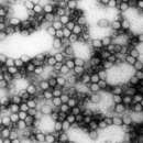
Elektronenmikroskop-bil faan en Flavivirus
Süstemaatik Hoodkategorii: Wiiren Kategorii: nian Famile: Flaviviridae Skööl: Flavivirus Taksonomii Genoom: (+)ssRNA Baltimore-klas: Skööl 4 Sümetrii: ikosaedrisk Wiirusskan: as diar Wedenskapelk nööm Flavivirus (ingelsk) FerwisangenFlavivirus as en skööl faan wiiren, diar faan tegen an magen üüb fögler an tetjdiarten auerdraanj wurd.
Di nööm komt faan't latiinsk flavus „güül“ uf, auer tu detdiar skööl uk det güülfiiberwiirus hiart.
Bütj güülfiiber wurd uk ööder kraankhaiden, so üs FSME, denguefiiber an zikafiiber faan sok wiiren ütjliaset.
Apoi virus – Aroa virus – Bagaza virus – Banzi virus – Bouboui virus – Bukalasa bat virus – Cacipacore virus – Carey Island virus – Cowbone Ridge virus – Dakar bat virus – Dengue virus – Edge Hill virus – Entebbe bat virus – Gadgets Gully virus – Ilheus virus – Israel turkey meningoencephalomyelitis virus – Japanese encephalitis virus – Jugra virus – Jutiapa virus – Kadam virus – Kedougou virus – Kokobera virus – Koutango virus – Kyasanur Forest disease virus – Langat virus – Louping ill virus – Meaban virus – Modoc virus – Montana myotis leukoencephalitis virus – Murray Valley encephalitis virus – Ntaya virus – Omsk hemorrhagic fever virus – Phnom Penh bat virus – Powassan virus – Rio Bravo virus – Royal Farm virus – Saboya virus – Sal Vieja virus – San Perlita virus – Saumarez Reef virus – Sepik virus – St. Louis encephalitis virus – Tembusu virus – Tick-borne encephalitis virus – Tyuleniy virus – Uganda S virus – Usutu virus – Wesselsbron virus – West Nile virus – Yaounde virus – Yellow fever virus – Yokose virus – Zika virus
Flavivirus as en skööl faan wiiren, diar faan tegen an magen üüb fögler an tetjdiarten auerdraanj wurd.
Di nööm komt faan't latiinsk flavus „güül“ uf, auer tu detdiar skööl uk det güülfiiberwiirus hiart.
Bütj güülfiiber wurd uk ööder kraankhaiden, so üs FSME, denguefiiber an zikafiiber faan sok wiiren ütjliaset.
पीत विषाणु (अंग्रेज़ी: Flavivirus) फ़्लैविविरिडेई विषाणु परिवार Flaviviridae के विषाणुओं की एक जाति है। इस प्राजाति में पश्चिमी नील विषाणु (West Nile virus), डेंगी विषाणु, टिक-बॉर्न मेनिनगो इंसेफ़लाइटिस विषाणु (tick-borne encephalitis virus), पीत जवर विषाणु, ज़ीका विषाणु और कई अन्य विषाणु होते हैं जो इंसेफ़लाईटिस, दिमागी बुखार जैसी खतरनाक बीमारियों का कारण बनते हैं। [2]
पीत विषाणु का नाम पीलिया या पीत ज्वर के विषाणू के नाम से पडा है जो कि इसी विषाणू परिवार का सदस्य है। पीत का संस्कृत में अर्थ पीला होता है। लैटिन भाषा में फ़्लैवी का अर्थ पीला होता है। इसी नाम से इसका जैविक नाम फ़्लैविवायरस पडा। इसका नाम पीले रंग से इसलिये जुडा हुआ है क्योंकि इसके शिकार पीली जान्डिस से पीडित होते हैं। [3]
फ़्लैवीवायरस में कई प्रकार की समानताएँ होती हैं: समान आकार (40–65 नैमी), समरूपता (घिरे हुए, विंशतिफलक न्युक्लियोकैप्सिड), न्यूक्लीक एसिड (पॉजिटिव सेन्स, एकल फँसा हुआ आरएनए लगभग 10,000–11,000 क्षार), और माइक्रोस्कोप में दृष्यता।
इनमें से अधिकांश विषाणु संक्रमित संधिपादों (arthropod) जैसे मच्छर इत्यादि के काटने से फैलती हैं। संधिपादों से फ़ैलने के कारण इन विषाणुओं को संधिविषाणु (arboviruses) भी कहते हैं। अर्थ्रोपॉड यूनानी भाषा का शब्द है जो अर्थ्रो (अर्थात संधि या जोड़) और पॉड यानि पैर से मिलकर बना है। अर्बोवायरस का अर्थ है अर्थ्रोपॉड जनित विषाणु जो कि इस परिभाषा के शब्दों के प्रथम अक्षरों से मिलकर बना है।[4] (arthro borne viruses)
इस विषाणु से संक्रमण का एक और जरिया है संक्रमित जानवरों व उनकी लाशों को छूना, संक्रमित खून से संपर्क, संक्रमित माँ से नवजात बच्चे को, संक्रमित जानवर के दूध पीने से इत्यादि। हालांकि ये विषाणु मानवों में सबसे ज्यादा संधिपादों के काटने से फैलते हैं।
समूह: ssRNA(+)
पीतविषाणु लगभग ५० नैनोमीटर के व्यास के खोल या कवच में घिरा होता है। आकार में यह विंशतिफलक या गोलाकार होता है। जीनोम लंबाई में १०-११केबी के रेखीय और अखण्ड होते हैं। [6]
पीत ज्वर का सबसे सफ़लतम टीका या दवाई पीत ज्वर 17डी टीका, 1937 में बनाया गया था। इस दवा ने महामारी को रोकने में महत्वपूर्ण भूमिका निभाई। जापानी इंसेफ़लाइटिस और अष्टपाद-जनित इंसेफ़लाइटिस विषाणु को मारने की प्रभावकारी दवा/टीका बीसवीं शताब्दी के मध्य तक बना ली गई थी।[7] पहले की दवा के दुष्प्रभावों की वजह से दूसरे पीढी की जापानी इंसेफ़लाइटिस की दवा बनाई गई जो ज्यादा सफल रही। इनका एशिया के विशाल जनसंख्या का इस खतरनाक बीमारी से निपटने में व्यापक इस्तेमाल किया जाता है। 95 प्रतिशत लोगों में टीकाकरण के 10 दिन के बाद इसका असर शुरू होता है और कम से कम 10 वर्ष तक रहता है (81 प्रतिशत मरीज़ों में प्रतिरक्षा 30 साल के बाद तक भी रही)। डब्ल्यूएचओ स्थानिक क्षेत्रों में लोगों को जन्म के 9वें और 12 महीनें के बीच नित्य टीकाकरण की सिफारिश करता है। 2013 में विश्व स्वास्थ्य संगठन ने कहा था ‘पीत ज्वर रोग के खिलाफ आजीवन प्रतिरक्षा के लिए टीकाकरण की एक खुराक ही काफी होती है’।[7] विश्वभर में घूमने वाले एडीज मच्छरों की वजह से सालाना लाखों लोग खतरनाक डेंगू के शिकार हो जाते हैं। चूंकि मच्छरों की अनगिनत संख्या पर नियंत्रण मुश्किल है इसलिये डेंगू से बचाव की विभिन्न दवाएँ अपने विकास के विभिन्न चरणों में हैं।
|accessdate=, |date= में तिथि प्राचल का मान जाँचें (मदद) पीत विषाणु (अंग्रेज़ी: Flavivirus) फ़्लैविविरिडेई विषाणु परिवार Flaviviridae के विषाणुओं की एक जाति है। इस प्राजाति में पश्चिमी नील विषाणु (West Nile virus), डेंगी विषाणु, टिक-बॉर्न मेनिनगो इंसेफ़लाइटिस विषाणु (tick-borne encephalitis virus), पीत जवर विषाणु, ज़ीका विषाणु और कई अन्य विषाणु होते हैं जो इंसेफ़लाईटिस, दिमागी बुखार जैसी खतरनाक बीमारियों का कारण बनते हैं।
पीत विषाणु का नाम पीलिया या पीत ज्वर के विषाणू के नाम से पडा है जो कि इसी विषाणू परिवार का सदस्य है। पीत का संस्कृत में अर्थ पीला होता है। लैटिन भाषा में फ़्लैवी का अर्थ पीला होता है। इसी नाम से इसका जैविक नाम फ़्लैविवायरस पडा। इसका नाम पीले रंग से इसलिये जुडा हुआ है क्योंकि इसके शिकार पीली जान्डिस से पीडित होते हैं।
फ़्लैवीवायरस में कई प्रकार की समानताएँ होती हैं: समान आकार (40–65 नैमी), समरूपता (घिरे हुए, विंशतिफलक न्युक्लियोकैप्सिड), न्यूक्लीक एसिड (पॉजिटिव सेन्स, एकल फँसा हुआ आरएनए लगभग 10,000–11,000 क्षार), और माइक्रोस्कोप में दृष्यता।
इनमें से अधिकांश विषाणु संक्रमित संधिपादों (arthropod) जैसे मच्छर इत्यादि के काटने से फैलती हैं। संधिपादों से फ़ैलने के कारण इन विषाणुओं को संधिविषाणु (arboviruses) भी कहते हैं। अर्थ्रोपॉड यूनानी भाषा का शब्द है जो अर्थ्रो (अर्थात संधि या जोड़) और पॉड यानि पैर से मिलकर बना है। अर्बोवायरस का अर्थ है अर्थ्रोपॉड जनित विषाणु जो कि इस परिभाषा के शब्दों के प्रथम अक्षरों से मिलकर बना है। (arthro borne viruses)
इस विषाणु से संक्रमण का एक और जरिया है संक्रमित जानवरों व उनकी लाशों को छूना, संक्रमित खून से संपर्क, संक्रमित माँ से नवजात बच्चे को, संक्रमित जानवर के दूध पीने से इत्यादि। हालांकि ये विषाणु मानवों में सबसे ज्यादा संधिपादों के काटने से फैलते हैं।
Flavivirus is a genus of positive-strand RNA viruses in the family Flaviviridae. The genus includes the West Nile virus, dengue virus, tick-borne encephalitis virus, yellow fever virus, Zika virus and several other viruses which may cause encephalitis,[3] as well as insect-specific flaviviruses (ISFs) such as cell fusing agent virus (CFAV), Palm Creek virus (PCV), and Parramatta River virus (PaRV).[4] While dual-host flaviviruses can infect vertebrates as well as arthropods, insect-specific flaviviruses are restricted to their competent arthropods.[5] The means by which flaviviruses establish persistent infection in their competent vectors and cause disease in humans depends upon several virus-host interactions, including the intricate interplay between flavivirus-encoded immune antagonists and the host antiviral innate immune effector molecules.[6]
Flaviviruses are named for the yellow fever virus; the word flavus means 'yellow' in Latin, and yellow fever in turn is named from its propensity to cause yellow jaundice in victims.[7]
Flaviviruses share several common aspects: common size (40–65 nm), symmetry (enveloped, icosahedral nucleocapsid), nucleic acid (positive-sense, single-stranded RNA around 10,000–11,000 bases), and appearance under the electron microscope.
Most of these viruses are primarily transmitted by the bite from an infected arthropod (mosquito or tick), and hence are classified as arboviruses. Human infections with most of these arboviruses are incidental, as humans are unable to replicate the virus to high enough titers to reinfect the arthropods needed to continue the virus life-cycle – humans are then a dead end host. The exceptions to this are the yellow fever virus, dengue virus and zika virus. These three viruses still require mosquito vectors but are well-enough adapted to humans as to not necessarily depend upon animal hosts (although they continue to have important animal transmission routes, as well).
Other virus transmission routes for arboviruses include handling infected animal carcasses, blood transfusion, sex, childbirth and consumption of unpasteurised milk products. Transmission from nonhuman vertebrates to humans without an intermediate vector arthropod however mostly occurs with low probability. For example, early tests with yellow fever showed that the disease is not contagious.
The known non-arboviruses of the flavivirus family reproduce in either arthropods or vertebrates, but not both, with one odd member of the genus affecting a nematode.[8]
Flaviviruses are enveloped and spherical and have icosahedral geometries with a pseudo T=3 symmetry. The virus particle diameter is around 50 nm.[9]
Flaviviruses have positive-sense, single-stranded RNA genomes which are non-segmented and around 10–11 kbp in length.[9] In general, the genome encodes three structural proteins (Capsid, prM, and Envelope) and seven non-structural proteins (NS1, NS2A, NS2B, NS3, NS4A, NS4B, NS5).[10] The genomic RNA is modified at the 5′ end of positive-strand genomic RNA with a cap-1 structure (me7-GpppA-me2).[11]

Flaviviruses replicate in the cytoplasm of the host cells. The genome mimics the cellular mRNA molecule in all aspects except for the absence of the poly-adenylated (poly-A) tail. This feature allows the virus to exploit cellular apparatuses to synthesize both structural and non-structural proteins, during replication. The cellular ribosome is crucial to the replication of the flavivirus, as it translates the RNA, in a similar fashion to cellular mRNA, resulting in the synthesis of a single polyprotein.[10]
Cellular RNA cap structures are formed via the action of an RNA triphosphatase, with guanylyltransferase, N7-methyltransferase and 2′-O methyltransferase. The virus encodes these activities in its non-structural proteins. The NS3 protein encodes a RNA triphosphatase within its helicase domain. It uses the helicase ATP hydrolysis site to remove the γ-phosphate from the 5′ end of the RNA. The N-terminal domain of the non-structural protein 5 (NS5) has both the N7-methyltransferase and guanylyltransferase activities necessary for forming mature RNA cap structures. RNA binding affinity is reduced by the presence of ATP or GTP and enhanced by S-adenosyl methionine.[11] This protein also encodes a 2′-O methyltransferase.

Once translated, the polyprotein is cleaved by a combination of viral and host proteases to release mature polypeptide products.[12] Nevertheless, cellular post-translational modification is dependent on the presence of a poly-A tail; therefore this process is not host-dependent. Instead, the poly-protein contains an autocatalytic feature which automatically releases the first peptide, a virus specific enzyme. This enzyme is then able to cleave the remaining poly-protein into the individual products. One of the products cleaved is a RNA-dependent RNA polymerase, responsible for the synthesis of a negative-sense RNA molecule. Consequently, this molecule acts as the template for the synthesis of the genomic progeny RNA.
Flavivirus genomic RNA replication occurs on rough endoplasmic reticulum membranes in membranous compartments. New viral particles are subsequently assembled. This occurs during the budding process which is also responsible for the accumulation of the envelope and cell lysis.
A G protein-coupled receptor kinase 2 (also known as ADRBK1) appears to be important in entry and replication for several viruses in Flaviviridae.[13]
Humans, mammals, mosquitoes, and ticks serve as the natural host. Transmission routes are zoonosis and bite.[9]
The positive sense RNA genome of Flavivirus contains 5' and 3' untranslated regions (UTRs).
The 5'UTRs are 95–101 nucleotides long in Dengue virus.[14] There are two conserved structural elements in the Flavivirus 5'UTR, a large stem loop (SLA) and a short stem loop (SLB). SLA folds into a Y-shaped structure with a side stem loop and a small top loop.[14][15] SLA is likely to act as a promoter, and is essential for viral RNA synthesis.[16][17] SLB is involved in interactions between the 5'UTR and 3'UTR which result in the cyclisation of the viral RNA, which is essential for viral replication.[18]
The 3'UTRs are typically 0.3–0.5 kb in length and contain a number of highly conserved secondary structures which are conserved and restricted to the flavivirus family. The majority of analysis has been carried out using West Nile virus (WNV) to study the function the 3'UTR.
Currently 8 secondary structures have been identified within the 3'UTR of WNV and are (in the order in which they are found with the 3'UTR) SL-I, SL-II, SL-III, SL-IV, DB1, DB2 and CRE.[19][20] Some of these secondary structures have been characterised and are important in facilitating viral replication and protecting the 3'UTR from 5' endonuclease digestion. Nuclease resistance protects the downstream 3' UTR RNA fragment from degradation and is essential for virus-induced cytopathicity and pathogenicity.
SL-II has been suggested to contribute to nuclease resistance.[20] It may be related to another hairpin loop identified in the 5'UTR of the Japanese encephalitis virus (JEV) genome.[21] The JEV hairpin is significantly over-represented upon host cell infection and it has been suggested that the hairpin structure may play a role in regulating RNA synthesis.
This secondary structure is located within the 3'UTR of the genome of Flavivirus upstream of the DB elements. The function of this conserved structure is unknown but is thought to contribute to ribonuclease resistance.
These two conserved secondary structures are also known as pseudo-repeat elements. They were originally identified within the genome of Dengue virus and are found adjacent to each other within the 3'UTR. They appear to be widely conserved across the Flaviviradae. These DB elements have a secondary structure consisting of three helices and they play a role in ensuring efficient translation. Deletion of DB1 has a small but significant reduction in translation but deletion of DB2 has little effect. Deleting both DB1 and DB2 reduced translation efficiency of the viral genome to 25%.[19]
CRE is the Cis-acting replication element, also known as the 3'SL RNA elements, and is thought to be essential in viral replication by facilitating the formation of a "replication complex".[22] Although evidence has been presented for an existence of a pseudoknot structure in this RNA, it does not appear to be well conserved across flaviviruses.[23] Deletions of the 3' UTR of flaviviruses have been shown to be lethal for infectious clones.
A conserved hairpin (cHP) structure was later found in several Flavivirus genomes and is thought to direct translation of capsid proteins. It is located just downstream of the AUG start codon.[24]
Subgenomic flavivirus RNA (sfRNA) is an extension of the 3' UTR and has been demonstrated to play a role in flavivirus replication and pathogenesis.[25] sfRNA is produced by incomplete degradation of genomic viral RNA by the host cells 5'-3' exoribonuclease 1 (XRN1).[26] As the XRN1 degrades viral RNA, it stalls at stemloops formed by the secondary structure of the 5' and 3' UTR.[27] This pause results in an undigested fragment of genome RNA known as sfRNA. sfRNA influences the life cycle of the flavivirus in a concentration dependent manner. Accumulation of sfRNA causes (1) antagonization of the cell's innate immune response, thus decreasing host defense against the virus[28] (2) inhibition of XRN1 and Dicer activity to modify RNAi pathways that destroy viral RNA[29] (3) modification of the viral replication complex to increase viral reproduction.[30] Overall, sfRNA is implied in multiple pathways that compromise host defenses and promote infection by flaviviruses.

The flaviviruses can be divided into two clades: one with vector-borne viruses and the other with no known vector.[31] The vector clade, in turn, can be subdivided into a mosquito-borne clade and a tick-borne clade. These groups can be divided again.[32]
The mosquito group can be divided into two branches: one branch contains neurotropic viruses, often associated with encephalitic disease in humans or livestock. This branch tends to be spread by Culex species and to have bird reservoirs. The second branch is the non-neurotropic viruses associated with human haemorrhagic disease. These tend to have Aedes species as vectors and primate hosts.
The tick-borne viruses also form two distinct groups: one is associated with seabirds and the other – the tick-borne encephalitis complex viruses – is associated primarily with rodents.
The viruses that lack a known vector can be divided into three groups: one closely related to the mosquito-borne viruses, which is associated with bats; a second, genetically more distant, is also associated with bats; and a third group is associated with rodents.
Evolutionary relationships between endogenised viral elements of Flaviviruses and contemporary flaviviruses using maximum likelihood approaches have identified that arthropod-vectored flaviviruses likely emerged from an arachnid source.[33] This contradicts earlier work with a smaller number of extant viruses showing that the tick-borne viruses emerged from a mosquito-borne group.[34]
Several partial and complete genomes of flaviviruses have been found in aquatic invertebrates such as the sea spider Endeis spinosa[35] and several crustaceans and cephalopods.[36] These sequences appear to be related to those in the insect-specific flaviviruses and also the Tamana bat virus groupings. While it is not presently clear how aquatic flaviviruses fit into the evolution of this group of viruses, there is some evidence that one of these viruses, Wenzhou shark flavivirus, infects both a crustacean (Portunus trituberculatus) Pacific spadenose shark (Scoliodon macrorhynchos) shark host,[37][36] indicating an aquatic arbovirus life cycle.
Estimates of divergence times have been made for several of these viruses.[38] The origin of these viruses appears to be at least 9400 to 14,000 years ago. The Old World and New World dengue strains diverged between 150 and 450 years ago. The European and Far Eastern tick-borne encephalitis strains diverged about 1087 (1610–649) years ago. European tick-borne encephalitis and louping ill viruses diverged about 572 (844–328) years ago. This latter estimate is consistent with historical records. Kunjin virus diverged from West Nile virus approximately 277 (475–137) years ago. This time corresponds to the settlement of Australia from Europe. The Japanese encephalitis group appears to have evolved in Africa 2000–3000 years ago and then spread initially to South East Asia before migrating to the rest of Asia.
Phylogenetic studies of the West Nile virus has shown that it emerged as a distinct virus around 1000 years ago.[39] This initial virus developed into two distinct lineages, lineage 1 and its multiple profiles is the source of the epidemic transmission in Africa and throughout the world. Lineage 2 was considered an Africa zoonosis. However, in 2008, lineage 2, previously only seen in horses in sub-Saharan Africa and Madagascar, began to appear in horses in Europe, where the first known outbreak affected 18 animals in Hungary in 2008.[40] Lineage 1 West Nile virus was detected in South Africa in 2010 in a mare and her aborted fetus; previously, only lineage 2 West Nile virus had been detected in horses and humans in South Africa.[41] A 2007 fatal case in a killer whale in Texas broadened the known host range of West Nile virus to include cetaceans.[42]
Omsk haemorrhagic fever virus appears to have evolved within the last 1000 years.[43] The viral genomes can be divided into 2 clades — A and B. Clade A has five genotypes, and clade B has one. These clades separated about 700 years ago. This separation appears to have occurred in the Kurgan province. Clade A subsequently underwent division into clade C, D and E 230 years ago. Clade C and E appear to have originated in the Novosibirsk and Omsk Provinces, respectively. The muskrat Ondatra zibethicus, which is highly susceptible to this virus, was introduced into this area in the 1930s.
In the genus Flavivirus there are 53 defined species:[44]
Species and strains sorted by vectors:
Mammalian tick-borne virus group
Seabird tick-borne virus group
The very successful yellow fever 17D vaccine, introduced in 1937, produced dramatic reductions in epidemic activity.
Effective inactivated Japanese encephalitis and Tick-borne encephalitis vaccines were introduced in the middle of the 20th century. Unacceptable adverse events have prompted change from a mouse-brain inactivated Japanese encephalitis vaccine to safer and more effective second generation Japanese encephalitis vaccines. These may come into wide use to effectively prevent this severe disease in the huge populations of Asia—North, South and Southeast.
The dengue viruses produce many millions of infections annually due to transmission by a successful global mosquito vector. As mosquito control has failed, several dengue vaccines are in varying stages of development. CYD-TDV, sold under the trade name Dengvaxia, is a tetravalent chimeric vaccine that splices structural genes of the four dengue viruses onto a 17D yellow fever backbone.[49][50] Dengvaxia is approved in five countries.[51]
An alternate approach to the development of flavivirus vaccine vectors is based on the use of viruses that infect insects. Insect-specific flaviviruses, such as Binjari virus, are unable to replicate in vertebrate cells. Nevertheless, recombinant viruses in which structural protein genes (prME) of Binjari virus are exchanged with those of dengue virus, Zika virus, West Nile virus, yellow fever virus, or Japanese encephalitis virus replicate efficiently in insect cells where high titers of infectious virus particles are produced. Immunization of mice with a Binjari vaccine bearing the Zika virus structural proteins protected mice from disease after challenge. A similar approach employs the insect-specific alphavirus Eilat virus as a vaccine platform. ... These new vaccine platforms generated from insect-specific flaviviruses and alphaviruses represent affordable, efficient, and safe approaches to rapid development of infectious, attenuated vaccines against pathogens from these two virus families.[52]
{{cite journal}}: CS1 maint: multiple names: authors list (link) {{cite journal}}: CS1 maint: multiple names: authors list (link) {{cite journal}}: CS1 maint: multiple names: authors list (link) {{cite journal}}: CS1 maint: multiple names: authors list (link) Flavivirus is a genus of positive-strand RNA viruses in the family Flaviviridae. The genus includes the West Nile virus, dengue virus, tick-borne encephalitis virus, yellow fever virus, Zika virus and several other viruses which may cause encephalitis, as well as insect-specific flaviviruses (ISFs) such as cell fusing agent virus (CFAV), Palm Creek virus (PCV), and Parramatta River virus (PaRV). While dual-host flaviviruses can infect vertebrates as well as arthropods, insect-specific flaviviruses are restricted to their competent arthropods. The means by which flaviviruses establish persistent infection in their competent vectors and cause disease in humans depends upon several virus-host interactions, including the intricate interplay between flavivirus-encoded immune antagonists and the host antiviral innate immune effector molecules.
Flaviviruses are named for the yellow fever virus; the word flavus means 'yellow' in Latin, and yellow fever in turn is named from its propensity to cause yellow jaundice in victims.
Flaviviruses share several common aspects: common size (40–65 nm), symmetry (enveloped, icosahedral nucleocapsid), nucleic acid (positive-sense, single-stranded RNA around 10,000–11,000 bases), and appearance under the electron microscope.
Most of these viruses are primarily transmitted by the bite from an infected arthropod (mosquito or tick), and hence are classified as arboviruses. Human infections with most of these arboviruses are incidental, as humans are unable to replicate the virus to high enough titers to reinfect the arthropods needed to continue the virus life-cycle – humans are then a dead end host. The exceptions to this are the yellow fever virus, dengue virus and zika virus. These three viruses still require mosquito vectors but are well-enough adapted to humans as to not necessarily depend upon animal hosts (although they continue to have important animal transmission routes, as well).
Other virus transmission routes for arboviruses include handling infected animal carcasses, blood transfusion, sex, childbirth and consumption of unpasteurised milk products. Transmission from nonhuman vertebrates to humans without an intermediate vector arthropod however mostly occurs with low probability. For example, early tests with yellow fever showed that the disease is not contagious.
The known non-arboviruses of the flavivirus family reproduce in either arthropods or vertebrates, but not both, with one odd member of the genus affecting a nematode.
Flavivirus (del lat. flavus: «amarillo») es un género de virus ARN pertenecientes a la familia Flaviviridae. Los Flavivirus son virus con envoltura, la simetría de la nucleocápside icosaedrica, y cuyo material genético reside en una única cadena de ARN de polaridad positiva.
Son los causantes de numerosas enfermedades en animales y humanos, siendo las más conocidas la fiebre amarilla, dengue y fiebre de Zika.
Todos los Flavivirus tienen en común un tamaño de entre 40 y 60 nanómetros, la envoltura, una nucleocápsida icosaédrica, el ácido nucleico (cadena única de ARN de sentido positivo, de aproximadamente 10 000 a 11 000 bases), y la apariencia en el microscopio electrónico.
Los Flavivirus tienen un genoma formado por una sola cadena de ARN (+) y se replica en el citoplasma de las células hospedadoras. El genoma del virus es idéntico a las moléculas de ARNm de la célula en todos sus aspectos excepto en la ausencia de cola poli-A. De este modo el virus puede explotar el aparato celular para sintetizar sus propias proteínas, tanto estructurales como no estructurales. El ribosoma celular es crucial en la replicación de los Flavivirus al traducir el ARN vírico de manera similar al ARNm celular, dando como resultado la síntesis de una sola poliproteína.
Una vez traducida, la poliproteína es subdividida en varios polipéptidos gracias a la acción de proteasas, tanto virales como celulares. Dado que la modificación post-traducción de las proteínas celulares es dependiente de la presencia de la cola poli-A, este proceso no puede depender de la célula hospedadora. En efecto, la poliproteína posee propiedades autocatalíticas que automáticamente libera el primer péptido, un enzima específico del virus. Este enzima es capaz de segmentar el resto de la proteína en péptidos individuales; uno de ellos es una polimerasa responsable de la síntesis de una molécula de ARN(-), la cual actúa de molde para la síntesis del genoma de los virus hijos.
A continuación, se ensamblan las nuevas partículas víricas. Ello ocurre durante la fase de construcción que es también responsable de la acumulación de la envoltura y de la lisis de la célula hospedadora.
Los Flavivirus se clasifican según el vector, el grupo de animales infectado y/o la zona geográfica de origen.
Flavivirus (del lat. flavus: «amarillo») es un género de virus ARN pertenecientes a la familia Flaviviridae. Los Flavivirus son virus con envoltura, la simetría de la nucleocápside icosaedrica, y cuyo material genético reside en una única cadena de ARN de polaridad positiva.
Son los causantes de numerosas enfermedades en animales y humanos, siendo las más conocidas la fiebre amarilla, dengue y fiebre de Zika.
Todos los Flavivirus tienen en común un tamaño de entre 40 y 60 nanómetros, la envoltura, una nucleocápsida icosaédrica, el ácido nucleico (cadena única de ARN de sentido positivo, de aproximadamente 10 000 a 11 000 bases), y la apariencia en el microscopio electrónico.
Flavivirukset (Flavivirus) ovat niveljalkaisten, yleensä hyttysten tai puutiaisten, levittämiä viruksia. Flaviviruksia tunnetaan tällä hetkellä 70 eri lajia, joista 13 aiheuttaa tautia ihmisessä. Flavivirusryhmään kuuluvat dengue-, keltakuume-, Japanin aivotulehdus-, Länsi-Niilin ja puutiaisaivotulehdusvirukset.[1]
Flavivirukset (Flavivirus) ovat niveljalkaisten, yleensä hyttysten tai puutiaisten, levittämiä viruksia. Flaviviruksia tunnetaan tällä hetkellä 70 eri lajia, joista 13 aiheuttaa tautia ihmisessä. Flavivirusryhmään kuuluvat dengue-, keltakuume-, Japanin aivotulehdus-, Länsi-Niilin ja puutiaisaivotulehdusvirukset.
Flavivirus (du latin flavus : jaune) est un genre de virus de la famille des Flaviviridae.
Ce groupe comprend entre autres :
Ce sont des virus à ARN positif simple brin. Ils mesurent entre 40 et 50 nm en moyenne et leur génome fait environ 10 000 paires de bases. Leur génome code 10 protéines : 3 structurales (C, prM, E) et 7 non structurales (NS1, NS2, ...)
Ces virus sont principalement transmis par les moustiques. Ils posent des problèmes de santé publique, notamment en zone urbaine (transmission par Aedes aegypti).
Ils ont un cycle de réplication semblable entre eux : l'entrée dans la cellule cible se fait grâce à la protéine E qui va se lier aux récepteurs cellulaires et entraîner une endocytose. Le virus est alors embarqué dans une vésicule endosomiale. Le pH acide de cette dernière va entraîner une fusion de l'enveloppe virale et de la membrane endosomiale, relargant alors le génome dans le cytosol[2]. Le génome ARN est alors directement traduit en une polyprotéine qui va donner les 10 protéines virales. La RNA-dependent-RNA-polymérase va alors copier le génome pour créer un brin « matrice » servant à la réplication de tous les ARN viraux. Les virions immatures formés se retrouvent dans le réticulum endoplasmique pour être liés aux proto protéines prM et E, puis vont dans l'appareil de Golgi où une furine va cliver les deux protéines pour permettre aux virions de prendre leur forme finale. Les virions matures sont alors transportés par des vésicules et passent dans le milieu extracellulaire par exocytose[3].
Flavivirus (du latin flavus : jaune) est un genre de virus de la famille des Flaviviridae.
Flavivirus adalah genus yang tergolong familia Flaviviridae. Genus ini meliputi virus Nil Barat, virus demam berdarah, virus demam kuning, virus Zika, dan beberapa virus lainnya yang dapat menyebabkan ensefalitis.
Flavivirus memiliki ukuran 40-65 nm.
Beberapa spesies virus dari genus ini diantaranya:
Flavivirus adalah genus yang tergolong familia Flaviviridae. Genus ini meliputi virus Nil Barat, virus demam berdarah, virus demam kuning, virus Zika, dan beberapa virus lainnya yang dapat menyebabkan ensefalitis.
Flavivirus memiliki ukuran 40-65 nm.
Flavivirus è un genere di virus a RNA a singolo filamento positivo (+)ssRNA appartenenti alla famiglia Flaviviridae.
Strutturalmente sono composti da pericapside, capside ed una sola molecola di RNA a polarità positiva. Il genoma dei flavivirus codifica 3 proteine strutturali e 7 non strutturali; con un senso di lettura 3' - 5'. Il capside virale è composto di tre proteine strutturali: proteine di rivestimento E, proteina del capside C, proteina di membrana M; la glicoproteina E svolge un ruolo centrale nella biologia delle infezioni ed è responsabile del legame e della penetrazione nella cellula bersaglio. Essa è il principale bersaglio della risposta immunitaria dell'ospite, essa è costituita da 500 aminoacidi con tre domini antigenici. La proteina capside C è una proteina strutturale coinvolta nell'assemblaggio del virione.
Prove di laboratorio suggeriscono che la maggior parte dei flavivirus si formano nelle cisterne del reticolo endoplasmatico rugoso, sono poi trasferiti all'apparato del Golgi e quindi rilasciati per esocitosi dalla superficie cellulare. Nella dengue il meccanismo scoperto è diverso, in questo caso i virioni si assemblano nel citoplasma e vengono liberati per gemmazione della membrana.
Il genere include 86 virus, di questi 73 sono raggruppati in 53 specie. Di questi flavivirus 40 sono conosciuti come patogeni per l'uomo e altri vertebrati. Essi determinano una varietà di malattie diverse con febbre, talvolta grave encefalite e/o febbre emorragica.[1]
I flavivirus hanno una propensione a diffondere ed emergere in nuove aree geografiche. Le origini di questa evoluzione senza precedenti restano da chiarire.[2]
Essi rappresentano una potenziale fonte di nuova comparsa di malattia. Tra i fattori capaci di determinare malattia sono i cambiamenti nell'uso del suolo e la deforestazione, cose che aumentano i movimenti della popolazione, l'urbanizzazione e l'aumento dei commerci. Inoltre, vi è una forte probabilità che anche il riscaldamento globale possa aumentare in modo significativo il rischio di comparsa della malattia e/o diffusione da flavivirus.[3]
I virioni sono sferici, dotati di un pericapside di 50 nm che ricopre un nucleocapside di 30 nm di diametro.[4] Le proteine del pericapside sono la E e la M. La proteina M viene prodotta durante l'assemblaggio nella via secretoria dalla lisi di un precursore tradotto dall'RNA del virus detto prM.[4][5] Le 90 glicoproteine dimeriche E, coinvolte nell'attacco e nella penetrazione nella cellula, sono disposte parallelamente alla superficie del virione, formando una struttura "a spina di pesce" a simmetria icosaedrica. Questa struttura inusuale è stata scoperta per mezzo della cristallografia a raggi X applicata al virus della dengue di tipo 2, al virus del Nilo occidentale e al virus dell'encefalite trasmessa da zecche, confermata da immagini ottenute per microscopia crioelettronica.[6][7][8] Il nucleocapside ha una struttura meno ordinata, a forma di gabbia, costituita dalle proteine C. In corrispondenza di esso si ha un'alta densità elettronica, dovuta a un doppio foglietto fosfolipidico che lo circonda.[6]
I flavivirus sono virus a ssRNA positivo. La molecola di RNA è lunga circa 11 kb e, come negli altri flaviviridi, è composta da un unico ORF delimitato alle estremità 3' e 5' da due regioni non codificanti (NCR o UTR), lunghe rispettivamente 400-700 e 100 nucleotidi, coinvolte nei processi di replicazione, traduzione e assemblaggio. Queste sequenze non codificanti hanno una struttura secondaria piuttosto conservata ma possono differire notevolmente per lunghezza e composizione in basi.[9][10][11] L'estremità 5' è dotata di un cappuccio di tipo I m7GpppAmp. L'estremità 3' solitamente non è poli-adenilata, a differenza degli mRNA cellulari,[12] e presenta una sequenza CU altamente conservata.[13]
Il virione lega la cellula attraverso le glicoproteine che interagiscono colle proteine di membrana della cellula. Il virus entra per endocitosi clatrina-dipendente.[14][15][16] Il pH acido all'interno della vescicola determina il rilascio del nucleocapside nel citoplasma,[16][17] cui segue l'uncoating che libera il genoma nella cellula. La replicazione dei flavivirus comporta una notevole trasformazione delle membrane intracellulari, con formazioni di vescicole derivate dal reticolo endoplasmatico perinucleare, sedi della traduzione e del successivo assemblaggio.[18] La replicazione avviene per mezzo di un intermediario di RNA a polarità negativa che fa da stampo per la sintesi della molecola di RNA positiva. L'ORF viene tradotto in un'unica grande poliproteina che a sua volta viene lisata nelle proteine strutturali e funzionali del virus da proteasi cellulari che dalla serin proteasi NS2B-NS3 virale. La gemmazione avviene dalle membrane intracellulari,[14][19][20] ma alcuni studi mostrano una gemmazione che prende luogo nella membrana citoplasmatica.[20] Poco prima che di essere rilasciato fuori dalla cellula per la via secretoria, il prM viene lisato nella proteine M caratteristica del virione maturo.[17]
Le proteine strutturali sono la proteina del nucleocapside C (11 kDa), le proteine del peplos E (50 kDa) e prM (26 kDa) nei virioni immaturi e M (8 kDa) in quelli maturi. La proteina E è una emoagglutinina che permette sia l'attacco con un recettore di membrana e l'entrata nella cellula, nonché la successiva fusione nel citosol.[13] La proteina precursore prM crea un complesso dimerico con la E (prM-E) la cui funzione sembra quella di permettere il corretto ripiegamento della E, in particolare di impedire cambiamenti nella conformazione durante la via secretoria, per via dell'ambiente acido.[19] Poco prima del rilascio, la furina cellulare lisa la glicoproteina prM in pr e M, questa'ultima distintiva dei virus infettivi e maturi.[21] Sette proteine strutturali sono sintetizzate nelle cellule infette: NS1 (46 kDa), NS2A (22 kDa), NS2B (14 kDa), NS3 (70 kDa), NS4A (16 kDa), NS4B (27 kDa) and NS5 (103 kDa).[13]
I flavivirus possono essere divisi in 2 cladi: il primo è dato dai virus trasmessi da un vettore e mentre il secondo clade è dato dai virus senza il vettore noto.[22] Il clade vettoriale a sua volta può essere suddiviso in un clade trasmesso dalle zanzare e un clade trasmesso dalle zecche. Questi gruppi possono essere ulteriormente divisi.[23]
Il gruppo delle zanzare può essere diviso in due rami: un ramo contiene i virus neurotropici, spesso associati alla malattia encefalitica nell'uomo o nel bestiame. Questo ramo tende ad essere diffuso dalle specie del genere Culex e ad avere animali serbatoi negli uccelli. Il secondo ramo è dato dai virus non neurotropici associati alla malattia emorragica nell'uomo. Questi tendono ad avere specie del genere Aedes come vettori e come ospiti i primati.
I virus trasmessi da zecche formano anche essi due gruppi distinti: il primo associato agli uccelli marini e l'altro - i virus complessi dell'encefalite da zecche - è associato principalmente ai roditori.
I virus che mancano di un vettore noto possono essere, invece, divisi in tre gruppi: uno strettamente correlato ai virus trasmessi dalle zanzare associati ai pipistrelli; un secondo, geneticamente più distante, anche esso associato ai pipistrelli; e un terzo gruppo è associato ai roditori.[24]
Inizialmente i flavivirus furono classificati come arbovirus appartenenti al gruppo B. In seguito, il genere è stato assegnato alla famiglia Togaviridae che conteneva anche il genere Alphavirus. Oggi il termine "arbovirus" non ha più alcun significato tassonomico.[25] Nel 1985, per via di differenze che riguardavano la replicazione, la morfogenesi e la struttura, il genere Flavivirus venne assegnato a una famiglia separata, Flaviviridae, avente come specie tipo il virus della febbre gialla.[26]
Specie:
Esiste un vaccino e la diagnosi è fatta tramite reazione a catena della polimerasi su liquor e sangue per dimostrare il genoma virale nel materiale biologico.
Altre manifestazioni cliniche e/o sintomi tipici provocate dai Flavivirus sono:[27]
Flavivirus è un genere di virus a RNA a singolo filamento positivo (+)ssRNA appartenenti alla famiglia Flaviviridae.
Strutturalmente sono composti da pericapside, capside ed una sola molecola di RNA a polarità positiva. Il genoma dei flavivirus codifica 3 proteine strutturali e 7 non strutturali; con un senso di lettura 3' - 5'. Il capside virale è composto di tre proteine strutturali: proteine di rivestimento E, proteina del capside C, proteina di membrana M; la glicoproteina E svolge un ruolo centrale nella biologia delle infezioni ed è responsabile del legame e della penetrazione nella cellula bersaglio. Essa è il principale bersaglio della risposta immunitaria dell'ospite, essa è costituita da 500 aminoacidi con tre domini antigenici. La proteina capside C è una proteina strutturale coinvolta nell'assemblaggio del virione.
Prove di laboratorio suggeriscono che la maggior parte dei flavivirus si formano nelle cisterne del reticolo endoplasmatico rugoso, sono poi trasferiti all'apparato del Golgi e quindi rilasciati per esocitosi dalla superficie cellulare. Nella dengue il meccanismo scoperto è diverso, in questo caso i virioni si assemblano nel citoplasma e vengono liberati per gemmazione della membrana.
Il genere include 86 virus, di questi 73 sono raggruppati in 53 specie. Di questi flavivirus 40 sono conosciuti come patogeni per l'uomo e altri vertebrati. Essi determinano una varietà di malattie diverse con febbre, talvolta grave encefalite e/o febbre emorragica.
I flavivirus hanno una propensione a diffondere ed emergere in nuove aree geografiche. Le origini di questa evoluzione senza precedenti restano da chiarire.
Essi rappresentano una potenziale fonte di nuova comparsa di malattia. Tra i fattori capaci di determinare malattia sono i cambiamenti nell'uso del suolo e la deforestazione, cose che aumentano i movimenti della popolazione, l'urbanizzazione e l'aumento dei commerci. Inoltre, vi è una forte probabilità che anche il riscaldamento globale possa aumentare in modo significativo il rischio di comparsa della malattia e/o diffusione da flavivirus.
Flavivirus (-i, n.) est genus virorum familiae Flaviviridarum. Quod comprehendit virus Nili Occidentalis, virus febris dengue, virus meningoencephalitidis ab ixodidis latae, virus febris flavae, virus Zikanum, et nonnulla alia vira quae encephalitem et alios morbos efficere possunt.[2]
Appellantur flavivira ex viro febris flavae, typo familiae. Nomen febris flavae ortum est, quia icterum flavum in aegris efficere solet.[3]
Flavivira nonnulla indicia una partiunt: magnitudinem (40–65 nm), symmetriam (involutam, icosahedralem nucleocapsidalem), acidum nucleicum (sensu positivo, RNA fili unius, circa 10 000–11 000 basium), et speciem in microscopio electronico visam.
Plurima ex his viris transmittuntur per ictum arthropodi infecti, (culicis vel ixodidi), atque ergo arthropod-borne virus, ex locutione Anglica, arbovira describuntur. Infectiones humanae ab his viris effectae usitate sunt leves, quia homines virus replicare plerumque non possunt ad titulos tam altos quam arthropoda iterum infici possunt, ut circulum vitae viralis alant; quam ob rem homines hostes cessationis appellantur. Exceptiones sunt febris flava, febris dengue, virusque Zikanum, quae tamen vectorum inter culices egent, sed hominibus tam accommodantur quam non necessario hostibus inter alia animalia nituntur (quamquam vias transmissionis magni momenti in animalibus iam habent).
Aliae viae transmissionis per arbovira sunt cadavera animalium infectorum tractata, transfusio sanguinis, partus, et consumptio operum lactis non pasteurizati. Transmissio ab animalibus ad homines sine arthropodo vectore intermediato haud probabilis putatur. Exempli gratia, prima febris flavae experimenta hunc morbum non contagiosum esse demonstraverunt.
Nota familiae flavivirorum non arbovira in arthropodis aut vertebratis, sed non in ambobus, regenerare possunt.
Flavivirus (-i, n.) est genus virorum familiae Flaviviridarum. Quod comprehendit virus Nili Occidentalis, virus febris dengue, virus meningoencephalitidis ab ixodidis latae, virus febris flavae, virus Zikanum, et nonnulla alia vira quae encephalitem et alios morbos efficere possunt.
Appellantur flavivira ex viro febris flavae, typo familiae. Nomen febris flavae ortum est, quia icterum flavum in aegris efficere solet.
Flavivira nonnulla indicia una partiunt: magnitudinem (40–65 nm), symmetriam (involutam, icosahedralem nucleocapsidalem), acidum nucleicum (sensu positivo, RNA fili unius, circa 10 000–11 000 basium), et speciem in microscopio electronico visam.
Plurima ex his viris transmittuntur per ictum arthropodi infecti, (culicis vel ixodidi), atque ergo arthropod-borne virus, ex locutione Anglica, arbovira describuntur. Infectiones humanae ab his viris effectae usitate sunt leves, quia homines virus replicare plerumque non possunt ad titulos tam altos quam arthropoda iterum infici possunt, ut circulum vitae viralis alant; quam ob rem homines hostes cessationis appellantur. Exceptiones sunt febris flava, febris dengue, virusque Zikanum, quae tamen vectorum inter culices egent, sed hominibus tam accommodantur quam non necessario hostibus inter alia animalia nituntur (quamquam vias transmissionis magni momenti in animalibus iam habent).
Aliae viae transmissionis per arbovira sunt cadavera animalium infectorum tractata, transfusio sanguinis, partus, et consumptio operum lactis non pasteurizati. Transmissio ab animalibus ad homines sine arthropodo vectore intermediato haud probabilis putatur. Exempli gratia, prima febris flavae experimenta hunc morbum non contagiosum esse demonstraverunt.
Nota familiae flavivirorum non arbovira in arthropodis aut vertebratis, sed non in ambobus, regenerare possunt.
Flavivirus is een familie van virussen. Het woord Flavivirus komt van het Latijn flavus (geel).
Er zijn vier geslachten binnen de familie:
Flavivirussen zijn positive stranded, lineair ssRNA-virussen met een genoom van gemiddeld zo'n 10.000 nucleotiden lang. Het genoom heeft een karakteristieke opbouw met achtereenvolgens:
Replicatie gebeurt door de productie van enkele negative stranded intermediairen, vanwaar dan de positive stranded genomen kunnen worden gekopieerd. Transmissie gebeurt parenteraal, wat voor de mens neerkomt op muggenbeten zowel overdag als 's nachts, o.a. door Aedes aegypti en Aedes albopictus, bloedtransfusies, intraveneus druggebruik en transmissie van moeder op kind.
Bronnen, noten en/of referenties
Flawiwirus jest rodzajem wirusów z rodziny Flaviviridae. Rodzaj ten obejmuje wirus Zachodniego Nilu, wirus dengi, wirus kleszczowego zapalenia mózgu, wirus żółtej gorączki, wirus Zika i kilka innych wirusów, które mogą prowadzić do zapalenia mózgu. Nazwa pochodzi od wirusa żółtej gorączki, który jest gatunkiem typowym dla rodzaju; słowo flavus oznacza żółty po łacinie.
Flawiwirusy posiadają rozmiary 40-60 nanometrów, symetrię ikozaedralną nukleokapsydu, otoczkę oraz pojedynczą nić RNA o dodatniej polarności.
Flavivírus (do latim flavus, amarelo) é um gênero de Vírus RNA de cadeia simples com sentido positivo (grupo IV), da família Flaviviridae. São transmitidos por mosquitos ou carrapatos. Além dele, o vírus da febre amarela e dengue são seus representantes mais conhecidos. Muitos flavivírus causam encefalite viral como o Vírus do Nilo Ocidental.[1]
Os Flavivírus partilham um tamanho comum (40-60 nanômetros), com envoltura simétrica, nucleocapsídeo icosaédrico e uma única fita positiva de RNA. O genoma de RNA contém aproximadamente 10,000 nucleotídeos ou 10 genes contidos em uma cadeia aberta de leitura que codifica uma poliproteína de 3391 aminoácidos (no caso da Dengue 2) que depois é clivada em três proteínas estruturais e sete proteínas não estruturais.[2]. Dentre as arboviroses, aquelas transmitidas por mosquitos, as causadas por Flavivírus são as mais importantes causadoras de surtos ou epidemias [2]
Transmitidos por carrapatos[1]:
Transmitidos por mosquitos[1]
Transmissão desconhecida:
Até o ano de 2000, eram dez as doenças causadas por flavivirus no Brasil: dengue 1, 2 e 4, Iguapé, Ilhéus, Rocio, encefalite de Saint Louis, Bussuquara, Cacipacoré e febre amarela.[3]
A identificação e caracterização molecular do Flavivírus são importantes para detectar o vírus circulante e alertar a vigilância epidemiológica em uma região. Assim, recentemente o vírus da febre amarela vem ganhando destaque na mídia brasileira, visto que vários casos vêm sendo catalogados na região Centro-Oeste, sobretudo, causando a preocupação da população em geral e providências das autoridades responsáveis pelo combate ao vírus [4].
Algumas complicações podem ser tratadas com ribavirina ou favipiravir(T-705 ou Avigan).[5]
Flavivírus (do latim flavus, amarelo) é um gênero de Vírus RNA de cadeia simples com sentido positivo (grupo IV), da família Flaviviridae. São transmitidos por mosquitos ou carrapatos. Além dele, o vírus da febre amarela e dengue são seus representantes mais conhecidos. Muitos flavivírus causam encefalite viral como o Vírus do Nilo Ocidental.
Flavivirus este un gen din familia Flaviviridae.Din categoria flavivirusurilor fac parte: virusul West Nile, virusul Denga, virusul meningoencefalitei acariene, virusul febrei galbene, precum și alte virusuri ce produc encefalită.
Flavivirusul are circa 40-60 nm, genomul fiind format din ARN monocatenar (circa 10000-10000 nucleotide).Spre deosebire de alphavirus, legarea 5’-3’, nu mai există.Virionul are o proteină C (capsidă) de circa 13 000 Da. Anvelopa este constituită dintr-un strat bilipidic, există o singură proteină E (circa 51000-59000 Da), și o proteină mai mică M neglucozilată, de circa 8500 Da.
Replicarea are loc în citoplasma celulei gazdă. Mecanismul de intrare a virusului în celula gazdă include o interacție între proteina E și receptorii celulari urmată de o fuziune post-atașare, fenomen care are loc în vacuolele intracitoplasmice.Genomul ARN își manifestă virulența doar dacă este introdu în citoplasma celulei.Datorită neexistenței legăturii 5’-3’, din aceasta cauză ARN servește de ARNm, pentru sinteza tuturor proteinelor virale.Virusul exploatează la maximum capacitățile celulei gazdă, pe care le utilizează pentru sintetizarea proteinelor structural și non-structurale.Rolul crucial în sinteza proteică îl au ribozomii celulei gazdă , care au un rol important în replicarea virusurilor, în sinteza ARNm,rezultatul fiind o unică poliproteină.Odată sintetizată poliproteina este clivată sub acțiunea proteazei, eliberând compuși polipeptidici.Poliproteina conține o enzimă autocatalitică , acțiunea sa având drept rezultat eliberarea unei peptide inițiale,o enzimă specifică ce va scinda lanțul poliproteic în produși individuali.Unul din acești compuși este polimeraza, responsabilă de sinteza molecului de (-) ARN, ce va servi drept matriță pentru sinteza ARN genomic.
Flavivirus este un gen din familia Flaviviridae.Din categoria flavivirusurilor fac parte: virusul West Nile, virusul Denga, virusul meningoencefalitei acariene, virusul febrei galbene, precum și alte virusuri ce produc encefalită.
Flavivirus (Flaviviridae) är en virusfamilj bestående av RNA-virus. Exempel på sjukdomar orsakade av virus tillhörande familjen är gula febern, hepatit C, TBE, japansk encefalit, zikafeber och denguefeber.
Flavivirus (Flaviviridae) är en virusfamilj bestående av RNA-virus. Exempel på sjukdomar orsakade av virus tillhörande familjen är gula febern, hepatit C, TBE, japansk encefalit, zikafeber och denguefeber.
Sarıhumma virüsü, maymun, fare, kobay ve sivrisineklerde kolayca üreyen küçük bir RNA virusudur.
Kanamalarla seyreden ve son derece öldürücü bir viral hastalığa neden olur. Isı ve dezenfektanlara dayanıklıdır. Aedes aegypti cinsi Sivrisineğin sokmasıyla bulaşarak, "sarıhumma"ya neden olur. Ateş, ağrı, bulantı, kusma yapar. Deride sararmaya neden olur. Afrika ve Güney Amerika'da yaygındır. Bulaşıcıdır, kontrol altına alınmadığında salgınlar görülür. Aşıyla koruma sağlanır. Sarı humma virüsü, 45-55 nm boyundadır ve genetik materyali RNA'dan oluşur. Siyahi insanlar doğuştan sarıhumma hastalığına karşı dirençlidir.
Flavivirus
Группа по БалтиморуIV: (+)оцРНК-вирусы
Флавивирус (лат. Flavivirus) — род арбовирусов из семейства Flaviviridae. Типовой вид — вирус жёлтой лихорадки. Преимущественно циркулируют между членистоногими (клещи, комары) и млекопитающими (приматы, летучие мыши, грызуны, скот). При укусе заражённого комара или клеща могут передаваться человеку, вызывая заболевания различной тяжести, варьирующей от бессимптомного протекания до угрожающих жизни геморрагических лихорадок и энцефалитов (лихорадка денге, лихорадка Западного Нила, лихорадка Зика, клещевой энцефалит, японский энцефалит, энцефалит Сент-Луис и др.).
По данным Международного комитета по таксономии вирусов (ICTV), на май 2016 г. в род включают 53 вида[2]:
Распространены преимущественно в тропических регионах:
Распространены в основном в регионах умеренного климата:
Выделены из различных млекопитающих, но не из членистоногих, цикл циркуляции неизвестен:
Флавивирус (лат. Flavivirus) — род арбовирусов из семейства Flaviviridae. Типовой вид — вирус жёлтой лихорадки. Преимущественно циркулируют между членистоногими (клещи, комары) и млекопитающими (приматы, летучие мыши, грызуны, скот). При укусе заражённого комара или клеща могут передаваться человеку, вызывая заболевания различной тяжести, варьирующей от бессимптомного протекания до угрожающих жизни геморрагических лихорадок и энцефалитов (лихорадка денге, лихорадка Западного Нила, лихорадка Зика, клещевой энцефалит, японский энцефалит, энцефалит Сент-Луис и др.).


