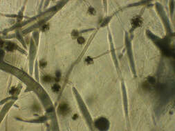
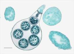
Description: English: Light microscopy of Saprolegnia showing the different stages of development for the oogonium of Saprolegnia. Showing a young, immature, and mature oogonium. The mature oogonium has eggs inside. Scale bar = 0.01mm. Date: 21 May 2014, 09:18:37. Source: Jon Houseman and Matthew Ford. Author: Jon Houseman. Other versions: Labeled. : This is a retouched picture, which means that it has been digitally altered from its original version. Modifications: Balance (Color, brightness, and contrast) and adjust background color.
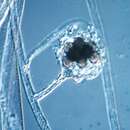
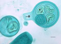
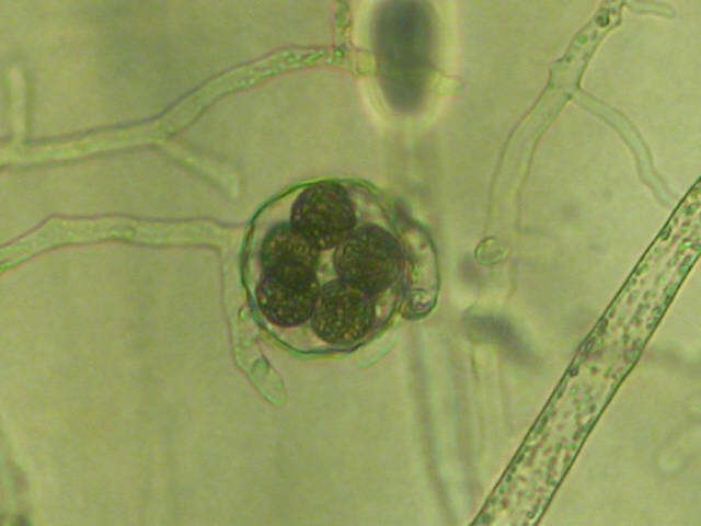
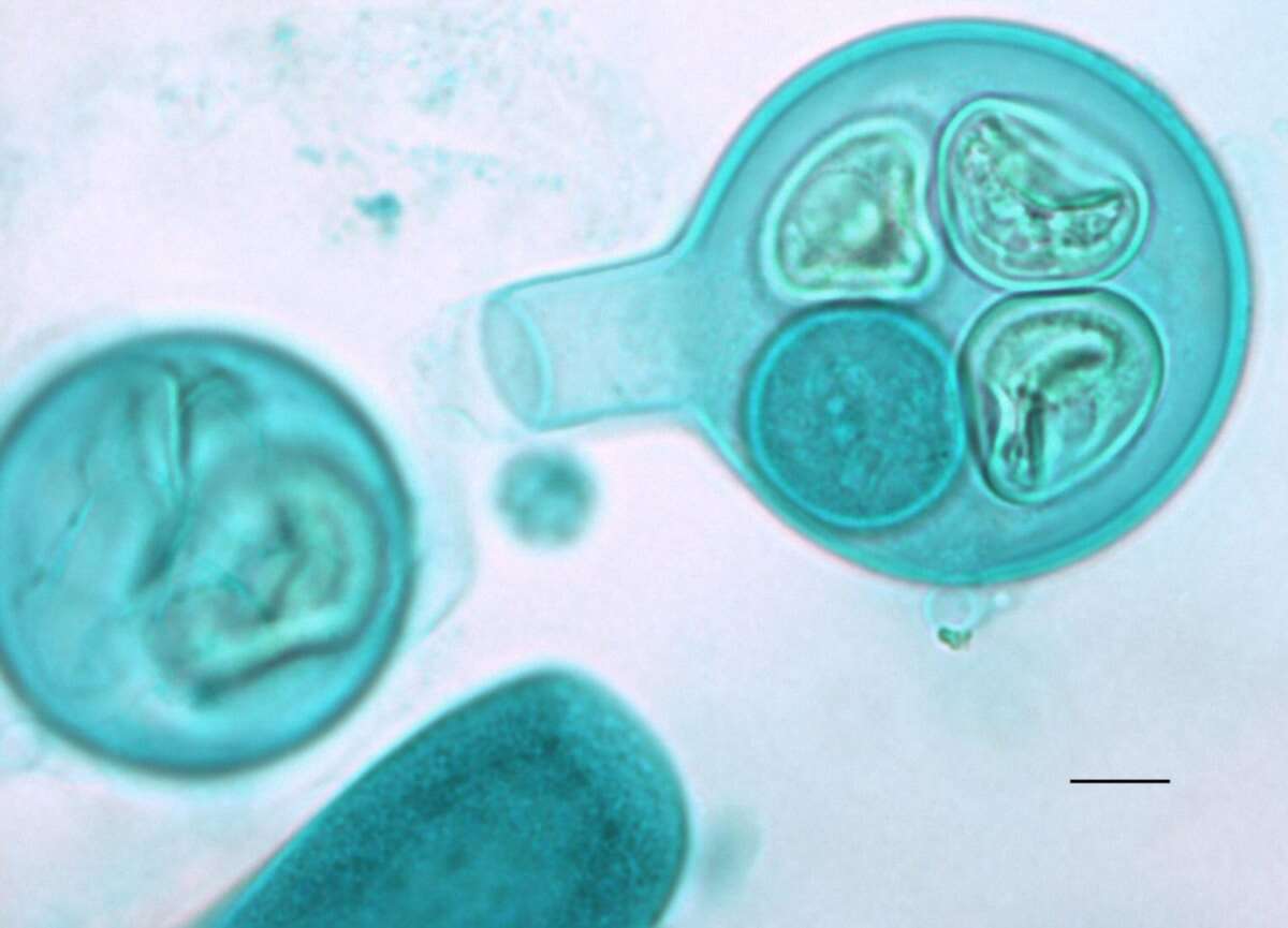
Description: English: Light microscopy of Saprolegnia with an oogonium and zygotes inside with a septum to enclose the entrance. A=Zygote, B=Septum. Scale bar = 0.01mm. Date: 26 May 2014, 09:17:05. Source: Jon Houseman and Matthew Ford. Author: Jon Houseman. Other versions: Original (unlabeled). : This is a retouched picture, which means that it has been digitally altered from its original version. Modifications: Balance (Color, brightness, and contrast) and adjust background color.
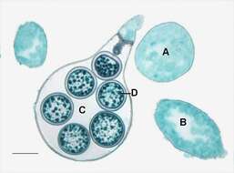
Description: English: Light microscopy of Saprolegnia showing the different stages of development for the oogonium of Saprolegnia. Showing a young, immature, and mature oogonium. The mature oogonium has eggs inside. A=Immature oogonium, B= Developing oogonium, C=Oogonium, D=Egg.Scale bar = 0.01mm. Date: 26 May 2014, 09:17:04. Source: Jon Houseman and Matthew Ford. Author: Jon Houseman. Other versions: Original (unlabeled). : This is a retouched picture, which means that it has been digitally altered from its original version. Modifications: Balance (Color, brightness, and contrast) and adjust background color.
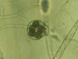
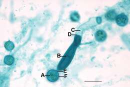
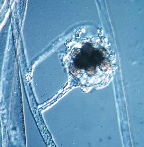
Description: English: Light microscopy of Saprolegnia showing a close up view of an oognium with zygotes inside that is in contact with a zoosporangium filled with zoospores (that are contained by a septum) and the vegetative hypha. Scale bar = 0.2mm. Date: 21 May 2014, 09:18:26. Source: Jon Houseman and Matthew Ford. Author: Jon Houseman. Other versions: Labeled. : This is a retouched picture, which means that it has been digitally altered from its original version. Modifications: Balance (Color, brightness, and contrast) and adjust background color.
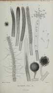
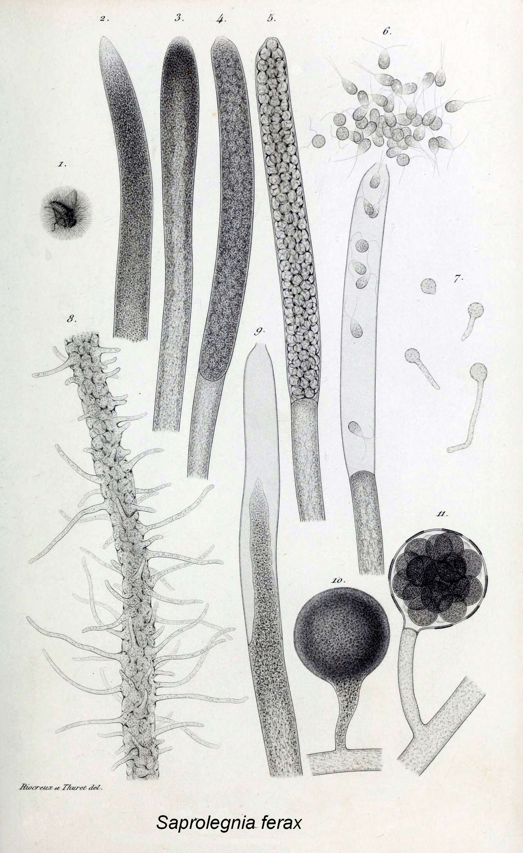
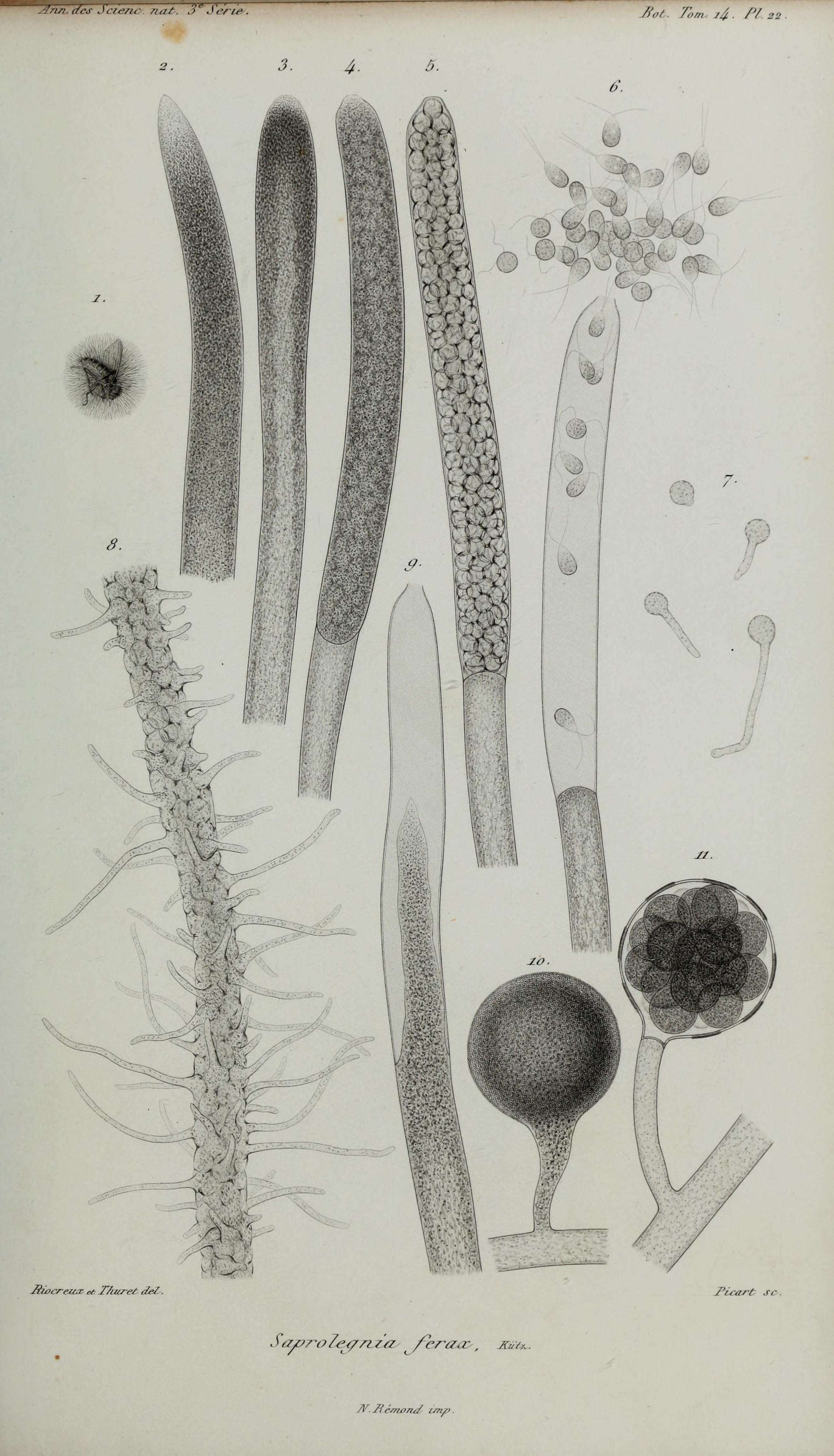
Description: English: Title: Annales des Sciences Naturelles Botaniques Identifier: annalesdesscienc3141850pa (find matches) Year: 1850 (1850s) Authors: Subjects: Publisher: Paris Contributing Library: Natural History Museum Library, London Digitizing Sponsor: BHL-SIL-FEDLINK View Book Page: Book Viewer About This Book: Catalog Entry View All Images: All Images From Book Click here to view book online to see this illustration in context in a browseable online version of this book. Text Appearing Before Image: c/^Jcuvu- n. Bot Tom 1&. /'/ Text Appearing After Image: Siocreux es TAï/reS de/ Picart S
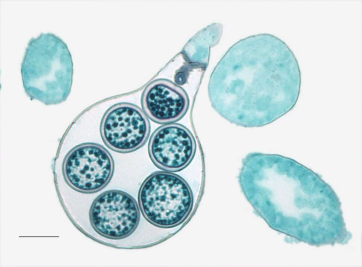
Description: English: Light microscopy of Saprolegnia with an oogonium and zygotes inside with a septum to enclose the entrance. Scale bar = 0.01mm. Date: 21 May 2014, 09:18:32. Source: Jon Houseman and Matthew Ford. Author: Jon Houseman. Other versions: Labeled. : This is a retouched picture, which means that it has been digitally altered from its original version. Modifications: Balance (Color, brightness, and contrast) and adjust background color.
Description: English: Light microscopy of Saprolegnia showing a close up view of an oognium with zygotes inside that is in contact with a zoosporangium filled with zoospores (that are contained by a septum) and the vegetative hypha. A=Zygote, B=Zoosporangium, C=Vegitative hypha, D=Zoosporangium septum, E=Oogonium septum, F=Oogonium. Scale bar = 0.2mm. Date: 26 May 2014, 09:17:20. Source: Jon Houseman and Matthew Ford. Author: Jon Houseman. Other versions: Original (unlabeled). : This is a retouched picture, which means that it has been digitally altered from its original version. Modifications: Balance (Color, brightness, and contrast) and adjust background color.
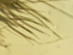
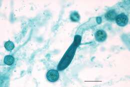
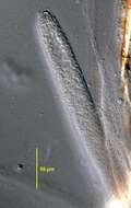
Description: English: Light microscopy of Saprolegnia showing an immature oogonium with its vegetative hypha. A=Developing oognium, B=Hypha. Scale bar = 0.02mm. Date: 26 May 2014, 09:17:04. Source: Jon Houseman and Matthew Ford. Author: Jon Houseman. Other versions: Original (unlabeled). : This is a retouched picture, which means that it has been digitally altered from its original version. Modifications: Balance (Color, brightness, and contrast) and adjust background color.
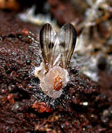
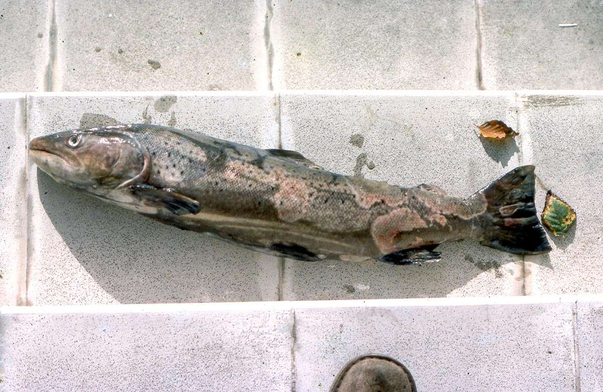
Description: Sea-trout suffering from UDN with secondary Saprolegnia infections. Date: 19 November 1977. Source: Own work. Author: Velela. Permission(Reusing this file): GFDL.
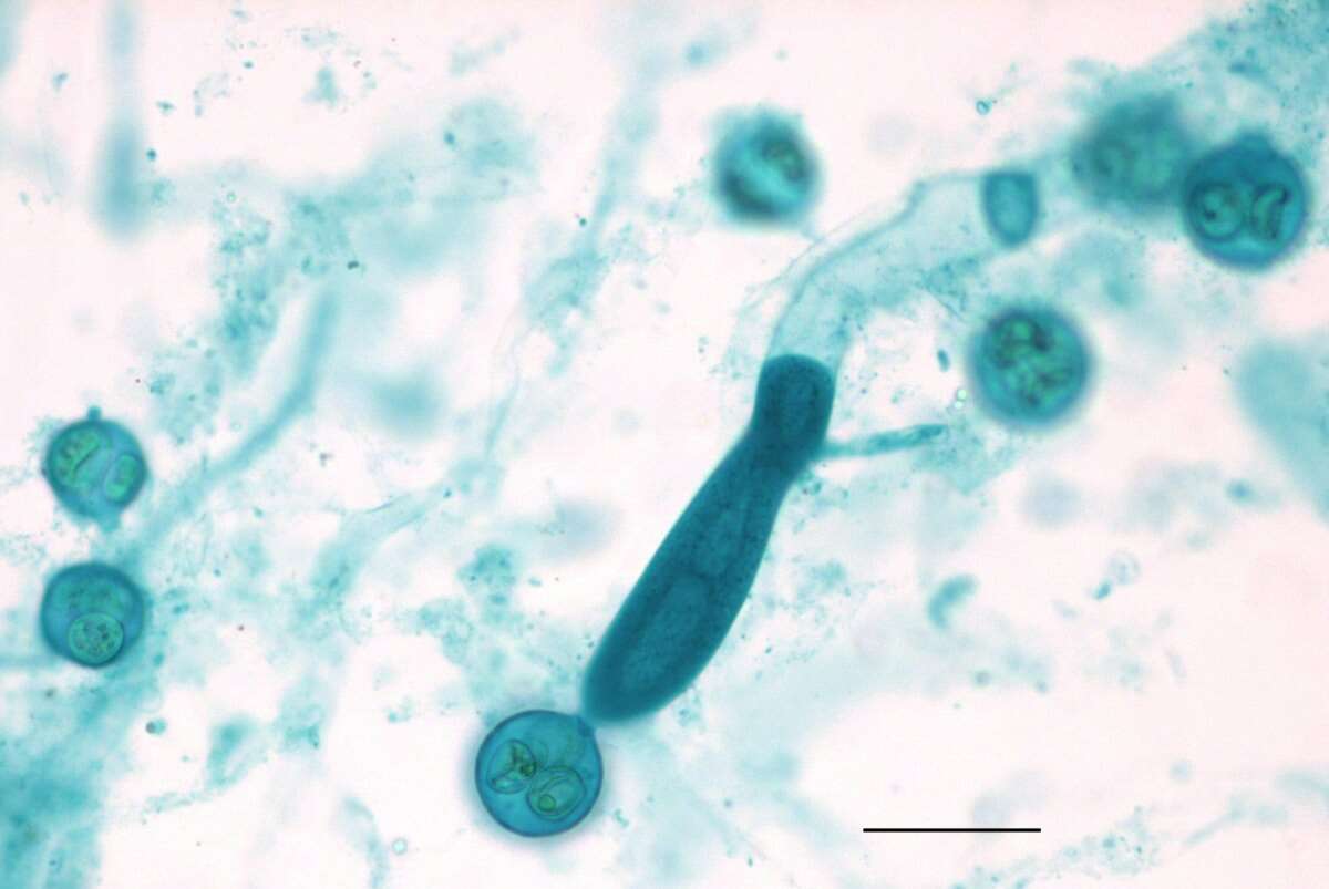
Description: English: Light microscopy of Saprolegnia showing an immature oogonium with its vegetative hypha. Scale bar = 0.02mm. Date: 21 May 2014, 09:18:43. Source: Jon Houseman and Matthew Ford. Author: Jon Houseman. Other versions: Labeled. : This is a retouched picture, which means that it has been digitally altered from its original version. Modifications: Balance (Color, brightness, and contrast) and adjust background color.
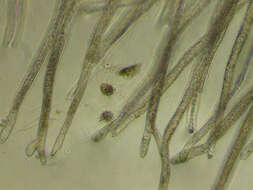
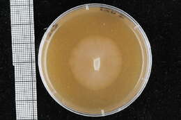
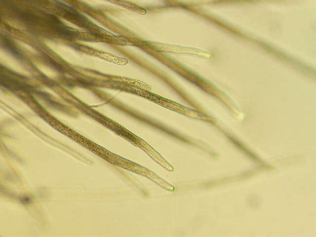
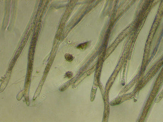
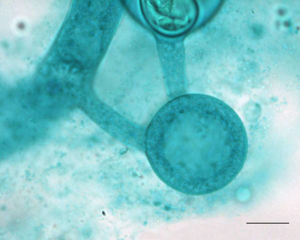
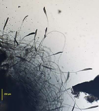
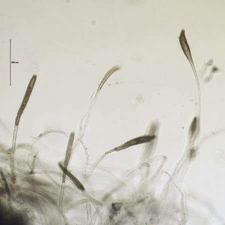
Portrait of zoosporangia of a water mold, probably a species of Saprolegnia. Growing on dead chironomid larvae in a culture vessel. April 2009. Brightfield.
Portrait of zoosporangia of a water mold, probably a species of Saprolegnia. Growing on dead chironomid larvae in a culture vessel. April 2009. Brightfield.


