-
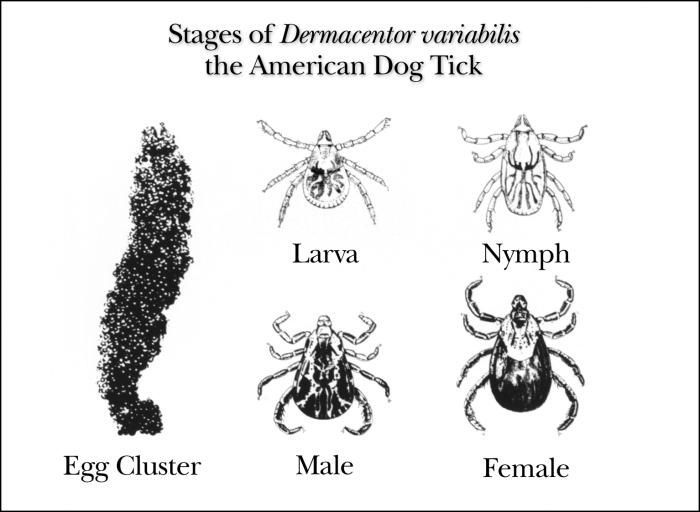
This illustration shows the growth stages of the American dog tick, Dermacentor variabilis, from eggs to adult insects.Created: 1975
-
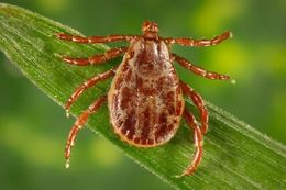
This photograph depicts a dorsal view of a male Rocky Mountain wood tick, Dermacentor andersoni. This tick specie is a known North American vector of Rickettsia rickettsii, which is the etiologic agent of Rocky Mountain spotted fever.Rocky Mountain spotted fever, like all rickettsial infections, is classified as a zoonosis. Zoonoses are diseases of animals that can be transmitted to humans. Many zoonotic diseases require a biological vector (e.g., a mosquito, tick, flea, or mite) in order to be transmitted from the animal host to the human host. In the case of Rocky Mountain spotted fever, ticks are the natural hosts, serving as both reservoirs and vectors of R. rickettsii. Ticks transmit the organism to vertebrates primarily by their bite. Less commonly, infections may occur following exposure to crushed tick tissues, fluids, or tick feces.See PHIL 10869, for a side-by-side comparative view of both a male and female D. andersoni tick.Created: 2008
-
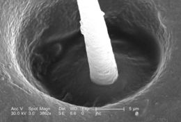
Under a high magnification of 3862X, this scanning electron micrograph (SEM) depicted the recessed base of a singe seta, or hair emanating from the dorsum of an unidentified male Dermacentor sp. tick found upon a cat in the suburbs of Decatur, Georgia, which measured approximately 3.5mm from its gnathosoma (i.e., capitulum), which is where its mouthparts are located, to the distal abdominal margin (PHIL 9961). PHIL 9959 revealed all this ticks legs, placing it into the Phylum Arthropoda, i.e., from jointed ( Arthro), and legs (poda), as well as the Class Arachnida, for theyve eight of these legs, unlike insects, which use six appendages to move about. Setae are chitinous exoskeletal adnexae which are sensorial in nature, sensing environmental changes in temperature, movement, i.e., wind, and chemistry, i.e., pheromones.Created: 2006
-
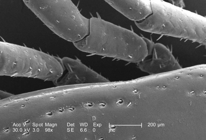
Under a relatively low magnification of 98X, this scanning electron micrograph (SEM) provided a closer view of this male Dermacentor sp. tick found upon a cat in the suburbs of Decatur, Georgia, which measured approximately 3.5mm from its gnathosoma (i.e., capitulum), which is where its mouthparts are located, to the distal abdominal margin (PHIL 9961). PHIL 9959 revealed all this ticks legs, placing it into the Phylum Arthropoda, i.e., from jointed ( Arthro), and legs (poda), as well as the Class Arachnida, for theyve eight of these legs, unlike insects, which use six appendages to move about. From proximal to distal location, each leg is comprised of a coxa, trochanter 1, trochanter 2, a femur, patella, tibia, a two-sectioned tarsus, and a two-part pretarsus, i.e., a pulvillus and claw. Here we see the trochantofemoral joints of the arachnids left 3rd and 4th legs, and the femoropatellar joints of its left 2nd and 3rd legs.Created: 2006
-
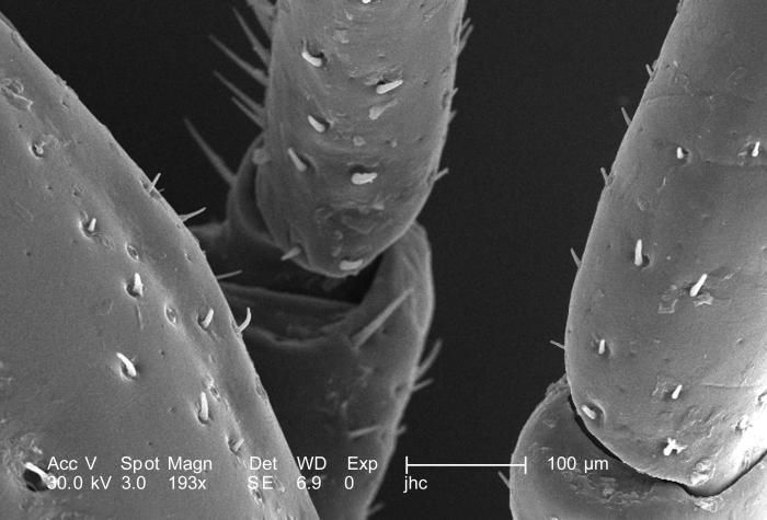
Under a relatively low magnification of 193X, this scanning electron micrograph (SEM) provided a closer view of this male Dermacentor sp. tick found upon a cat in the suburbs of Decatur, Georgia, which measured approximately 3.5mm from its gnathosoma (i.e., capitulum), which is where its mouthparts are located, to the distal abdominal margin (PHIL 9961). PHIL 9959 revealed all this ticks legs, placing it into the Phylum Arthropoda, i.e., from jointed ( Arthro), and legs (poda), as well as the Class Arachnida, for theyve eight of these legs, unlike insects, which use six appendages to move about. From proximal to distal location, each leg is comprised of a coxa, trochanter 1, trochanter 2, a femur, patella, tibia, a two-sectioned tarsus, and a two-part pretarsus, i.e., a pulvillus and claw. Here we see the trochantofemoral joint of the arachnids left 3rd leg, and the femoropatellar joint of its left 2nd leg.Created: 2006
-
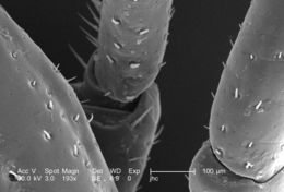
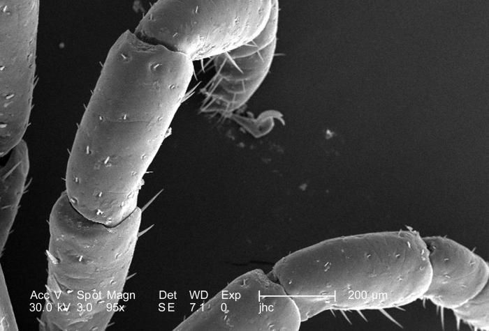
Under a relatively low magnification of 95X, this scanning electron micrograph (SEM) provided a closer view of this male Dermacentor sp. tick found upon a cat in the suburbs of Decatur, Georgia, which measured approximately 3.5mm from its gnathosoma (i.e., capitulum), which is where its mouthparts are located, to the distal abdominal margin (PHIL 9961). PHIL 9959 revealed all this ticks legs, placing it into the Phylum Arthropoda, i.e., from jointed ( Arthro), and legs (poda), as well as the Class Arachnida, for theyve eight of these legs, unlike insects, which use six appendages to move about. From proximal to distal location, each leg is comprised of a coxa, trochanter 1, trochanter 2, a femur, patella, tibia, a two-sectioned tarsus, and a two-part pretarsus, i.e., a pulvillus and claw. Here we see the femur, patella, and tibia of arachnids left 2nd and 3rd legs.Created: 2006
-
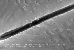
Under a magnification of 3206X, four times greater than PHIL 9965, this scanning electron micrograph (SEM) depicted a dorsal view of an unidentified male Dermacentor sp. tick found upon a cat in the suburbs of Decatur, Georgia, which measured approximately 3.5mm from its gnathosoma (i.e., capitulum), which is where its mouthparts are located, to the distal abdominal margin (PHIL 9961). Note in PHIL 9959 and 9960, that the entire dorsum of this ticks abdomen is covered by its tough scutum, or shield, categorizing it as a male. In female Ixodid-species ticks, the scutum only partially covers the dorsal abdomen. Revealed in this image is the base of the hypostome, which is one of the ticks mouthparts that acts to pierce the host skin surface, thereby, anchoring the tick to the host as it obtains its blood meal. See PHIL 9964 for another view of the foliated hypostomal surface, which begins here as small scales.Created: 2006
-
Under a magnification of 3206X, four times greater than PHIL 9965, this scanning electron micrograph (SEM) depicted a dorsal view of an unidentified male Dermacentor sp. tick found upon a cat in the suburbs of Decatur, Georgia, which measured approximately 3.5mm from its gnathosoma (seen here) (i.e., capitulum), which is where its mouthparts are located, to the distal abdominal margin (PHIL 9961). Note in PHIL 9959 and 9960, that the entire dorsum of this ticks abdomen is covered by its tough scutum, or shield, categorizing it as a male. In female Ixodid-species ticks, the scutum only partially covers the dorsal abdomen. Revealed in this image is the base of the hypostome, which is one of the ticks mouthparts that acts to pierce the host skin surface, thereby, anchoring the tick to the host as it obtains its blood meal. See PHIL 9964 for another view of the foliated hypostomal surface.Created: 2006
-
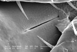
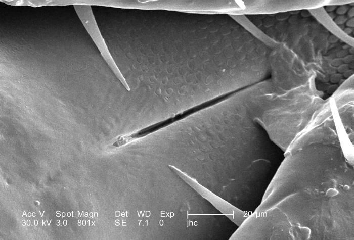
Under a magnification of 801X, this scanning electron micrograph (SEM) depicted a dorsal view of an unidentified male Dermacentor sp. tick found upon a cat in the suburbs of Decatur, Georgia, which measured approximately 3.5mm from its gnathosoma (seen here) (i.e., capitulum), which is where its mouthparts are located, to the distal abdominal margin (PHIL 9961). Note in PHIL 9959 and 9960, that the entire dorsum of this ticks abdomen is covered by its tough scutum, or shield, categorizing it as a male. In female Ixodid-species ticks, the scutum only partially covers the dorsal abdomen. Seen clearly in this image is the base of the hypostome, which is one of the ticks mouthparts that acts to pierce the host skin surface, thereby, anchoring the tick to the host as it obtains its blood meal. See PHIL 9964 for another view of the foliated hypostomal surface.Created: 2006
-
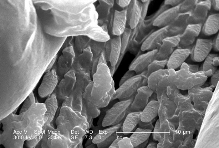
Under a magnification of 3043X, approximately 8 times greater than PHIL 9963, this scanning electron micrograph (SEM) depicted a dorsal view of an unidentified male Dermacentor sp. tick found upon a cat in the suburbs of Decatur, Georgia, which measured approximately 3.5mm from its gnathosoma (i.e., capitulum), which is where its mouthparts are located, to the distal abdominal margin (PHIL 9961). Note in PHIL 9959 and 9960, that the entire dorsum of this ticks abdomen is covered by its tough scutum, or shield, categorizing it as a male. In female Ixodid-species ticks, the scutum only partially covers the dorsal abdomen. Seen clearly in this image is the foliate covering of the ticks skin-piercing hypostome, which is located in what appears to be a trough between its pedipalps.Created: 2006
-
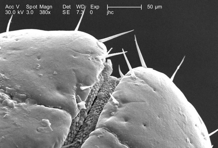
Under a magnification of 380X, approximately 4 times greater than PHIL 9962, this scanning electron micrograph (SEM) depicted a dorsal view of an unidentified male Dermacentor sp. tick found upon a cat in the suburbs of Decatur, Georgia, which measured approximately 3.5mm from its gnathosoma (i.e., capitulum), which is where its mouthparts are located, to the distal abdominal margin (PHIL 9961). Note in PHIL 9959 and 9960, that the entire dorsum of this ticks abdomen is covered by its tough scutum, or shield, categorizing it as a male. In female Ixodid-species ticks, the scutum only partially covers the dorsal abdomen. Seen clearly in this image is the ticks pedipalps, between which is located its skin-piercing hypostome and chelicerae. Note PHIL 9964 for an additional view of this region under a greater magnification.Created: 2006
-
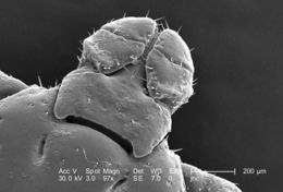
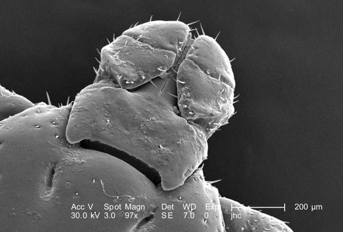
Under a magnification of 97X, this scanning electron micrograph (SEM) depicted a dorsal view of an unidentified male Dermacentor sp. tick found upon a cat in the suburbs of Decatur, Georgia, which measured approximately 3.5mm from its gnathosoma (seen here) (i.e., capitulum), which is where its mouthparts are located, to the distal abdominal margin (PHIL 9961). Note in PHIL 9959 and 9960, that the entire dorsum of this ticks abdomen is covered by its tough scutum, or shield, categorizing it as a male. In female Ixodid-species ticks, the scutum only partially covers the dorsal abdomen. Seen clearly in this image are the ticks pedipalps between which is located its skin-piercing hypostome and chelicerae. Note PHIL 9963 and 9964 for two additional views of this region under successively greater magnification.Created: 2006
-
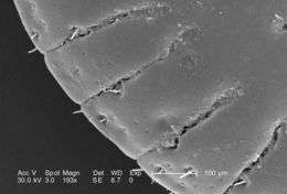
Under a magnification of 193X, four times that of PHIL 9960, this scanning electron micrograph (SEM) depicted a dorsal view of an unidentified male Dermacentor sp. tick found upon a cat in the suburbs of Decatur, Georgia, which measured approximately 3.5mm from its gnathosoma (i.e., capitulum), which is where its mouthparts are located, to the distal abdominal margin. Note here, and more so in PHIL 9959 and 9960, that the entire dorsum of this ticks abdomen is covered by its tough scutum, or shield, categorizing it as a male. In female Ixodid-species ticks, the scutum only partially covers the dorsal abdomen. Seen clearly in this image are the ridges that run along the distal abdominal border, and are known as festoons, and are one of the identifying characteristics used to classify hard, or Ixodid ticks.Created: 2006
-
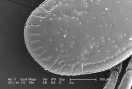
Under a low magnification of 49X, twice that of PHIL 9959, this scanning electron micrograph (SEM) depicted a dorsal view of an unidentified male Dermacentor sp. tick found upon a cat in the suburbs of Decatur, Georgia, which measured approximately 3.5mm from its gnathosoma (i.e., capitulum), which is where its mouthparts are located, to the distal abdominal margin. Note that the entire dorsum of this ticks abdomen is covered by its tough scutum, or shield, categorizing it as a male, whereas, in female Ixodid-species ticks, the scutum only partially covers the dorsal abdomen. The ridges running along the distal abdominal border are known as festoons.Created: 2006
-
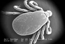
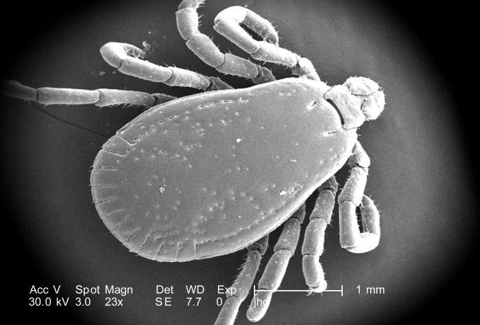
Under a low magnification of 23X, this scanning electron micrograph (SEM) depicted a dorsal view of an unidentified male Dermacentor sp. tick found upon a cat in the suburbs of Decatur, Georgia, which measured approximately 3.5mm from its gnathosoma (i.e., capitulum), which is where its mouthparts are located, to the distal abdominal margin. Note that the entire dorsum of this ticks abdomen is covered by its tough scutum, or shield, categorizing it as a male, whereas, in female Ixodid-species ticks, the scutum only partially covers the dorsal abdomen. The ridges running along the distal abdominal border are known as festoons.Created: 2006
-
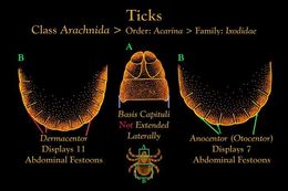
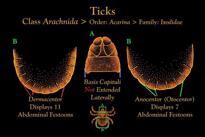
This illustration depicts the festoons found on the abdomen of Dermacentor and Anocentor Ixodidae hard ticks.Created: 1976
-
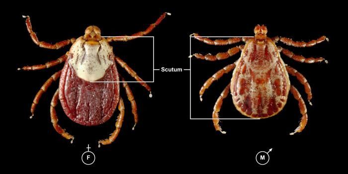
This photograph depicts a dorsal view of both a female (Lt), and male (Rt) Rocky Mountain wood tick, Dermacentor andersoni. This tick specie is a known North American vector of Rickettsia rickettsii, which is the etiologic agent of Rocky Mountain spotted fever. Note the smaller size of the females scutum compared to the males larger scutum. From the Latin word for shield, the scutum, i.e., dorsal shield, covers only a small part of the females dorsal surface, thereby, enabling her abdomen to expand, becoming engorged during feeding. The males scutum covers his entire dorsal surface, and is mottled with brown markings overall, while the females small scutum sports an almost entirely a cream-colored gray surface.Created: 2008
-
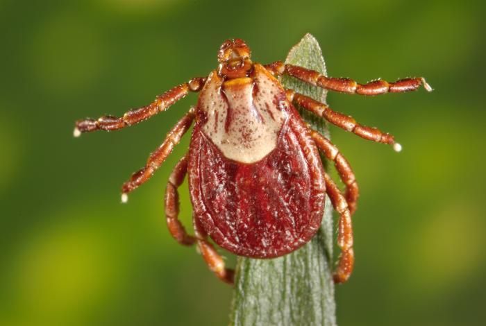
This photograph depicts a dorsal view of a male Rocky Mountain wood tick, Dermacentor andersoni. This tick specie is a known North American vector of Rickettsia rickettsii, which is the etiologic agent of Rocky Mountain spotted fever.Rocky Mountain spotted fever, like all rickettsial infections, is classified as a zoonosis. Zoonoses are diseases of animals that can be transmitted to humans. Many zoonotic diseases require a biological vector (e.g., a mosquito, tick, flea, or mite) in order to be transmitted from the animal host to the human host. In the case of Rocky Mountain spotted fever, ticks are the natural hosts, serving as both reservoirs and vectors of R. rickettsii. Ticks transmit the organism to vertebrates primarily by their bite. Less commonly, infections may occur following exposure to crushed tick tissues, fluids, or tick feces.See PHIL 10869, for a side-by-side comparative view of both a male and female D. andersoni tick.Created: 2008
-
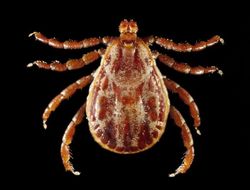
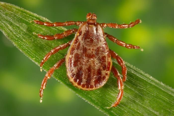
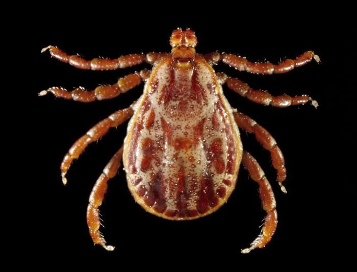
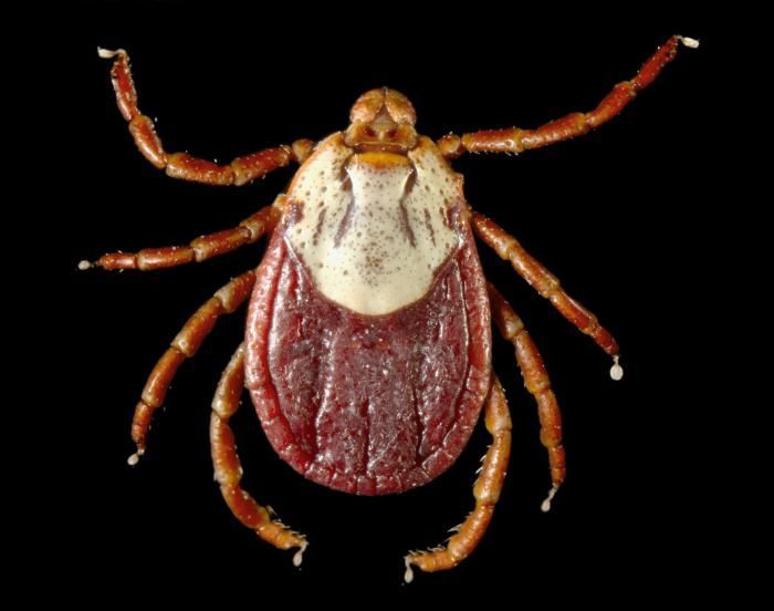
This photograph depicts a dorsal view of a female Rocky Mountain wood tick, Dermacentor andersoni. This tick specie is a known North American vector of Rickettsia rickettsii, which is the etiologic agent of Rocky Mountain spotted fever.Dermacentor andersoni is found in the Rocky Mountain states and in southwestern Canada. The life cycle of this tick may require up to 2 to 3 years for completion. Adult wood ticks feed primarily on large mammals, while the larvae and nymphs feed on small rodents. See PHIL 10869, for a side-by-side comparative view of both a male and female D. andersoni tick.Created: 2008
-
This photograph depicts a dorsal view of both a female (Lt), and male (Rt) Rocky Mountain wood tick, Dermacentor andersoni. This tick specie is a known North American vector of Rickettsia rickettsii, which is the etiologic agent of Rocky Mountain spotted fever. Note the smaller size of the females scutum compared to the males larger scutum. From the Latin word for shield, the scutum, i.e., dorsal shield, covers only a small part of the females dorsal surface, thereby, enabling her abdomen to expand, becoming engorged during feeding. The males scutum covers his entire dorsal surface, and is mottled with brown markings overall, while the females small scutum sports an almost entirely a cream-colored gray surface.Created: 2008
-
This photograph depicts a dorsal view of a male Rocky Mountain wood tick, Dermacentor andersoni. This tick specie is a known North American vector of Rickettsia rickettsii, which is the etiologic agent of Rocky Mountain spotted fever.Rocky Mountain spotted fever, like all rickettsial infections, is classified as a zoonosis. Zoonoses are diseases of animals that can be transmitted to humans. Many zoonotic diseases require a biological vector (e.g., a mosquito, tick, flea, or mite) in order to be transmitted from the animal host to the human host. In the case of Rocky Mountain spotted fever, ticks are the natural hosts, serving as both reservoirs and vectors of R. rickettsii. Ticks transmit the organism to vertebrates primarily by their bite. Less commonly, infections may occur following exposure to crushed tick tissues, fluids, or tick feces.See PHIL 10869, for a side-by-side comparative view of both a male and female D. andersoni tick.Created: 2008
-
This photograph depicts a dorsal view of a female Rocky Mountain wood tick, Dermacentor andersoni. This tick specie is a known North American vector of Rickettsia rickettsii, which is the etiologic agent of Rocky Mountain spotted fever.Dermacentor andersoni is found in the Rocky Mountain states and in southwestern Canada. The life cycle of this tick may require up to 2 to 3 years for completion. Adult wood ticks feed primarily on large mammals, while the larvae and nymphs feed on small rodents. See PHIL 10869, for a side-by-side comparative view of both a male and female D. andersoni tick.Created: 2008