-
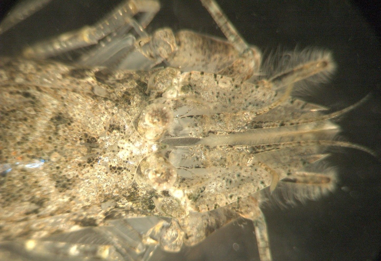
In this underside (ventral) view of the head the subchelate first pereopods can be clearly seen. In this dorsal view of the head the small rostrum and the single median spine behind it can be seen. The arrangement of the eyestalks can also be clearly seen.
-
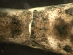
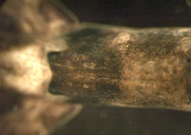
The ventral side of abdominal segment 6 has a median groove, visible as the light-colored area in this photo.
-
-
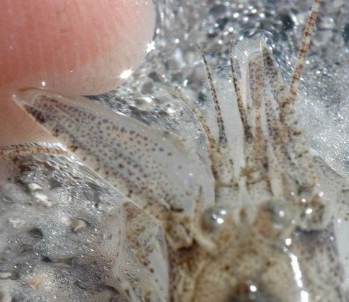
The antennal scale of the second antennae, being held out by my finger in this photo, is more than twice as long as wide and the spine on the end (left side of scale) is longer than the flatter lamella (right side of end of scale)
-
The upper posterolateral margin of abdominal segment 5 has no spines, as seen in this dorsal view. Anterior is to the right, and segment 6 is to the left.
-
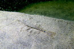
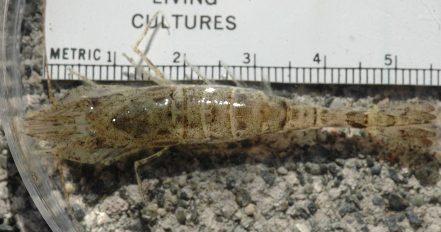
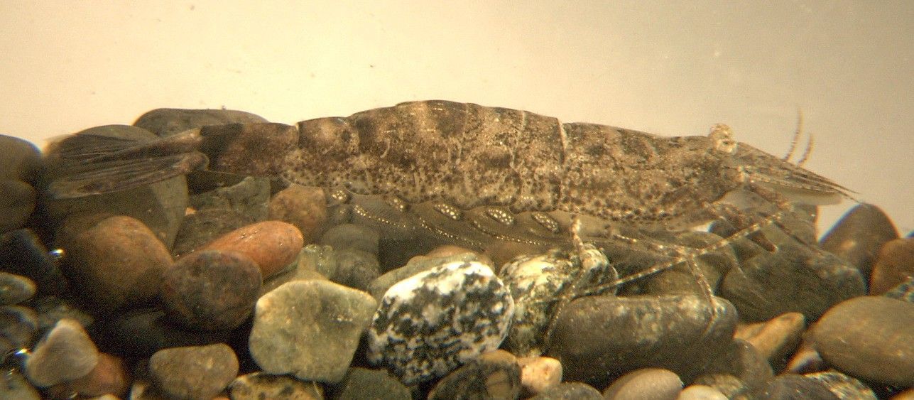
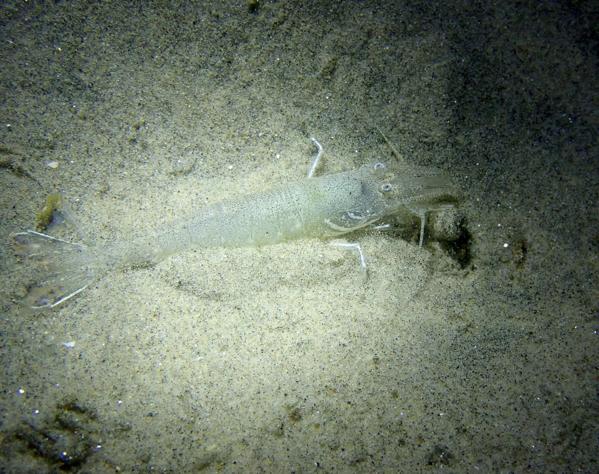
A view of the whole, live animal ffom which the parts above were photographed. Photo by Dave Cowles, July 2005
-
Abdominal segment 6 has a dorsal median groove (sulcus) but no median ridge. Anterior is to the right and the tailfan is to the left.
-
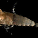
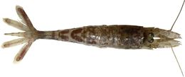
(Crangon alaskensis from 100 m depth, San Juan Channel, WA. About 5 cm long. (Photo by: Dave Cowles, July 2000)
-
The ventral sides of abdominal segments 5 (right) and 6 (left) are both smooth and clear, with no median groove (sulcus). This view is an oblique view of the right pleura and the ventral side from the right side of the shrimp. The base of leg 5 can be seen at the right.
-
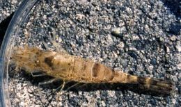
Adult female Neocrangon communis from 308 m depth near the southwest coast of Kamchatka (Photo by: Andrey Gontchar )
-
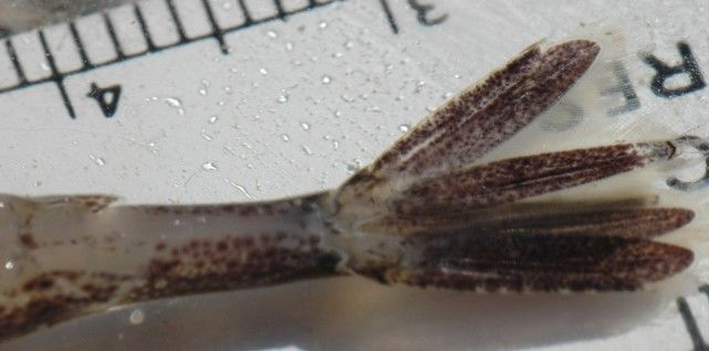
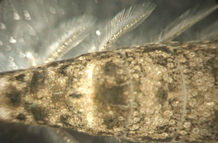
The feathery exopods of the pleopods are typically held out to the side. They are used for swimming and likely also for burrowing.
-
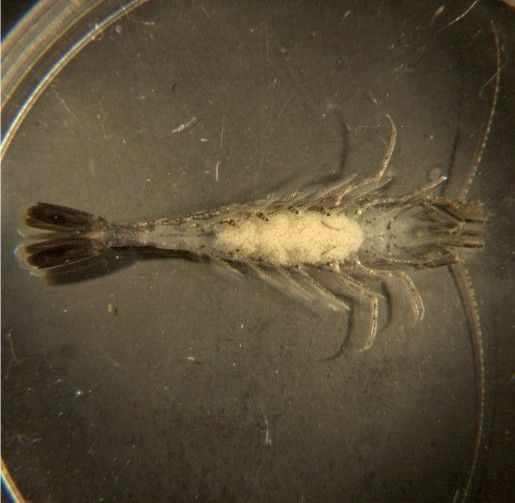
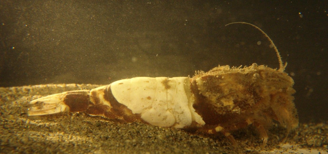
This individual was carrying a large batch of white eggs.
-
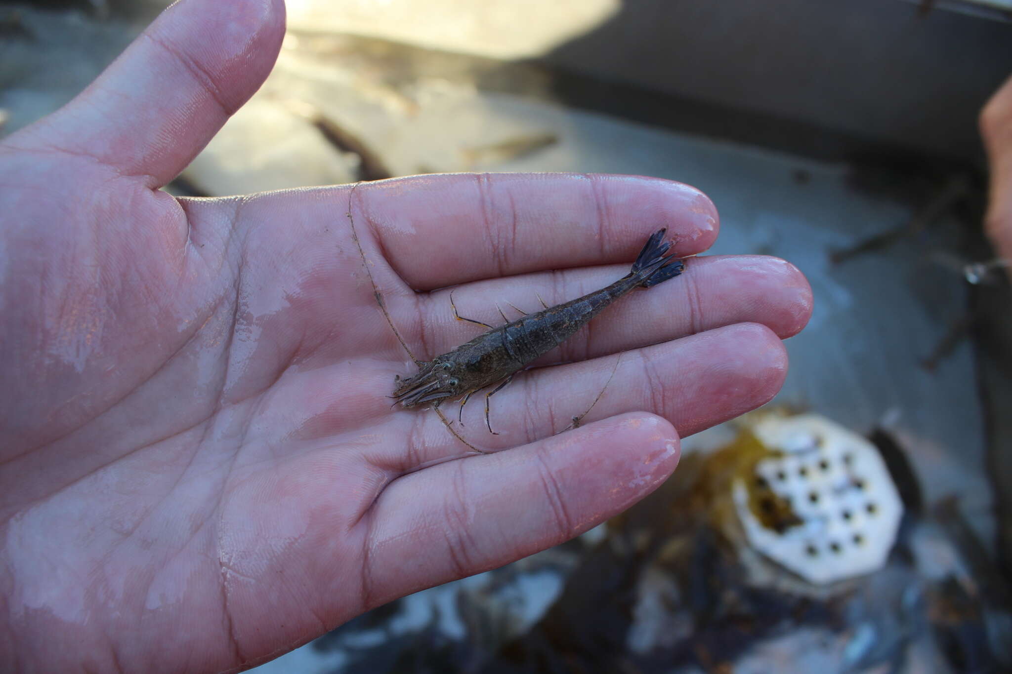
Crangon alba captured in Padilla Bay tide flats (Photos by: Dave Cowles, July 2008)
-
Crangon alba captured in Padilla Bay tide flats (Photos by: Dave Cowles, July 2008)
-
This ventral view of the head shows that the first pair of pereopods, held closely below the head in this view, is subchelate.
-
In this side view of the carapace, the short rounded rostrum and two median dorsal spines can be seen. The dorsal profile of the carapace is highest at the posterior median spine and descends anteriorly all the way to the rostrum in a female, while in the male the dorsal profile remains high until the anterior median spine, then descends to the rostrum. From this view and the animal's size I conclude that this is a female. Also note that pereopod 5 (the last walking leg) does not have a broad and flattened dactyl. On the side of the carapace can be seen the light-brown submedian spine, and a hepatic spine just below and forward of it.
-
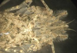
The many spines and setae and the disruptive coloration of the carapace of this species makes it hard to see structures up close. This is a dorsal view of the carapace, with the head facing right. The two eyes can be seen on the right. Between them is the small rostrum, then the mid-dorsal ridge runs back to the left. The first mid-dorsal spine is visible in the light area at the left of the picture. In front of the mid-dorsal spine and directly behind the eyes on each side is a single sub-median spine on each side, colored light brown and just to the left of center in this view.
-
This side view of abdominal segments 2-4 shows that there are no ventrally-directed spines on segments 1-3 and no prominant longitudinal mid-dorsal ridge on segments 3-5. The first and third pleura have a depression that the larger 2nd pleuron fits over.
-
This dorsal view of the head shows the antennal scale (2nd antenna). Note that the lamella of the scale extends past the antennal spine.
-
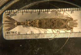
Metacrangon munita, about 4 cm long, captured at 75 m depth in the San Juan Channel (Photo by Dave Cowles, July 2008 )
-
-
-
-