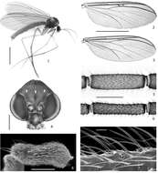
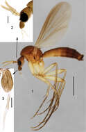
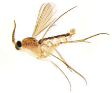
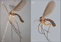
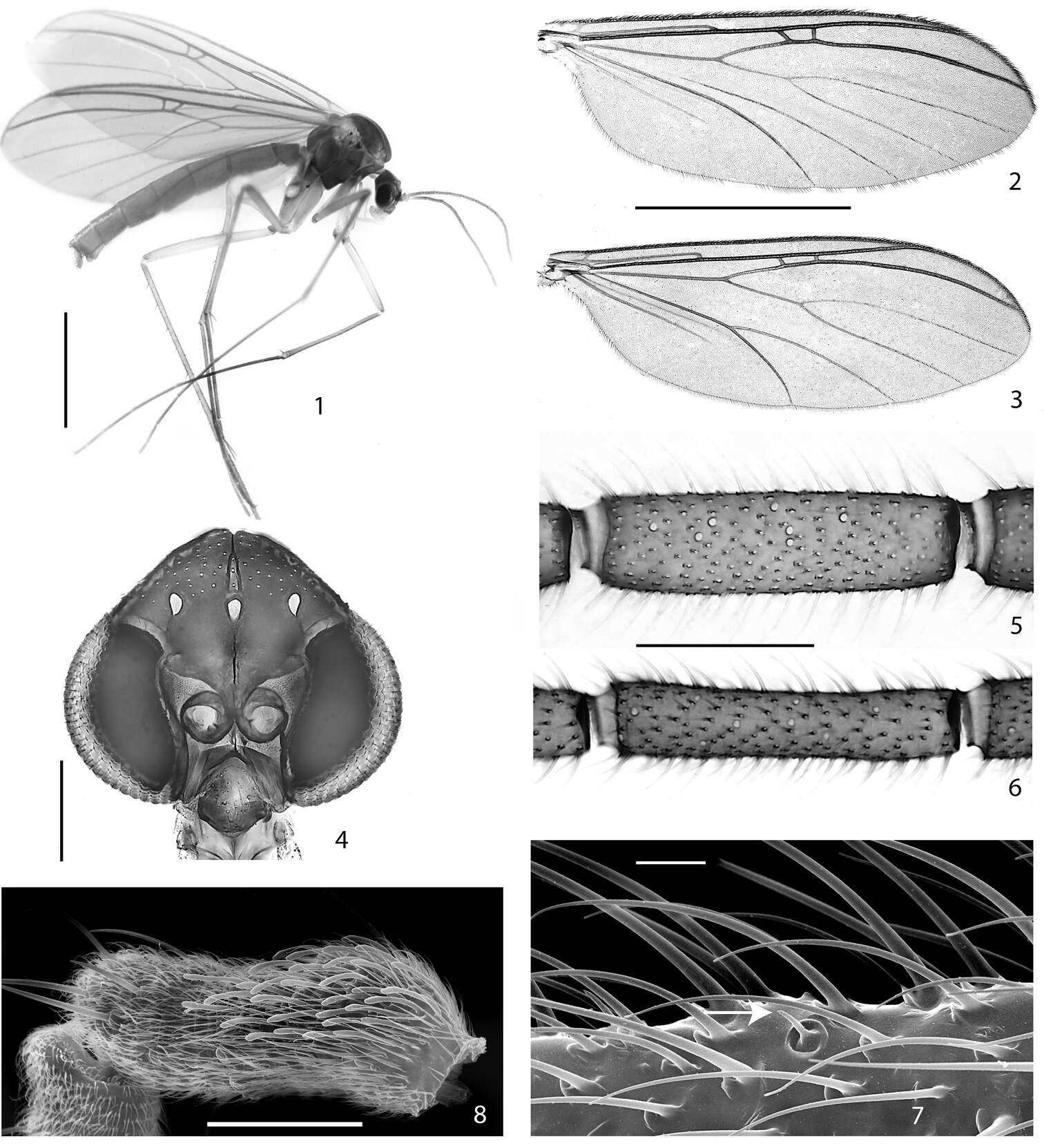
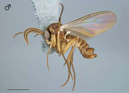
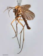
Figures 1–3.Cordyla australica sp. n. 1 male habitus 2 head with antennae and maxillary palpi, closer view 3 three apical segments of maxillary palpus. Scale bar = 1 mm (1), 0.2 mm (2) and 0.1 mm (3).
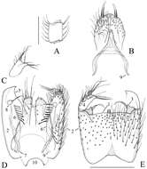
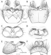
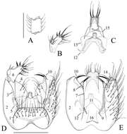
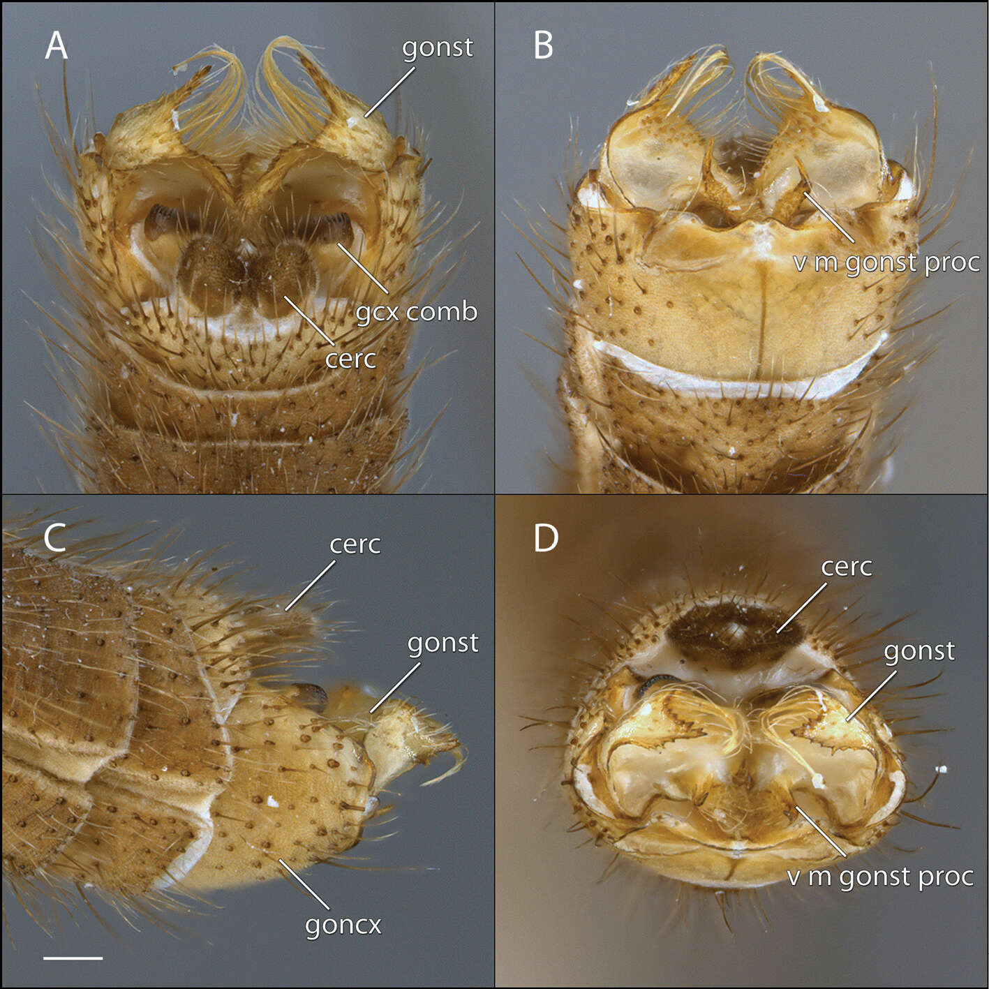
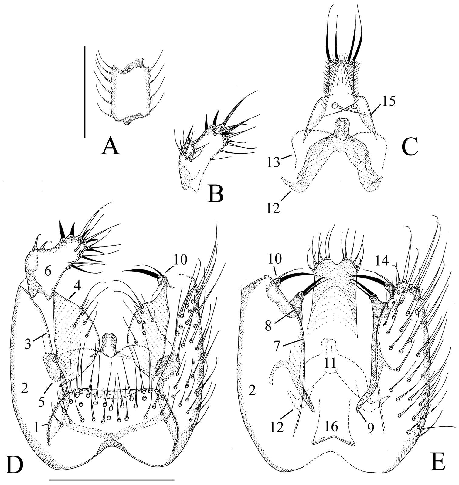
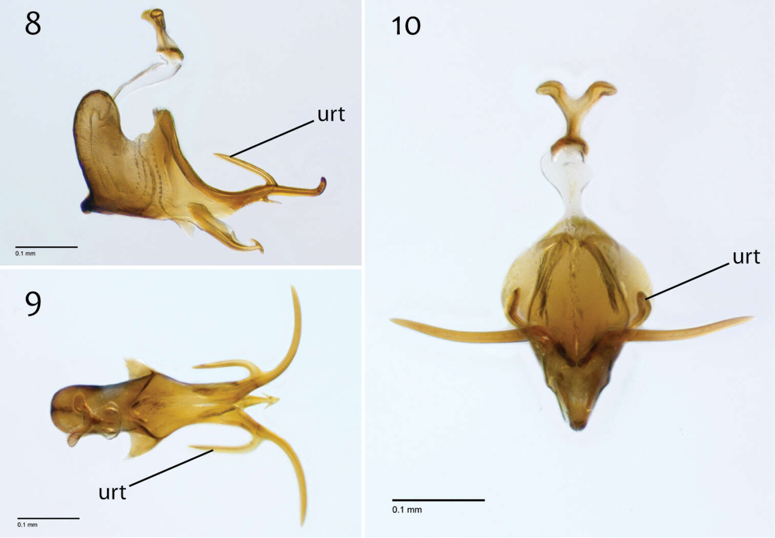
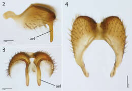
Figures 14–20.Terminalia of Acomopterella martinovskyi, male. 14 Terminalia in dorsal view, tergite IX removed 15 Gonocoxites in ventral view 16 Epiproct and hypoproct, dorsal view 17 Abdominal segments VII & VIII and terminalia in lateral view 18–19 Posterior view of terminalia 20 Cercal setae in detail. Length of scale bar = 100 µm (for 14–15, 18–19), 50 µm (for 16, 20), 0,5 mm (for 17).
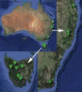
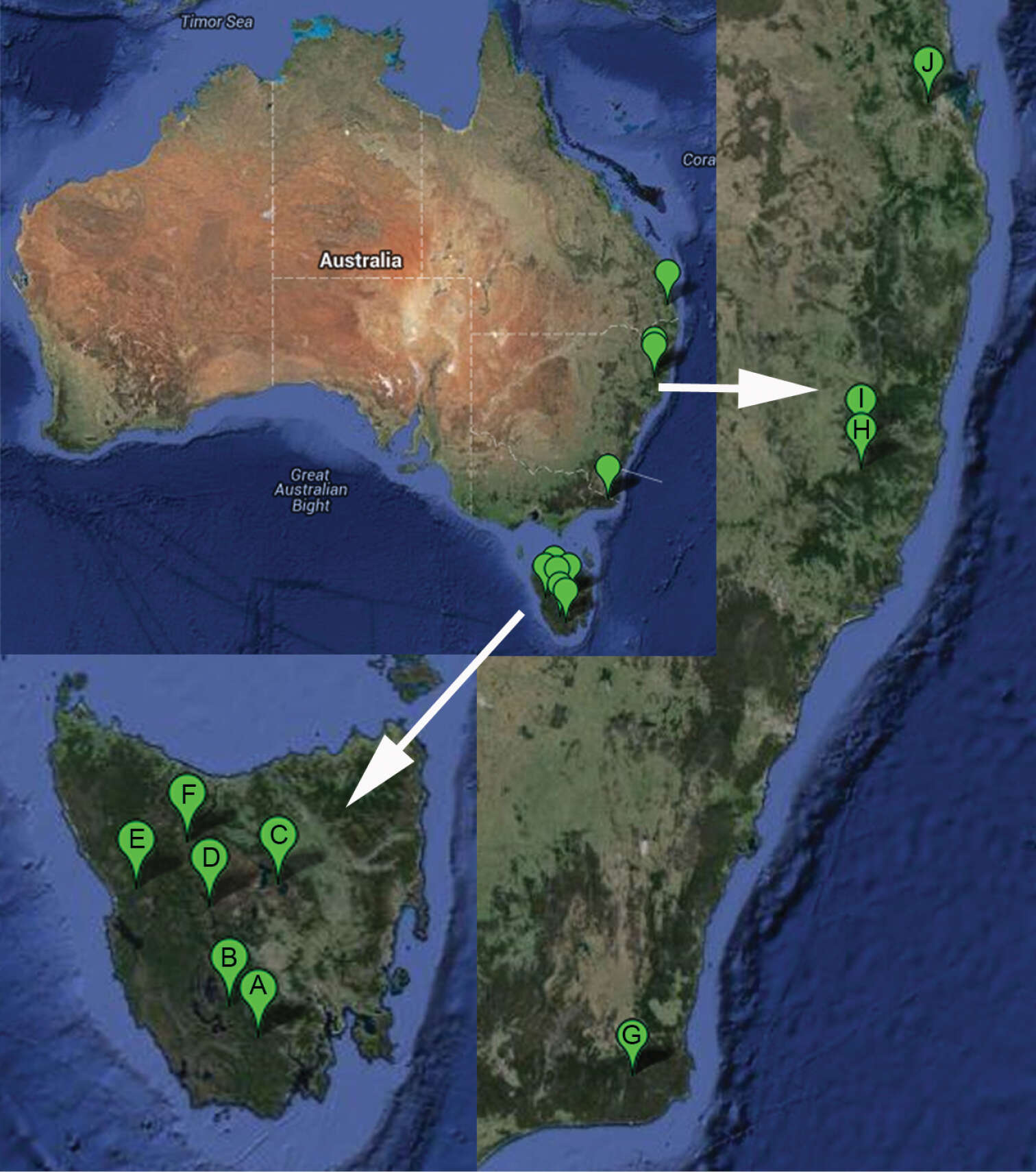
Figure 15.Collecting localities of Cordyla australica sp. n. in the continental Australia and Tasmania. A Tasmania, Warra long-term ecological research site B Tasmania, Southwest National Park C Tasmania, Central Plateau D Tasmania, King William Creek E Tasmania, Ewart creek F Tasmania, Cradle Mountain G Victoria, Coopracambra National Park H New South Wales, Werrikimbe National Park I New South Wales, Carrai State Forest J Queensland, Brisbane Forest Park.
Figures 14–20.Terminalia of Acomopterella martinovskyi, male. 14 Terminalia in dorsal view, tergite IX removed 15 Gonocoxites in ventral view 16 Epiproct and hypoproct, dorsal view 17 Abdominal segments VII & VIII and terminalia in lateral view 18–19 Posterior view of terminalia 20 Cercal setae in detail. Length of scale bar = 100 µm (for 14–15, 18–19), 50 µm (for 16, 20), 0,5 mm (for 17).
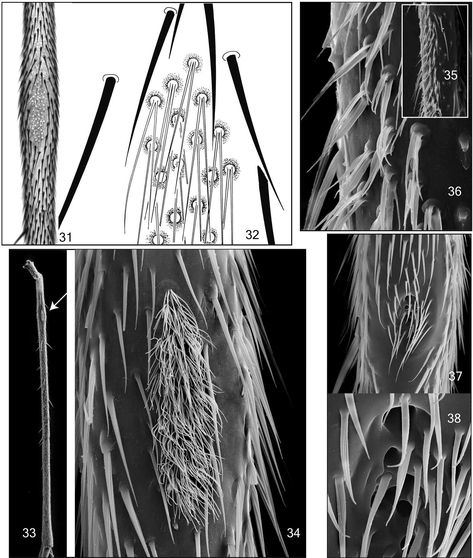
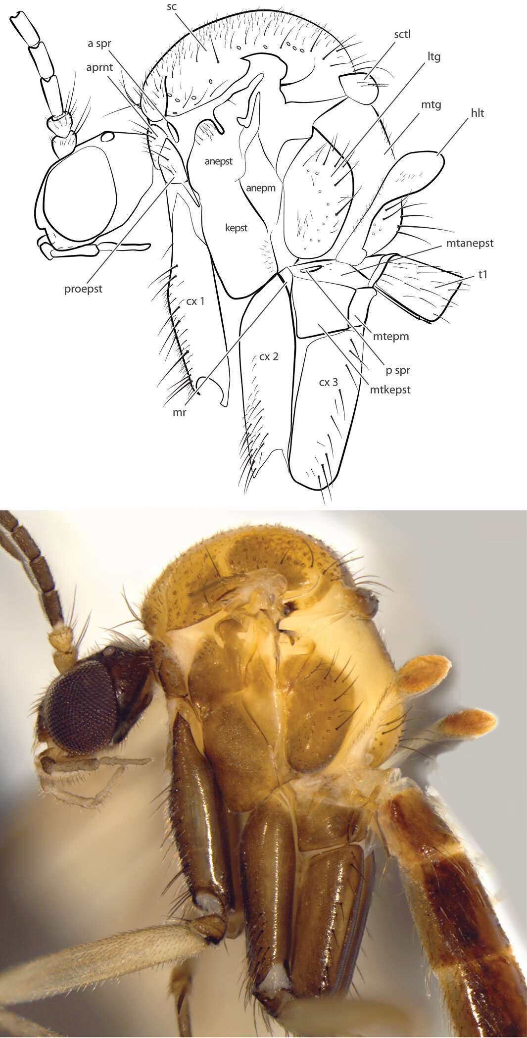
Figures 31–38.Mid tibial organ. Not on the same scale. 31–32 Acomopterella yoshiwae sp. n. 33–34 Acomopterella martinovskyi 33 General outer view of mid tibia, the arrowhead points to the tibial organ 35–36 Speolepta leptogaster 37–38 Ectrepesthoneura hirta.
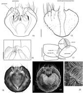
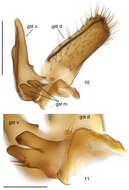
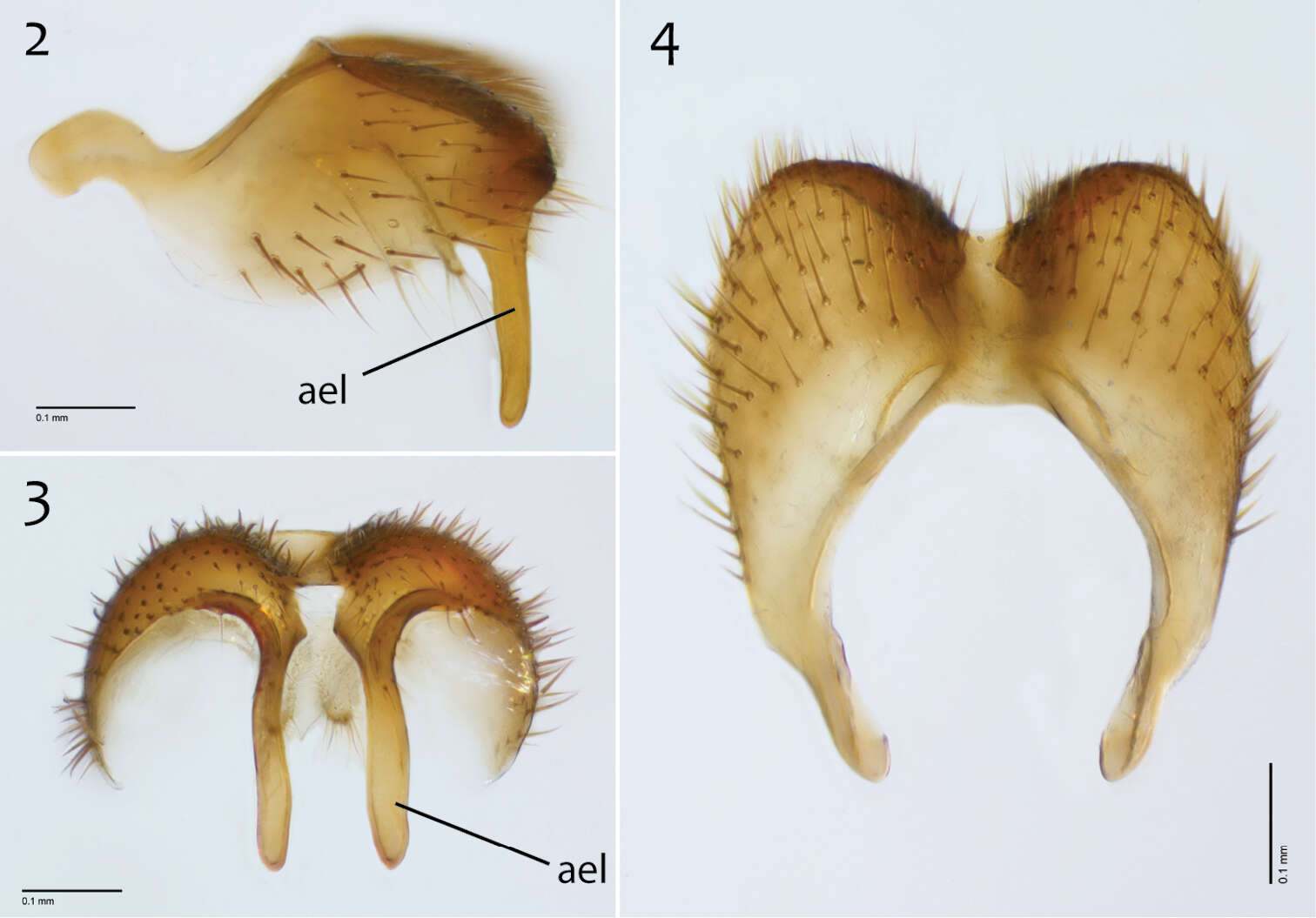
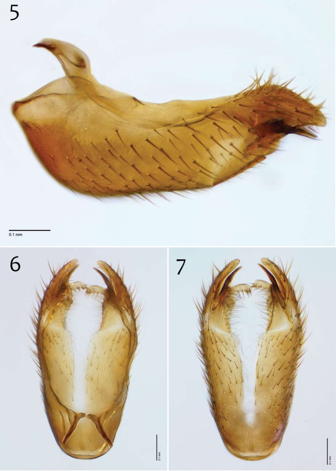
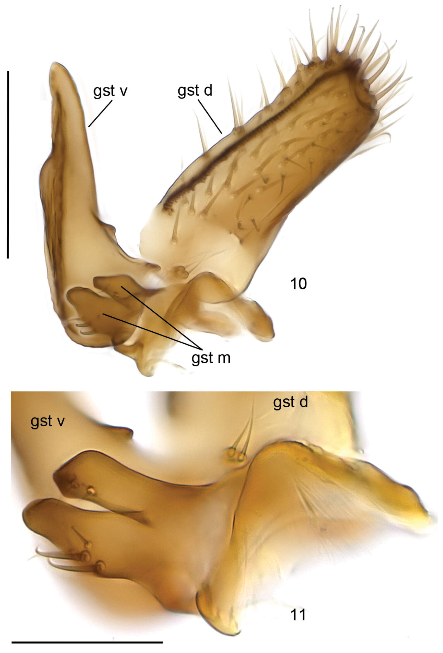
Figures 10–11.Cordyla australica sp. n., gonostylus. 10 internal view 11 lobes of medial branch of gonostylus. Scale bar = 0.1 mm (10) and 0.05 mm (11).
Jan Ševčík, Heikki Hippa, Rodzay Abdul Wahab
Zookeys
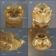
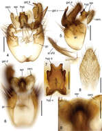
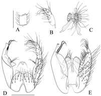
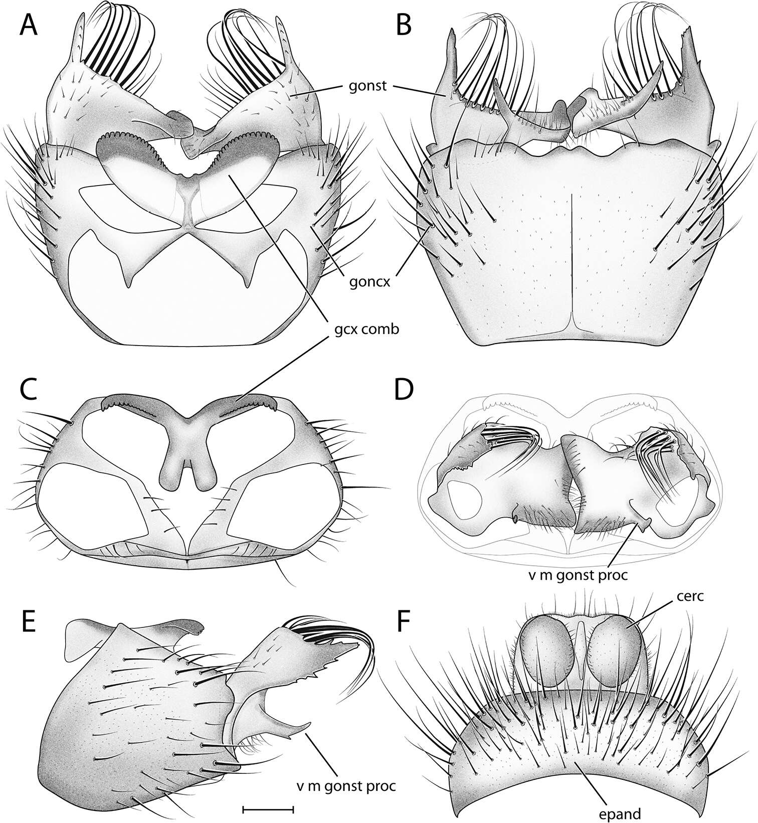
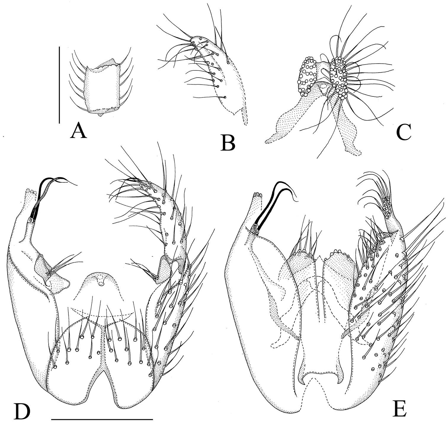
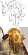
Figure 5.Manota macrothrix sp. n. (holotype). A Antennal flagellomere 4, lateral view B Gonostylus, dorsal view C Aedeagus and hypoproct, ventral view D Hypopygium, ventral view E Hypopygium, dorsal view. Scale 0.1 mm.