These parasites are usually not preyed on directly, but are ingested from host to host.
Toxocara canis is smaller than most of the other species in the family Ascarididae. It has a complete gut in the form of a simple tube. It is a "round worm" implying the shape of the outer layer to be round (if seen in a cross section ). Depending on the host the worm gets into T. canis will have different number of larval stages. Most worms have three larval stages before becoming infective.
Toxocara canis is dioecious having morphology distinctly different for the male and female. Males, 4-6 cm long, are smaller than females. The male's posterior end is curved ventrally and the tail is bluntly pointed. The male has a single tubular testis. He also has simple spicules, which allows for direct sperm transfer. The female worms are generally around 6.5 cm but can be as long as 15 cm long. In the female the vulva is about one-third the body length from the anterior end. The ovaries are very large and extensive. The uteri contain up to 27 million eggs at a time.
Both males and females have three prominent lips. Each lip has a dentigerous ridge. The lateral hypodermal cords are visible with the naked eye. No gubernacullum is present. In both sexes there are prominent cervical alae. The eggs are brownish and almost spherical. The eggs measure 75-90 micrometers. The eggs are embryonated when laid and have surficial pits. These eggs are very resistant to various weather and chemical conditions.
Range length: 4 to 15 cm.
Other Physical Features: ectothermic ; heterothermic ; bilateral symmetry
Sexual Dimorphism: female larger; sexes shaped differently
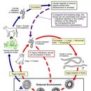
The eggs of T. canis are excreted in the feces of an infected canid host. The embryonated eggs can live in the feces for up to three weeks. The feces are often deposited in soil or sandy areas. A host must ingest the eggs for the life cycle to continue. If ingested, the new habitat becomes the internal organs of the host. The gut is the first area T. canis larvae reside. If the host has not been previously infected, hatched juveniles go throught the circulation to the lungs, then back to the gut. If in a canid host, they take up residence in the intestine and develop into adults. If hosts have been previously "immunized" junveniles go to the body tissues and become dormant as if they were in a paratenic host. Often the infectious larvae stay in the mammary glands until a pregnancy where they are passed on to a nursing pup. If in a human or other non-canid host the larvae will wonder throughout the organs. These wandering larvae are called visceral larva migrans. They may travel to the eyes, lungs, brain, heart, muscles, liver, and other organs. Here they do not develop further but can cause severe local reactions.
Habitat Regions: terrestrial
Terrestrial Biomes: desert or dune ; savanna or grassland ; chaparral ; forest ; rainforest ; scrub forest ; mountains
Other Habitat Features: urban ; suburban ; agricultural
Toxcocara canis has a worldwide distribution. It is prevalent in all locations that have domestic dogs, puppies, and other canids. Toxocara canis is also found in places that have other various mammals such as mice, pigs, birds, and foxes, but these hosts are only paratenic hosts. Hosts are terrestrial mammals and therefore T. canis is mainly found in terrestrial terrain.
Biogeographic Regions: nearctic ; palearctic ; oriental ; ethiopian ; neotropical ; australian
Other Geographic Terms: cosmopolitan
The location of T. canis in hosts is in the small intestine. There they feed on intestinal contents. The adults have a specialized anaerobic metabolism. This specialized metabolism gives the adult worms an extra ATP. Adult T. canis worms are very host specific.
Pharyngeal glands and intestinal epithelium produce digestive enzymes to feed on the hosts’ body fluids. Extracellular digestion begins within the lumen and is finished intracellularly.
Animal Foods: body fluids
Primary Diet: carnivore (Eats body fluids)
Toxocara canis is a canid parasite. Humans acquire the parasite as accidental hosts. In the tissues of all dogs, in many birds, and other mammals, the larval form of T. canis is found. The dog or canid host is the definitive host and only there will T. canis develop further than the larval stage. The name for the disease when in T. canis is in a host is Toxocariasis. Many animals such as mice, rabbits, and monkeys can serve as paratenic hosts.
Ecosystem Impact: parasite
Species Used as Host:
Toxocara canis is widespread, causing disease in many mammals including humans. Many humans are infected with T. canis larvae. The larvae can cause serious damage to the human paratenic host. It can be found in wealthy and well-developed countries just as much as poor and under-developed places. In the United States about 98% of puppies and 20% of adult dogs are infected with T. canis. The means the risk of exposure to humans in the United States is very high. Most cases go unreported or are unrecognized. All small mammals can be paratenic hosts especially small children. The disease is most common in children between the ages one and three. Ingesting embryonated eggs from the feces of dogs and other canids spreads the diease. Often pet owners take their dog for a walk in the park. During the walk the dog may deposit egg-bearing feces in the park soil or sand. The next day an unsuspecting parent brings their small child to play in the park. The young child is at an age where everything is picked up and tasted, including the contaminated soil. The eggs of T. canis are most commonly ingested this way. The eggs once excreted from the definitive host can survive for 10-20 days in the external environment. This means even long after the dog has been in the park humans can still be infected. In Britain, one study showed that the climate conditions there allow for some T. canis eggs to survive in the soil for up to three years!
Many of the wandering larvae end up in the brain causing serious reactions, which can lead to death of the host. The most common place in the body of infection is the liver, but it can be found in every organ. The amount of damage is related to the number of juveniles in the body of the host. One of the more serious results of visceral larva migrans is blindness. The worms that infect the eye are called ocular larval migrans. Blindness occurs from the infection when a larva becomes trapped in the blood vessels at the back of the eye.
Negative Impacts: injures humans (causes disease in humans ); causes or carries domestic animal disease
Toxocara canis is a canid parasite. Humans acquire the parasite as accidental hosts. In the tissues of all dogs, in many birds, and other mammals, the larval form of T. canis is found. The dog or canid host is the definitive host and only there will T. canis develop further than the larval stage. The name for the disease when in T. canis is in a host is Toxocariasis. Many animals such as mice, rabbits, and monkeys can serve as paratenic hosts.
Regardless of the path T. canis larvae take to get to the canid intestine once there the third stage larvae molt into adults. The adult worm remains in the intestine and produces an enormous number of eggs each day. Not until the fifth day post-infection do the eggs begin to appear in the canid feces.
Toxocara canis has a complex life cycle. Similar to other nematodes, T. canis is not infectious immediately when it leaves the definitive host. It needs to grow and develop into the stage that is infectious, ensheathed L3. Only this stage can infect other definitive hosts. There is strong evidence of two molts taking place inside the developing eggs, before the eggs even hatch. The molting process involves a separation of the cuticle from the epidermis. This causes a formation of the new cuticle, which is arising from the outermost surface of the epidermis. It also includes the shedding of the old cuticle.
US Federal List: no special status
CITES: no special status
Nematodes within the Secernentea have phasmids, which are unicellular glands. Phasmids likely function as chemoreceptors. Females may produce pheromones to attract males.
Nematodes in general have papillae, setae and amphids as the main sense organs. Setae detect motion (mechanoreceptors), while amphids detect chemicals (chemoreceptors).
Communication Channels: tactile ; chemical
Other Communication Modes: pheromones
Perception Channels: tactile ; chemical
If a worm gets into an improper host such as humans the juveniles migrate through the body. The juveniles begin a typical tissue migration. They do not undergo development nor do they complete the normal migration, instead they will randomly wander through the body. Visceral larva migrans (VLM) is the resulting disease.
Toxocara canis infection is largely preventable. Worming pets often, with worming agents (such as antihelmintics: fenbendazole, piperazine, and Dichlorvos) from a veterinarian will reduce the possibility of human infection. This drugs also help in human treatment. Also careful and prompt disposal of dog feces will help. Humans should also wash their hands and the hands of children after handling dogs or dog feces, and especially before handling food. Lastly, parents and childcare givers need to watch out for toddlers eating soil and try to prevent it.
Females may produce a phermomone to attract males. The male coils around a female with his curved area over the female genital pore. The gubernaculum, made of cuticle tissue, guides spicules which extend through the cloaca and anus. Males use spicules to hold the female during copulation. Nematode sperm are amoeboid-like and lack flagella. The adult worm remains in the intestine and produces an enormous number of eggs each day. Not until the fifth day post-infection do the eggs begin to appear in the canid feces. There is strong evidence of two moults taking place inside the developing eggs.
Key Reproductive Features: sexual ; fertilization (Internal ); oviparous
Parental Investment: pre-fertilization (Provisioning); pre-hatching/birth (Provisioning: Female)
Infection with larvae of the nematode (roundworm) Toxocara canis, which occurs worldwide, is the most common cause of toxocariasis in humans (less frequently, it is caused by T. cati, a parasite of cats). Both dogs and cats can acquire their respective nematode parasites at any age by ingesting eggs or paratenic hosts ("transport hosts"). The most widely recognized source of infection by Toxocara in humans is ingestion of contaminated soil, often by toddlers, but infection is also possible via the consumption of partial or whole paratenic hosts, such as raw livers of domestic animals (chickens, ducks, cows, and pigs), as well as earthworms. Uncooked vegetables have also been reported as a possible source of infection, especially those from farms that utilize animal or human excrement as fertilizer. One additional possible source of infection reported is contact with embryonated eggs on a dog’s hair coat. (Lee et al. 2010 and references therein)
Toxocara canis completes its life cycle in dogs, with humans acquiring the infection as accidental hosts. Unembryonated eggs are shed in the feces of the definitive host. Eggs embryonate and become infective in the environment. Following ingestion by dogs, the infective eggs hatch and larvae penetrate the gut wall. In younger dogs, the larvae migrate through the lungs, bronchial tree, and esophagus; adult worms develop and oviposit in the small intestine. In older dogs, patent infections can also occur, but larval encystment in tissues is more common (an infection is "patent" when direct evidence of the organism can be detected, e.g., in the patient’s feces or blood, regardless of whether symptoms have appeared). Encysted stages are reactivated in female dogs during late pregnancy and infect by the transplacental and transmammary routes the puppies, in whose small intestine adult worms become established. Puppies are a major source of environmental egg contamination. Toxocara canis can also be transmitted through ingestion of paratenic hosts: eggs ingested by small mammals (e.g., rabbits) hatch and larvae penetrate the gut wall and migrate into various tissues where they encyst. The life cycle is completed when dogs eat these hosts and the larvae develop into egg-laying adult worms in the small intestine. Humans are accidental hosts who become infected by ingesting infective eggs in contaminated soil or infected paratenic hosts. After ingestion, the eggs hatch and larvae penetrate the intestinal wall and are carried by the circulation to a wide variety of tissues (liver, heart, lungs, brain, muscle, eyes). While the larvae do not undergo any further development in these sites, they can cause severe local reactions that are the basis of toxocariasis. The two main clinical presentations of toxocariasis are visceral larva migrans and ocular larva migrans. Diagnosis is usually made by serology or the finding of larvae in biopsy or autopsy specimens.
Škrkavka psí (Toxocara canis, Werner 1782) je celosvětově rozšířený červ z kmene Nematoda, který parazituje u psů a psovitých masožravců. Dospělí jedinci T. canis jsou odděleného pohlaví, bělavě žluté barvy, měří 9–18 cm a vyskytují se ve střevě definitivního hostitele. Škrkavky způsobují záněty střeva, migrující larvy škrkavek vyvolávají záněty v dalších orgánech. Zatímco u starších psů nevyvolávají většinou žádné klinické příznaky, u štěňat mohou způsobit závažné onemocnění končící úhynem. Jako paratenický hostitel slouží škrkavkám řada obratlovců včetně člověka a někteří bezobratlí. U lidí mohou larvy škrkavek způsobit vážné onemocnění zvané larvální toxokaróza.
Škrkavka psí představuje jednoho z nejběžnějších parazitů psů a vzhledem k možnosti přenosu z feny na štěňata se doporučuje preventivní odčervení feny a novorozených štěňat od 2.–3. týdne po narození. Mezi účinná anthelmintika používaná k léčbě psů patří například preparáty na bázi pyrantelu, fenbendazolu nebo mebendazolu.
Škrkavky psí jsou oblé hlístice, jejichž tělo je na obou koncích zašpičatělé a pokryté kroužkovanou kutikulou nažloutlé barvy. Samci měří 9–13 × 0,2–0,25 cm a samičky 10–18 × 0,25–0,3 cm.[1] Přední konec škrkavek je opatřen latelárními (bočními), poměrně širokými křidélky (alae) dlouhými 2,0–2,5 mm a širokými jen 0,2 mm. Samec má na ocasním konci kónusovitý prstovitý výběžek.[2] Vajíčka jsou oválná až kulovitá, silnostěnná s granulovaným povrchem, v čerstvém stavu obsahují jednu velkou tmavě šedou blastomeru, která vyplňuje celý obsah vajíčka. Velikost se pohybuje od 72 do 85 μm.[1]
Systematika a taxonomie, zejména na úrovni čeledí, není u škrkavek dosud ustálena. V rámci nadčeledi Ascaridoidea preferují někteří autoři dělení na čeledi Anisakidae a Ascarididae, která obsahuje rody Ascaris, Parascaris, Toxocara, Toxascaris a další méně známé rody.[3][4] Dle jiného zdroje[5] se nadčeleď Ascaridoidea člení na více čeledí: Anisakidae, Ascarididae a Toxocaridae (nově vytvořená čeleď) atd., z čehož plyne, že škrkavka psí (Toxocaridae) patří do jiné čeledě než třeba Ascaris lumbricoides (Ascarididae).
Dospělé škrkavky žijí v tenkém střevě definitivního hostitele, kde se živí střevním obsahem. Zde rovněž dochází k pohlavnímu rozmnožování. Oplozená vajíčka produkovaná samičkami škrkavek jsou vylučována trusem do vnějšího prostředí, kde se postupně rýhují. Ve vajíčku se rýhováním blastomery vyvíjí postupně larva 1. generace (larva L1). Ta se ve vajíčku dvakrát svléká až do stádia larvy 3. generace tzv. larvy L3. Toto stádium je již infekční. Vývoj larvy ve vajíčku trvá v závislosti na teplotě vnějšího prostředí 2–5 týdnů. Při teplotách pod 8 °C se larvy nevyvíjí a cyklus je přerušen, avšak vajíčka jsou nadále životná a při vzestupu teploty vývoj opět pokračuje. Teploty nad 35 °C způsobují značnou mortalitu (úmrtnost) larev. Vysoké teploty (zhruba nad 50 °C) a teploty pod bodem mrazu vajíčka s larvami devitalizují (usmrtí).[6]
Hostitel se nakazí pozřením vajíček obsahující larvy L3, které se dostanou do střeva. Larvy L3 se v tenkém střevě uvolní z vajíčka a pronikají skrze stěnu střevní do krevních kapilár a migrují organismem hostitele. Obecně se škrkavky rodu Toxocara vyznačují tzv. entero-hepato-pulmonálním typem migrace (larvy migrují ze střeva přes játra do plic). Larvy L3 se krevním řečištěm dostávají ze střeva do jater, a odtud žilním oběhem do pravého srdce. Ze srdce jsou larvy unášeny krví do plic, kde se usazují a postupně se vyvíjejí. Při průniku plicní tkáni larvy poškozují plicní sklípky a sliznici dolních cest dýchacích. Postupně se dostávají do průdušnice, jsou vykašlány a se slinami opět polknuty. Larvy se tak dostávají zpět do tenkého střeva, kde se naposledy svlékají a pohlavně dospívají. Do střeva se larvy dostávají zhruba za 10 dní po pozření vajíček hostitelem. Dospělci škrkavek ve střevě kopulují a samičky následně produkují vajíčka. Takto popsaný cyklus je označován jako tracheální migrace. Při tzv. somatické migraci se larvy dostávají z plic do velkého krevního oběhu a jsou krví roznášeny do všech orgánů, nejčastěji játra, ledviny, střevo, mozek, svalovina, podkoží. Zde se usazují, opouzdřují a mohou zůstat velmi dlouhou dobu životaschopné. Bylo prokázáno, že infekce mladých psů velkým počtem vajíček vede převážně k somatické migraci, zatímco nízký počet vajíček snáze dokončí vývoj jako pohlavně dospělé škrkavky ve střevě. U dospělých a starších zvířat dochází především k somatické migraci, a tím lze vysvětlit skutečnost, že se u těchto kategorií psů se škrkavky ve střevě vyskytují minimálně.[1]
Opouzdřené larvy v různých orgánech dospělých psů mají velký význam při přenosu škrkavek na štěňata. U gravidních (březích) fen dochází k aktivaci těchto larev, které prostupují přes placentu do plodu – transplacentární infekce. Štěňata se tudíž mohou narodit infikovaná. Rovněž dochází u fen po porodu k přestupu larev do mateřského mléka (laktogenní infekce) a štěňata se infikují při kojení.
S ohledem na fakt, že molekulární biologie je relativně mladý, ale velmi progresivní obor, je rovněž řada molekulárních aspektů týkajících se T. canis stále předmětem intenzivního výzkumu. V porovnání s medicínsky významnějšími helminty jako je Schistosoma spp. či Ancylostoma spp., je značná část genů a proteinů T. canis dosud neprobádána. Prvním helmintem vůbec, u něhož byl popsán celý genom (udělena Nobelova cena za fyziologii a medicínu v roce 2002), je volně žijící půdní hlístice Caenorhabditis elegans. Její genom obsahuje celkem 19 000 genů.[7] Odhaduje se, že genom T. canis je zhruba stejně velký jako u škrkavky dětské (Ascaris lumbricoides), jejíž genom je asi 3 x větší než právě C. elegans. Zatímco buňky škrkavky dětské obsahují celkem 24 chromozómů, jaderná DNA T. canis je seskupena do 18 chromozómů.[8]
Hlavní pozornost parazitologů je zaměřena na tzv. exkrečně-sekreční produkty (antigeny) larválních stádií T. canis, jelikož tyto látky mají velký význam při vývoji vakcíny či nových imunodiagnostických postupů u larvální toxokarózy člověka. Exkrečně-sekreční produkty vylučují larvy během migrace hostitelským organizmem. Jedná se především proteolytické enzymy, které pomáhají parazitu při pohybu v tkáních. U škrkavky psí bylo popsán nejméně 6 různých mucinů,[9] 4 typy lektinů typu C,[8] cathepsin-Z-like proteázy (cysteinové proteázy)[10] a další různé proteiny.[8] Při vývoji molekulárních vakcín (DNA vakcíny) byl vytipován gen pro protein myosin, který se jeví jako vhodný kandidát pro přípravu DNA vakcíny.[11] Nicméně žádná komerčně dostupná vakcína proti T. canis dosud neexistuje.
Vzhledem k tomu, že člověk žil odjakživa ve společenství se psem, je T. canis rozšířena po celém světě. Během objevitelských plaveb a osídlování nových území byl vždy přítomen vedle člověka i pes. Následkem kolonizací jsou pak četné populace zdivočelých psů a koček v místech, kde se přirozeně žádní savci nikdy nevyskytovali. Proto jsou škrkavky přítomny i na všech ostrovech v Tichém oceánu, včetně Galapág.[8]
Mezi definitivní hostitele, u nichž škrkavky ve střevě pohlavně dospívají a množí se, patří všechna plemena psa domácího a ostatní zástupci čeledi psovitých (Canidae). Kromě psů je v odborných pracích nejčastěji popisována škrkavka psí u lišek obecných (Vulpes vulpes) a lišek polárních (Alopex lagopus).[12][13]
Přestože kočky obecně nejsou tímto druhem infikovány,[1] existují důkazy o nálezech T. canis larva migrans v ledvinách, játrech a plicích.[14] Kočka v tomto případě figurovala jako paratenický hostitel. Někteří autoři však referují dokonce o přítomnosti dospělých škrkavek T. canis ve střevech experimentálně infikovaných koček.[15] Faktem nicméně zůstává, že kočka nepatří mezi definitivní hostitele T. canis.
Škrkavky se klinicky manifestují zejména u štěňat. U dospělých psů je sice vysoká (až 100 %) prevalence, nicméně infekce škrkavkami probíhá u nich většinou bez klinických příznaků. Nejvíce nebezpečné jsou pro štěňata již transplacentární a laktogenní infekce, které mohou vést k úhynu během prvních dnů života. Od 2. až 3. týdne stáří štěňat se škrkavky nacházejí již ve střevě.
Migrující larvy škrkavek v plicích vyvolávají zánět plic, který se projevuje kašlem a výtokem z nosu. Přítomnost dospělých škrkavek ve střevě štěňat může způsobit totální neprůchodnost střeva a vést až k ruptuře (prasknutí) střeva. Postižená štěňata mají zvětšené, bolestivé tzv. škrkavkové břicho. Dochází k častému zvracení, průjmu. Z dalších nespecifických příznaků lze pozorovat vyhublost, nechutenství, apatie, matnou srst, křeče až epileptické záchvaty, nebo také svědění a kopřivku. Slabé infekce probíhají většinou bez výraznějších klinických příznaků.[1][16] Někdy můžeme pozorovat u psů přechodné nechutenství či průjem střídaný se zácpou. Prognóza je u dospělých psů dobrá. V případě výskytu zjevných klinických příznaků (např. škrkavkové břicho) u štěňat je prognóza dubiózní až špatná.[17]
Významným patogenetickým faktorem při infekci T. canis je toxin askaridin. Tento toxin, který působí inhibičně na nervové synapse, je produkován dospělými škrkavkami a ve zvýšené míře se uvolňuje při rozkladu uhynulých škrkavek ve střevě hostitele. Askaridin je zodpovědný za křeče střevní svaloviny a může vést až k úhynu psa. V případech masivních infekcí po podání anthelmintik může dojít k rozkladu červů ve střevě a tím uvolnění značného množství askaridinu.[1] V důsledku toho může dojít ke křečím vedoucím až k úhynu odčervovaného psa.
Vajíčky s vyvinutou larvou L3 se může nakazit celá řada obratlovců (nejčastěji drobní hlodavci nebo drůbež), včetně člověka. Vajíčka mohou pozřít i bezobratlí živočichové (např. žížala). U všech těchto hostitelů však dochází pouze k somatické migraci a larvy zůstávají opouzdřeny v různých orgánech. Škrkavky tedy nedokončí svůj vývoj a do střeva se nikdy nedostanou. Tyto hostitele označujeme jako paratenické. Význam paratenických hostitelů z hlediska parazita samotného spočívá především v tom, že slouží jako zdroj infekce. Psi a psovité šelmy se mohou nakazit právě pozřením infikovaného drobného hlodavce.[6] Při experimentálních infekcích u potkanů, kterým bylo aplikováno do úst cca 500 vajíček s infekční larvou, bylo zjištěno, že nejvíce larev se nacházelo v plicích, játrech, pod kůží, méně pak již v mozku a v ledvinách.[18]
Člověk jakožto paratenický hostitel se může nakazit pozřením vajíček T. canis s infekčními larvami L3 z prostředí nebo přímo larvami od jiného paratenického hostitele (konzumace tepelně neupraveného masa a orgánů zvířat). Podobně jako u ostatních paratenických hostitelů škrkavky v lidském těle migrují různými tkáněmi, ale nikdy nedospějí. Onemocnění, které takto migrující larvy vyvolávají se nazývá larvální toxokaróza.[8] V odborné literatuře se pro označení migrujících larev v organismu užívá pojem larva migrans. Larvy T. canis jsou krevním oběhem roznášeny do různých orgánů, kde vyvolávají zánětlivou reakci a vznik granulomů. Larvy u člověka se mohou lokalizovat v játrech, plicích nebo v mozku. V tomto případě se užívá pojem larva migrans visceralis. Larvy Toxocara sp. mají rovněž vysokou afinitu k oku (larva migrans ocularis).[8] Přítomnost larev L3 v oku může narušit sítnici a vede ke ztrátě zraku.[8][16] Na základě epidemiologických dat je patrné, že larvální toxokaróza u lidí je vždy ve formě orgánové, oční anebo bezpříznakové.[8] Zatímco u dětí do pěti let je typická orgánová forma, u starších dětí (nad pět let) a dospělých převažuje forma oční.[8] Při epidemiologickém hodnocení je třeba odlišit skutečný počet nemocných od tzv. séroprevalence, což je počet jedinců v dané populaci, u nichž byly zjištěny protilátky proti T. canis. Ne u všech séropozitivních jedinců přitom muselo dojít ke klinickému onemocnění. Například v Česku se uvádí séroprevalence okolo 18–20 %, ale skutečný počet pacientů nemocných toxokarózou je daleko nižší a většina onemocnění probíhá bez příznaků.[19] Séroprevalence u dětí ve věku od 1 do 11 let z různých států USA v letech 1971–1973 se pohybovala od 4,6 do 7,3 %.[20] V tropických oblastech, zejména v rozvojových zemích, dosahuje séroprevalence daleko vyšších hodnot. V některých zemědělských oblastech Taiwanu činila séroprevalence u lidí 46 %,[21] na ostrově Bali až 63,2 %.[22] Ve francouzské ostrovní kolonii Réunion v Indickém oceánu byly zjištěny protilátky proti T. canis dokonce u 92,8 % tamních obyvatel.[22]
Zdrojem vajíček jsou výkaly psů, zejména pak štěňat. Vajíčka se s trusem psů dostávají do půdy, kde se kumulují. V půdě vydrží životaschopná až 3 roky.[1] Ve vnějším prostředí jsou vajíčka škrkavek rozptýlena pomocí vody, činností žížal a mechanickým přenosem na zobácích a nohou ptáků. Parateničtí hostitelé, kteří pozřou vajíčka T. canis, se také podílejí na šíření škrkavek v prostředí.[8]
Kontaminace půdy je závažným problémem městských travnatých ploch, především městských parků.[23] Ve většině velkých měst se provádějí odběry vzorků půdy z městských parků a zahrad či dětských hřišť s cílem zjistit přítomnost vajíček T. canis nebo T. cati. Například v sedmdesátých létech 20. století bylo při odběrech z londýnských parků zjištěny vajíčka T. canis v 35 % vzorcích půdy.[6] V brazilském Rio de Janeiru bylo téměř 42 % vzorků půdy z veřejných prostranství pozitivních na T. canis.[23] V hlavním městě Peru Limě byla dokonce 80 % pozitivita T. canis v půdě.[24]
Podle řady studií je séroprevalence T. canis u lidí na venkově většinou vyšší než u obyvatel žijících ve městech.[25][26][27][22] Z tohoto důvodu, je riziko nákazy větší u člověka na venkově než u lidí z městských aglomerací. Proto také kontaminace půdy na venkově může být velmi významná. Vajíčka se kumulují v půdách na travnatých prostorách ve vesnicích, na zahrádkách a dvorcích, polích či kompostech. Nejpravděpodobnějším vysvětlením tohoto faktu je, že na venkově je veterinární prevence na nižší úrovni. To znamená, že zdaleka ne všichni psi a kočky (v případě T. cati) na venkově jsou pravidelně odčervováni.[27]
Hlavními faktory ovlivňující životaschopnost vajíček a larev škrkavek v prostředí je teplota a vlhkost. Bylo prokázáno, že v půdě, kde se drží konstantní vlhkost, vajíčka vydrží déle životaschopná než na pískovištích, kde vajíčka při slunečných dnech rychle vysychají, a tudíž se inaktivují. Vajíčka jsou citlivá pouze na teploty pod bodem mrazu a vydrží i relativně vysoké teploty (kolem 40 °C). Při teplotách –15 °C vajíčka ztrácejí životaschopnost a larvy hynou. Přesto však v kanadském Montrealu, kde teploty v zimě klesají na -26 °C je 32,5 % vzorků půdy pozitivních.[6]
Jeden z dalších faktorů, který ovlivňuje výskyt vajíček škrkavek v prostředí, je přítomnost mikroskopických hub, respektive plísní. Bylo již prokázáno u jiných hlístic, že výskyt plísní v půdě má negativní vliv na vajíčka. Hyfy plísní buď vajíčka parazitů hubí nebo brzdí jejich vývoj.[28] Proto má výzkum vztahu plísní a hlístic v půdě zásadní význam jakožto potenciální způsob biologického boje proti parazitům. Jedním z druhů plísní, které mají inhibiční účinek na škrkavku psí, je druh Fusarium pallidoroseum. Předpokládá se, že mycelia této plísně pronikají do vajíček T. canis a narušují tak jejich další vývoj.[29] Vajíčka škrkavky psí ničí rovněž plíseň Paecilomyces lilacinus.[30]
Štěňata se nejčastěji nakazí transplacentární nebo laktogenní cestou.[1] Díky somatické migraci tak v těle každého infikovaného štěněte larvy zůstanou opouzdřeny v tkáních a mladý pes se tak stává latentním (skrytý, bezpříznakový) nosičem škrkavek. U gravidních nebo laktujících fen se tyto larvy aktivují a krevním oběhem se dostávají do plodu feny či po porodu do mléka. Dospělí psi i mladí psi se také mohou nakazit pozřením škrkavkami infikovaného paratenického hostitele.
Člověk se může nakazit nejčastěji z vajíček s larvami pocházejících z kontaminované půdy. Pojídání půdy (geofágie) u malých dětí či retardovaných osob nebo olizovaní špinavých rukou od hlíny je nejčastější příčinou infekce.[31][8] Proto je taky největší počet případů larvální toxokarózy u dětí do pěti let.[1][8] Rizikovým faktorem v domácnostech je kontakt s laktující fenou či štěňaty. Rovněž veterinární lékaři, veterinární technici a ošetřovatelé zvířat jsou vystaveni vyššímu riziku nákazy.[8] Nebylo však prokázáno, že by chovatelé psů a veterinární personál byli častěji nakaženi T. canis než zbytek populace. Například američtí autoři uvádějí srovnatelnou séroprevalenci T. canis mezi zaměstnanci veterinární nemocnice a populací lidí, kteří nechovají psy.[32] Další studie provedená v Rakousku zase naznačuje, že riziko infekce může souviset i s jinými faktory. Během roku 1999 byla odebrána krev na stanovení přítomnosti protilátek proti T. canis celkem 585 plně zdravým dospělým obyvatelům Štýrska. Autoři se zaměřili na 4 rizikové skupiny lidí dle povolání. Podle jejich výsledků byla nejvyšší séroprevalence (44 %) zjištěna u farmářů, za nimi se pak umístili veterináři (27 %), zaměstnanci jatek (25 %) a nakonec myslivci (17 %). U negativní kontroly, jež představovala běžné obyvatele města Graz, činila prevalence pouhé 2 %. Z toho vyplývá, že největšímu riziku nákazy T. canis jsou vystaveni zemědělci, tedy lidé, kteří žijí a pracují na venkově.[27] Zdá se být zřejmé, že riziko infekce pro dospělého člověka nesouvisí příliš s faktem, zda je člověk chovatelem psa či je s nimi často v kontaktu, ale spíše v jakém prostředí žije a pracuje. To ostatně dokládají i vyšší séroprevalence (protilátky v krvi, nemusí být klinicky nemocní) u populací lidí žijících na venkově než ve městech.[25][26][22]
Diagnostika u psů je založena především na koprologickém a klinickém vyšetření. Při koprologickém vyšetření lze najít typická oválná až kulatá silnostěnná vajíčka (o velikosti 72–85 μm) s granulovaným povrchem a tmavou blastomerou vyplňující celý obsah vajíčka.[1] Je třeba odlišit vajíčka Toxascaris leonina, která se může u psů rovněž vyskytovat. Vajíčka T. leonina měří 75–85 μm, nemají na rozdíl od T. canis výrazně granulovaný povrch, jejich blastomera je světlejší, menší a uložena excentricky.[1][2][16] Dospělé škrkavky mohou odcházet samovolně trusem nebo je pes může vyzvracet. Vedle klinických příznaků (viz kapitola Klinické příznaky u psů) a koprologie může v diagnostice pomoci i hematologické vyšetření s nálezem eosinofilie.
U lidí je diagnostika založena především na laboratorních metodách (hematologie, biochemie, sérologie, biopsie) a důsledném klinickém vyšetření. Orgánová forma larvální toxokarózy se diagnostikuje sérologickými a hematologickými metodami. Podezření na škrkavky naznačuje nález eosinofilie a hyperglobulinemie či zvětšení jater, avšak teprve sérologický test potvrzuje diagnózu. Ze sérologických testů se nejčastěji používá ELISA, méně pak Western blot, při kterých se zjišťuje přítomnost specifických protilátek (a to buď IgG nebo IgE) proti T. canis. Jako antigen se v těchto metodách používá výše zmiňovaný exkrečně-sekreční produkt larev L3 získaný jejich kultivací v tkáňovém médiu.[33]
V případě oční toxokarózy má zásadní význam oční vyšetření s nálezem larvy či granulomů na sítnici. Sérologické vyšetření u oční formy není spolehlivé. Podle jedné studie pouze u 45 % pacientů s klinicky diagnostikovanou oční formou byly detekovány specifiké protilátky proti T. canis.[34]
Hlavním terapeutickým a zároveň preventivním opatřením při tlumení škrkavek u psů je pravidelná dehelmintizace. Zatímco dospělé psy stačí odčervovat jen v případě pozitivního koprologického vyšetření, u nejmladších kategorií štěňat a gravidních fen je periodická dehelmintizace nevyhnutelná.[35] U fen, u kterých dochází k aktivaci encystovaných larev během gravidity, doporučují někteří autoři podat preventivně anthelmintika již před krytím (pářením) a dále pak 3. týden po narození štěňat.[1] Jiní autoři doporučují odčervení feny denně pomocí fenbendazolu od 40. dne gravidity (tedy dva týdny před porodem) do 2. týdne po porodu.[16] U novorozených štěňat je doporučené schéma odčervení: 2., 4., 6., 8. týden po narození, dále každé dva měsíce až do věku 6 měsíců.[35] Opakované odčervení ve dvoutýdenním intervalu je důležité, protože anthelmintikum působí v organismu jen po dobu 24 hodin a pouze na dospělé škrkavky ve střevě hostitele.[36] Larvy, které v okamžiku aplikace léčiva migrující tkáněmi, tak nejsou zasaženy. Ve velkých chovech psů je třeba dbát na dodržování hygienických standardů (čištění kotců apod.) a zabránit psům v pojídání drobných hlodavců.
Pravidelným odčervením štěňat i dospělých psů se zároveň snižuje kontaminace prostředí vajíčky.[6] Stejně tak odklízení psích výkalů nebo odchyt toulavých psů ve městech má preventivní účinek a vede ke snížení kontaminace půdy.
K odčervení psů se používá celá řada anthelmintik s různou účinností, většinou ve formě tablet, pasty, spot-on nebo v injekční formě.
Přehled všech registrovaných anthelmintik používaných proti T. canis[37] Účinná látka Název přípravku pyrantel Banminth® pasta fenbendazol Panacur® tbl., Fenbion® tbl. flubendazol Flubenol® mebendazol Telmin® KH selamektin Stronghold® ivermektin Ivomec® nitroskanát Lopatol® tbl., Troscan® praziquantel+pyrantel+febantel Drontal® Plus, Dehinel® Plus,Výsledky účinností různých anthelmintik se liší mezi jednotlivými studiemi. V Německu srovnávali různá anthelmintika u psů a koček v terénních podmínkách. Stanovená účinnost u pyrantelu 75,5 %, piperazinu 82,8 %, fenbendazolu 93,8 % a mebendazolu 89 % ukazuje, že dříve a často používané účinné látky pyrantel a piperazin vykázaly nižší účinnost než méně často aplikované benzimidazoly.[38] Při studii v Pákistánu provedené u psů masivně infikovaných T. canis byly testovány látky pyrantel, levamisol a ivermektin. Bylo zjištěno, že zatímco levamisol a ivermektin vykazovaly 100 % účinnost, pyrantel účinkoval jen z 95 %.[39] V Česku byla sledována účinnost různých anthelmintik proti T. canis u štěňat v chovné stanici Policie ČR. Při použití kombinace praziquantel, febantel, pyrantel (Drontal Plus®) byla účinnost 76 %, účinnost levamizolu (Decaris®) 52,9 %, zatímco účinnost mebendazolu (Telmin KH®) byla 100 %. Po aplikaci oxibendazolu s niklosamidem (Polyverkan®) byla účinnost 58,6 % a po podání fenbendazolu (Fenbion®) 80 %.[40]
Rovněž makrocyklické laktony působí proti škrkavkám. Jedná se předvším o milbemycin[41] či moxidektin[42][43][44].
Terapie u lidí je rovněž založena na aplikaci anthelmintik, případné symptomatické léčbě. Lékem první volby jsou benzimidazoly albendazol, mebendazol nebo thiabendazol.[8][22] Dle jedné pediatrické učebnice je doporučována dávka 400 mg albendazolu dvakrát denně po dobu pěti dní.[8] V případě oční formy je často vedle anthelminitcké léčby vyžadován i chirurgický zákrok či laserová terapie. K potlačení alergických příznaků se podávají kortikosteroidy. Kromě některých pacientů s oční či mozkovou formou (larvy v mozku) je prognóza při adekvátní léčbě larvální toxokarózy všeobecně příznivá.[22]
česky
anglicky
Škrkavka psí (Toxocara canis, Werner 1782) je celosvětově rozšířený červ z kmene Nematoda, který parazituje u psů a psovitých masožravců. Dospělí jedinci T. canis jsou odděleného pohlaví, bělavě žluté barvy, měří 9–18 cm a vyskytují se ve střevě definitivního hostitele. Škrkavky způsobují záněty střeva, migrující larvy škrkavek vyvolávají záněty v dalších orgánech. Zatímco u starších psů nevyvolávají většinou žádné klinické příznaky, u štěňat mohou způsobit závažné onemocnění končící úhynem. Jako paratenický hostitel slouží škrkavkám řada obratlovců včetně člověka a někteří bezobratlí. U lidí mohou larvy škrkavek způsobit vážné onemocnění zvané larvální toxokaróza.
Škrkavka psí představuje jednoho z nejběžnějších parazitů psů a vzhledem k možnosti přenosu z feny na štěňata se doporučuje preventivní odčervení feny a novorozených štěňat od 2.–3. týdne po narození. Mezi účinná anthelmintika používaná k léčbě psů patří například preparáty na bázi pyrantelu, fenbendazolu nebo mebendazolu.
Toxocara canis (deutsch: Hundespulwurm) ist neben Toxascaris leonina der häufigste Spulwurm, der bei Hunden zu finden ist. Der Mensch ist ein Fehlwirt, das heißt, die Larven können nicht geschlechtsreif werden. Die Larven können Krankheitsbilder verursachen, die in Abhängigkeit von der Lokalisation der Infektion als Larva migrans cutanea (bei Befall der Haut) oder als Larva migrans visceralis (Verdauungstrakt) bezeichnet werden.
Die Infektion verläuft oft über Monate unbemerkt und ruft erst bei Massenbefall Symptome hervor. Am häufigsten sind Infektionen beim Jungtier, diese können auch tödlich sein. Reinfektionen verlaufen schwächer oder symptomlos, dennoch werden Eier ausgeschieden.
Die Art tritt bei verschiedenen Arten der Hunde auf, darunter dem Haushund, dem Wolf (Canis lupus), dem Goldschakal (C. aureus), dem Dingo (C. dingo), dem Kojoten (C. latrans), dem Rotfuchs (Vulpes vulpes), dem Polarfuchs (V. lagopus), und dem Fennek (Megalotis zerda).[1] Sie kann bei Haushunden sowie bei wildlebenden Hunden gemeinsam mit Toxascaris leonina als Doppelinfektion vorkommen.[1]
Adulte Würmer sind glatt und rundlich, der Körperbau ist typisch fadenwurmartig. Sie erreichen eine Länge von 8 bis 18 cm. Die Eier sind dickwandig und erreichen einen Durchmesser von 75 µm.
Die Eier benötigen eine Reifezeit von 10 bis 15 Tagen, in dieser Zeit bilden sich im Ei die Larven 1–3. Hunde oder Nagetiere nehmen die reifen Eier mit der Nahrung auf. Am häufigsten fungieren Mäuse als paratenische Wirte. Sie können die Larven beherbergen, in ihnen findet aber keine Weiterentwicklung statt.
Frisst nun der Hund infizierte Nagetiere oder nimmt die reifen Eier auf, gelangen diese in den Darm. Die Larve 3 wandert über die Pfortader (Vena portae) in die Leber und dann in die Lunge ein, von dort gelangen sie in die Lunge und Luftröhre und werden ausgehustet oder erneut verschluckt. Die Körperwanderung dauert etwa 10 Tage. In dieser Zeit erfolgen mehrere Häutungen zur Larve 4. 25 bis 30 Tage nach der Infektion entwickelt sich im Dünndarm aus der Larve der adulte Wurm; die Weibchen produzieren unreife Eier, die über den Darm ausgeschieden werden. Darüber hinaus besteht die Möglichkeit der somatischen Wanderung (Körperwanderung), vor allem bei älteren, immunkompetenten Hunden. Hierbei wandern die Larven in andere Organe (vor allem die Skelettmuskulatur), werden eingekapselt und legen ein Ruhestadium ein, bei dem sie mehrere Jahre infektiös bleiben können.[2] Bei dieser Körperwanderung kann die Milchdrüse besiedelt werden und mit einsetzendem Säugen kommt es zur Infektion der Hundewelpen über die Muttermilch. Eine weitere Infektionsmöglichkeit für Hunde ist die direkte Darminfektion mit der Larve 4, auf die das Immunsystem des Hundes nicht reagiert. Für diese transplantatorische Infektion sind vor allem säugende Hündinnen prädisponiert, die sich beim Ablecken ihrer Welpen anstecken. Bei trächtigen Hündinnen wird auch der Fötus befallen (intrauterine Infektion), die Larven wandern hierbei chemotaktisch durch verschiedene Wachstumsfaktoren (vor allem TGF-β) aktiviert über die Plazenta ein und von dort in die Leber der Feten. Nach der Geburt der Welpen wandern die Larven weiter über die Lunge und in den Darm.[3]
Zur Behandlung von Infektionen mit Toxocara canis beim Menschen werden Albendazol oder Mebendazol als Antiparasitika eingesetzt.[4]
Behandelt wird ein Spulwurmbefall beim Hund mit Flubendazol, Fenbendazol, Febantel, Milbemycinoxim, Pyrantel oder Avermectinen. Vor allem bei Welpen empfehlen sich Entwurmungen im zweiwöchigen Abstand bis zu einem Alter von zwölf Wochen. Mit den Wirkstoffen Emodepsid und Fenbendazol können auch die wandernden Larven im Tier bekämpft werden.[3] Emodepsid[5] und Selamectin[6] können auch zur Verhinderung der transplazentaren Übertragung des Spulwurms eingesetzt werden.
Toxocara canis (deutsch: Hundespulwurm) ist neben Toxascaris leonina der häufigste Spulwurm, der bei Hunden zu finden ist. Der Mensch ist ein Fehlwirt, das heißt, die Larven können nicht geschlechtsreif werden. Die Larven können Krankheitsbilder verursachen, die in Abhängigkeit von der Lokalisation der Infektion als Larva migrans cutanea (bei Befall der Haut) oder als Larva migrans visceralis (Verdauungstrakt) bezeichnet werden.
Die Infektion verläuft oft über Monate unbemerkt und ruft erst bei Massenbefall Symptome hervor. Am häufigsten sind Infektionen beim Jungtier, diese können auch tödlich sein. Reinfektionen verlaufen schwächer oder symptomlos, dennoch werden Eier ausgeschieden.
Toxocara canis (also known as dog roundworm) is a worldwide-distributed helminth parasite of dogs and other canids. The name is derived from the Greek word "toxon," meaning bow or quiver, and the Latin word "caro," meaning flesh.[1] They live in the small intestine of the definitive host. In adult dogs, the infection is usually asymptomatic but may be characterized by diarrhea. By contrast, massive infection with Toxocara canis can be fatal in puppies, causing diarrhea, vomiting, an enlarged abdomen, flatulence, and poor growth rate.[2][3]
As paratenic hosts, a number of vertebrates, including humans, and some invertebrates can become infected. Humans are infected, like other paratenic hosts, by ingestion of embryonated T. canis eggs.[4] The disease (toxocariasis) caused by migrating T. canis larvae results in two syndromes: visceral larva migrans and ocular larva migrans.[5] Owing to transmission of the infection from the mother to her puppies, preventive anthelmintic treatment of newborn puppies is strongly recommended. Several anthelmintic drugs are effective against adult worms, for example pyrantel, fenbendazole, and selamectin.[6]
T. canis is dioecious, having morphology distinctly different between the male and female. Male worms measure 4 to 6 cm (1.5" to 2.3"), typically smaller than female worms who measure at 6.5 to 15 cm (2.6" to 5.9"). The male's posterior end is curved ventrally and the tail is bluntly pointed. The male has a single tubular testis.[7] They also have simple spicules, which allow for direct sperm transfer. In the female, the vulva is about one-third the body length from the anterior end. The ovaries are very large and extensive. The uteri contain up to 27 million eggs at a time.[7]
Both males and females have three prominent lips. Each lip has a dentigerous ridge. The lateral hypodermal chords are visible with the naked eye. No gubernaculum is present. In both sexes there are prominent cervical alae. The adult T. canis has a round body with spiky cranial and caudal parts, covered by yellow cuticula. Toxocara canis is gonochoristic. The cranial part of the body contains two lateral alae (length 2 to 3.5 mm, width 0.1 mm). The eggs are brownish and almost spherical.T. canis eggs have oval or spherical shapes with granulated surfaces, are thick-walled, and measure from 72 to 85 μm.[2] The eggs are very resistant to various weather and chemical conditions typically found in soil.[8]
Eggs are deposited in feces of dogs, becoming infectious after 2–4 weeks.[9] Dogs ingest infectious eggs, allowing the eggs to hatch and the larval form of the parasite to penetrate through the gut wall. In dogs under 3 months of age, the larvae hatch in the small intestine, get into the bloodstream, migrate through the liver, and enter the lungs. Once in the lungs, the larvae crawl up the trachea. The larvae are then coughed up and swallowed, leading back down to the small intestine, where they mature to adulthood. This process is called tracheal migration. In dogs older than 3 months of age, the larvae hatch in the small intestine and enter the bloodstream, where they are carried to somatic sites throughout the body (muscles, kidney, mammary glands, etc.) where they become encysted second stage larvae. This process is called somatic migration. At the height of pregnancy, the encysted eggs in an infected female dog will migrate from the mother to the developing fetus, where they will reside in the liver. After parturition, the larvae migrate from the pup's liver to the lungs to undergo tracheal migration. Alternatively, the migrating larvae in the mother may encyst within the mammary glands, becoming active during lactation and passing directly to the nursing puppy via the milk. Larvae transmitted in this manner do not migrate once they are within the small intestine of the puppy; they will develop directly into the adult stage in the small intestine.[1] Once infected, a female dog will usually harbor sufficient larvae to subsequently infect all of her litters, even if she never again encounters an infection. A certain amount of the female dog's dormant larvae penetrate into the intestinal lumen, where molting into adulthood takes place again, thus leading to a new release of eggs containing L1 larvae.[9]
Another possible route of infection is the ingestion of paratenic hosts that contain encysted larvae from egg consumption, allowing the parasite to escape from the paratenic host and grow to adulthood within the small intestine of its definitive host, the dog.
Four modes of infection are associated with this species. These modes of infection include direct transmission, prenatal transmission, paratenic transmission, and transmammary transmission.[10][1]
Transmammary transmission occurs when the suckling pup becomes infected by the presence of L3 larvae in the milk during the first three weeks of lactation. There is no migration in the pup via this route.[10]
L2 larvae may also be ingested by a variety of animals like mice or rabbits, where they stay in a dormant stage inside the animals' tissue until the intermediate host has been eaten by a dog, where subsequent development is confined to the gastrointestinal tract.[11][12]
Consumption of eggs from feces-contaminated items is the most common method of infection for humans especially children and young adults under the age of 20 years.[13] Although rare, being in contact with soil that contains infectious eggs can also cause human infection, especially handling soil with an open wound or accidentally swallowing contaminated soil, as well as eating undercooked or raw meat of an intermediate host of the parasite such as lamb or rabbit.[11]
Humans can be infected by this roundworm, a condition called toxocariasis, just by stroking an infected dog's fur and accidentally ingesting infective eggs that may be present on the dog's fur. When humans ingest infective eggs, diseases like hepatomegaly, myocarditis, respiratory failure and vision problems can result depending on where the larvae are deposited in the body.[13] In humans, this parasite usually grows in the back of the eye, which can result in blindness, or in the liver or lungs.[14] However, a 2004 study showed, of 15 infected dogs, only seven had eggs in their coats, and no more than one egg was found on each dog. Furthermore, only 4% of those eggs were infectious.[15] Given the low concentration of fertile eggs on infected dogs' coats (less than 0.00186% per gram), it is plausible that such eggs were transferred to the dog's coat by contact with fecal deposits in the environment, making dog coats the passive transport host vehicle.[15] However, although the risk of being infected by petting a dog is extremely limited, a single infected puppy can produce more than 100,000 roundworm eggs per gram of feces.[16]
Humans suffering from visceral infection of T. canis, the drugs albendazole (preferred), mebendazole and thiabendazole are highly effective. For other treatments, refer to the disease pages: visceralis larva migrans and ocularis larva migrans.
Anthelminthic drugs are used to treat infections in dogs and puppies for adult worms. Treatment protocol will vary based on the dog's age, production level and activity level. There are different treatment paths for puppies, pregnant bitches, lactating bitches, dogs with increased risk of infection, professional dogs, and dogs sharing homes with young children or immunocompromised individuals.
Puppies: from the age of two weeks, then every 14 days up to two weeks after weaning with fenbendazole/febantel, flubendazole, pyrantel, or nitroscanate, followed by monthly treatments for up to six months of age.
Pregnant bitches: to prevent transmission to the puppies, pregnant females can be given macrocyclic lactones on the 40th and 55th day of pregnancy or genbendazole daily from the 40th day of pregnancy continuing until the 14th day postpartum.
Lactating bitches: should be treated concurrently with the first treatment of puppies.
Dogs with increased risk of infection: i.e. those used in sports, competitions, shows, or those kept in kennels can be given two treatments 4 weeks before and 2–4 weeks after the event.
Professional dogs: i.e. therapy, rescue, or police dogs: 12 times a year, if excretion of worm eggs is to be excluded.
Dogs sharing homes with young children or immunocompromised individuals: 12 times a year, if excretion of worm eggs is to be excluded. [17]
There are several ways to prevent a T. canis infection in both dogs and humans. Regular deworming by a veterinarian is important to stop canine re-infections, especially if the dog is frequently outdoors.[9] Removing dog feces from the yard using sealed disposable bags will help control the spread of T. canis. Good practices to prevent human infections include: washing hands before eating and after disposing of animal feces, teaching children not to eat soil, and cooking meat to a safe temperature in order to kill potentially infectious eggs.[11][13]
{{cite web}}: CS1 maint: bot: original URL status unknown (link) Toxocara canis (also known as dog roundworm) is a worldwide-distributed helminth parasite of dogs and other canids. The name is derived from the Greek word "toxon," meaning bow or quiver, and the Latin word "caro," meaning flesh. They live in the small intestine of the definitive host. In adult dogs, the infection is usually asymptomatic but may be characterized by diarrhea. By contrast, massive infection with Toxocara canis can be fatal in puppies, causing diarrhea, vomiting, an enlarged abdomen, flatulence, and poor growth rate.
As paratenic hosts, a number of vertebrates, including humans, and some invertebrates can become infected. Humans are infected, like other paratenic hosts, by ingestion of embryonated T. canis eggs. The disease (toxocariasis) caused by migrating T. canis larvae results in two syndromes: visceral larva migrans and ocular larva migrans. Owing to transmission of the infection from the mother to her puppies, preventive anthelmintic treatment of newborn puppies is strongly recommended. Several anthelmintic drugs are effective against adult worms, for example pyrantel, fenbendazole, and selamectin.
El parásito Toxocara canis es un helminto de distribución mundial que parasita perros y otros cánidos. Los ejemplares adultos de T. canis son unisexuales (muestran dimorfismo sexual), miden desde 9 a 18 cm, son de coloración blanca a amarillenta, y se encuentran en el intestino de sus hospedadores definitivos. En los perros adultos, la infestación es normalmente asintomática, mientras que la infestación masiva de T. canis en los cachorros puede ser mortal.[1][2]Como hospedadores paraténicos, se incluyen ciertos vertebrados, incluyendo el hombre, y algunos invertebrados. Los humanos, como otros posibles hospedadores, pueden ser infestados por ingestión de huevos de T. canis.[3] La enfermedad, toxocariasis, es causada por la migración de las larvas a diversos órganos del cuerpo, causando dos posibles síndromes, conocidos como larva migrans ocular y larva migrans visceral, según los órganos invadidos sean los ojos, o el corazón o el hígado, respectivamente.[4]Debido a la posibilidad de transmisión de los parásitos de las hembras a los cachorros, los tratamientos preventivos con antihelmínticos son recomendados en crías recién nacidas.
Tratamiento
Varios medicamentos son efectivos contra los gusanos adultos, como el pirantel, fenbendazol, selamectina, etc.[5]
Los ejemplares adultos de T. canis tienen un cuerpo redondeado con púas craneales y caudales, cubierto por una cutícula amarillenta. En la parte lateral del cuerpo se encuentran dos aletas de longitud entre 2 y 2,5 mm y ancho 0,2 mm. Los ejemplares adultos machos miden de 9 a 13 cm de largo y de 0,2 a 0,25 cm de ancho, mientras que las hembras entre 10-18 × 0,25-0,3 cm. Los huevos tienen forma ovalada o esférica con una superficie rugosa, y miden de 72 a 85 μm.[1]
Los parásitos de esta especie pueden infestar a sus hospedadores de cuatro maneras diferentes.[6] La forma básica es la típica para todos los ascarídidos, la ingestión de los huevos que contienen la segunda forma larval (L2) del desarrollo, que permanece infectiva, a una temperatura y humedad óptimas, cuatro semanas después de que las heces hayan sido depositadas en el medio. Después de la ingestión, el huevo eclosiona en el intestino delgado y la larva viaja por el torrente sanguíneo hacia el hígado y los pulmones, siguiendo la ruta conocida como entero-hepática-pulmonar. El tercer estadio larvario (L3) tiene lugar en los pulmones, desde donde la larva vuelve por la tráquea hacia los intestinos, donde los dos últimos estadios larvarios tienen lugar.
Esta forma de transmisión es habitual en perros mayores de hasta tres meses de edad. En perros de mayor edad este tipo de migración larval ocurre menos frecuentemente y es prácticamente inexistente a partir de los seis meses. En su lugar, la forma L2 viaja a un variado número de órganos como el hígado, pulmones, cerebro, corazón y músculos esqueléticos, así como a las paredes del tracto gastrointestinal.
En hembras preñadas, la transmisión parental ocurre cuando las larvas comienzan a movilizarse a partir de la tercera semana previa al parto, aproximadamente, y migran a los pulmones del feto donde se desarrollan hasta la fase L3 justo en antes del nacimiento. En los cachorros recién nacidos, el ciclo se completa cuando las larvas migran a través de la tráquea hasta el lumen intestinal, donde el último estado larvario del desarrollo tiene lugar. Una vez la hembra ha sido parasitada, ésta alberga suficientes larvas para infestar a todas sus camadas, aunque nunca vuelva a infestarse. Una cierta cantidad de larvas aletargadas penetrarán en el lumen del intestino, donde se desarrollarán hasta adultos, liberando nuevos huevos que contienen la forma L1. Por otro lado, los cachorros lactantes pueden ser parasitados por la presencia de formas L3 en la leche durante las tres primeras semanas de lactancia, aunque las larvas no migrarán una vez dentro del intestino del cachorro cuando la transmisión ha seguido esta vía. Por lo anterior, es posible que un cachorro nazca ya infestado con el parásito, sin necesidad de tener contacto con otro individuo de la especie.
La forma L2 también puede ser ingerida por otras especies animales donde permanecerá en estado de letargo en el interior de los tejidos de los animales infestados hasta que estos sean comidos por un perro, donde los estados posteriores del desarrollo quedarán confinados al tracto gastrointestinal.
Toxocara es uno de los parásitos más comunes en perros y gatos, se puede reconocer a los toxocaras adultos por tener un gran parecido a una «pasta de fideo», aun así, los huevos son microscópicos y puede haber muchos de ellos en excremento aparentemente normal.
La desparasitación del individuo se recomienda cada seis meses con el fin de prevenir y eliminar al parásito en caso de existir. Para mascotas se recomienda también desparasitar a los propietarios o personas que tengan contacto con el perro. Es recomendable después de la desparasitación, el analizar el excremento de la mascota mediante un análisis coproparasitoscópico con el fin de asegurarse que el parásito ha sido eliminado.
El parásito Toxocara canis es un helminto de distribución mundial que parasita perros y otros cánidos. Los ejemplares adultos de T. canis son unisexuales (muestran dimorfismo sexual), miden desde 9 a 18 cm, son de coloración blanca a amarillenta, y se encuentran en el intestino de sus hospedadores definitivos. En los perros adultos, la infestación es normalmente asintomática, mientras que la infestación masiva de T. canis en los cachorros puede ser mortal.Como hospedadores paraténicos, se incluyen ciertos vertebrados, incluyendo el hombre, y algunos invertebrados. Los humanos, como otros posibles hospedadores, pueden ser infestados por ingestión de huevos de T. canis. La enfermedad, toxocariasis, es causada por la migración de las larvas a diversos órganos del cuerpo, causando dos posibles síndromes, conocidos como larva migrans ocular y larva migrans visceral, según los órganos invadidos sean los ojos, o el corazón o el hígado, respectivamente.Debido a la posibilidad de transmisión de los parásitos de las hembras a los cachorros, los tratamientos preventivos con antihelmínticos son recomendados en crías recién nacidas.
Tratamiento
Varios medicamentos son efectivos contra los gusanos adultos, como el pirantel, fenbendazol, selamectina, etc.
Koerasolge (Toxocara canis) on üks levinumaid koerlaste parasiite. Lahksuguline helmint (nugiuss) on täiskasvanuna 9–18 cm pikk, kollakasvalge ja leidub enamasti lõpp-peremehe soolestikus. Täiskasvanud koertel põhjustab solge tavaliselt asümptomaatilist haigust, seevastu võib kutsikatel tugev infektsioon olla surmav.[1][2]
Peremeesorganismiks on üldiselt selgroogsed, harva ka selgrootud [3]. Inimesed haigestuvad fekaal-oraalsel teel koerasolkme munadega. Koerasolkme põhjustatud toksokariaasil on kaks vormi: vistseraalne rändvastne ja okulaarne rändvastne [4]. Vältimaks haiguse kandumist emalt vastsündinud kutsikatele on ennetatav anthelmintne ravi rangelt soovituslik. Efektiivsete anthelmintsete ravimite alla kuuluvad püranteel, fenbendasool, selamektiin [5].
Täiskasvanud solkme ümmargune keha on kaetud kollase kutiikulaga, millel on võimalik eristada ogalist kraniaalset ja kaudaalset osa. Isane uss on 9–13 cm pikk ja 0,2–0,25 cm lai ning emane uss vastavalt 10–18 cm pikk ja 0,25–0,3 cm lai. T. canis munad on 27–85 μm pikad ning omavad ovaalset või sfäärilist kuju, teralist pinda ning paksu seina.[1]
Koerad võivad nakatuda toksokariaasi enamasti kahel moel: nakatunud ema kaudu vahetult kas enne või pärast sündi, või keskkonnast süües teise looma solkme munadega nakatunud väljaheiteid, nakatunud hiirt või muud looma. Koera organismi sattunud munad kooruvad ning neist saavad vastsed, kes on võimelised läbima seedetrakti seinu. Noortel koerlastel liigub solge vereringe kaudu alveoolidesse ja trahheasse ning sealt köhimise kaudu neelatakse nugiuss tagasi seedetrakti ja maksa. Soolestikus kasvavad noored ussid täiskasvanuks ja hakkavad omakorda mune munema.[6]
Inimene on solkmetele parateenseks ehk mitteadekvaatseks peremeheks, kelles parasiidi vastsed ehk larvid migreeruvad eri elunditesse ja kehaosadesse, kuid ei arene täiskasvanuks. Mitteadekvaatsete peremeeste puhul eelistavad vastsed rännata silmadesse ja kesknärvisüsteemi, kus võivad elada ja säilitada nakatamisvõime kuni peremehe elu lõpuni, kahjustades seal kudesid ja elundeid. Ka üksainuke larv on inimesele tugevalt patogeenne.[7]
Inimeste jaoks, kes kannatavad koerasolkme sisikonnainfektsiooni all, kasutatakse ravimitena albendasooli, mebendasooli ja tiabendasooli. Koerte ja kutsikate täiskasvanud solkme ravimiseks kasutatakse anthelmintikume. Parim ravi kutsikatele on püranteelpamoaadiga, mis takistab vastsete reproduktsiooni ja haiguse põhjustamist.[8] Lisaks sellele kasutatakse looma nahale määrimiseks selamektiini, mis vereringesse imendudes ravib efektiivselt ka lestadest, sügelistest ja kirpudest põhjustatud haigusi.[7]
Toksokariaasi nakatumist pole võimalik täielikult vältida, mistõttu soovitatakse loomadele teha regulaarset ussitõrjet: kuuekuuseks saamiseni iga kuu, edasi neli korda aastas. Roojaproov võib olla heaks meetodiks nakkuse tuvastamisel.[7] Inimeste puhul on heaks ennetuseks regulaarne kätepesu, liha kuumutamine õigel temperatuuril ning jälgimine, et väikelapsed ei sööks mulda.[9][10][11][12]
Koerasolge (Toxocara canis) on üks levinumaid koerlaste parasiite. Lahksuguline helmint (nugiuss) on täiskasvanuna 9–18 cm pikk, kollakasvalge ja leidub enamasti lõpp-peremehe soolestikus. Täiskasvanud koertel põhjustab solge tavaliselt asümptomaatilist haigust, seevastu võib kutsikatel tugev infektsioon olla surmav.
Peremeesorganismiks on üldiselt selgroogsed, harva ka selgrootud . Inimesed haigestuvad fekaal-oraalsel teel koerasolkme munadega. Koerasolkme põhjustatud toksokariaasil on kaks vormi: vistseraalne rändvastne ja okulaarne rändvastne . Vältimaks haiguse kandumist emalt vastsündinud kutsikatele on ennetatav anthelmintne ravi rangelt soovituslik. Efektiivsete anthelmintsete ravimite alla kuuluvad püranteel, fenbendasool, selamektiin .
Koiran suolinkainen (Toxocara canis) on sukkulamatoihin kuuluva koiran loiseläin, jota tavataan koko maailmassa. Aikuinen suolinkainen on 9-18 sentin pituinen, kellanvalkoinen mato, joka elää koiran suolistossa. Madon toukkavaiheet tunkeutuvat koiran verenkiertoon ja kudoksiin, esimerkiksi maksaan, keuhkoihin ja aivoihin, ja voivat jäädä sinne lepovaiheeseen. Toukat voivat elää myös ihmisessä ja aiheuttaa vakavia oireita.
Koiran suolinkainen on varsin yleinen koirilla Keski-Euroopassa mutta jokseenkin harvinainen Pohjoismaissa. Koirat saavat toukkatartunnan yleensä jo emältään sikiövaiheessa istukan kautta tai pentuna imetyksen yhteydessä. Toisaalta koirat voivat saada tartunnan munista syödessään loista kantavan eläimen ulostetta taikka oksennusta. Peruspiirteiltään koiran suolinkaisen elämänkaari on samanlainen kuin ihmisen suolinkaisella. Koiran suolinkainen esiintyy myös luonnonvaraisissa ketuissa.
Suolinkainen aiheuttaa pienillä koiranpennuilla hengitysvaikeuksia ja sierainvuotoa, hieman isommilla pahoinvointia ja ripulointia, vatsan turvotusta, anemiaa, kasvun hidastumista sekä joskus sappiteiden tukkeutumisia ja harvoin aivo-oireita. Aikuisilla koirilla on harvoin oireita. Koiran suolinkaisen häätöön voidaan käyttää lääkkeitä, joissa tehoaineena on flubendatsoli.
Koiran suolinkainen voi elää myös ihmisessä ja aiheuttaa vakavan sairauden. Se ei kuitenkaan kasva aikuiseksi, eikä ihmisen suolessa siis esiinny lisääntymiskykyisiä matoja.
Mato joutuu ihmiseen munina, jotka ovat peräisin koiran ulosteesta, ja toukka tunkeutuu suolen läpi verenkiertoon ja kudoksiin käyden läpi samat kehitysvaiheet kuin koirassakin. Sairauden oireita voivat olla mahakivut, kuumeilu, huonovointisuus, hengityksen vinkuminen ja yskä. Vaellustoukat saattavat päätyä aivoihin ja aiheuttaa epileptisiä kohtauksia. Pahimmillaan suolinkainen sokeuttaa ihmisen, mikäli toukka päätyy näköhermoon taikka verkkokalvolle aiheuttaen tulehduksen. Tartunnan syytä voi olla joskus vaikea määrittää, mikäli infektio on ollut vähäinen eikä vasta-ainetutkimuksista löydy reaktiivisia aineita. Näkökyvyn menetys saattaa pahimmillaan olla pysyvä ja useimmiten toispuoleista.
Suomessa koiran suolinkainen on jokseenkin harvinainen muihin Euroopan maihin nähden; ihmistartuntoja on vähän. Pitkä talvi ja jäinen maa rajoittavat suolinkaisen leviämistä. Sulana pysyvät maat sen sijaan ovat otollisia niin koiran kuin ihmisen suolinkaisen munille, jotka saattavat esimerkiksi maassa ryömineen lapsen vaatteissa ja käsissä kulkeutua kotiin, elintarvikkeisiin, suuhun ja elimistöön.
Vuonna 2002 Suomessa tehdyssä tutkimuksessa suolinkaisen munia löydettiin ulosteesta noin kolmella prosentilla koirista.
Koiran suolinkainen (Toxocara canis) on sukkulamatoihin kuuluva koiran loiseläin, jota tavataan koko maailmassa. Aikuinen suolinkainen on 9-18 sentin pituinen, kellanvalkoinen mato, joka elää koiran suolistossa. Madon toukkavaiheet tunkeutuvat koiran verenkiertoon ja kudoksiin, esimerkiksi maksaan, keuhkoihin ja aivoihin, ja voivat jäädä sinne lepovaiheeseen. Toukat voivat elää myös ihmisessä ja aiheuttaa vakavia oireita.
Koiran suolinkainen on varsin yleinen koirilla Keski-Euroopassa mutta jokseenkin harvinainen Pohjoismaissa. Koirat saavat toukkatartunnan yleensä jo emältään sikiövaiheessa istukan kautta tai pentuna imetyksen yhteydessä. Toisaalta koirat voivat saada tartunnan munista syödessään loista kantavan eläimen ulostetta taikka oksennusta. Peruspiirteiltään koiran suolinkaisen elämänkaari on samanlainen kuin ihmisen suolinkaisella. Koiran suolinkainen esiintyy myös luonnonvaraisissa ketuissa.
Suolinkainen aiheuttaa pienillä koiranpennuilla hengitysvaikeuksia ja sierainvuotoa, hieman isommilla pahoinvointia ja ripulointia, vatsan turvotusta, anemiaa, kasvun hidastumista sekä joskus sappiteiden tukkeutumisia ja harvoin aivo-oireita. Aikuisilla koirilla on harvoin oireita. Koiran suolinkaisen häätöön voidaan käyttää lääkkeitä, joissa tehoaineena on flubendatsoli.
Toxocara canis
Toxocara canis, aussi appelé couramment ascaris du chien, est une espèce de nématodes dont l'adulte parasite l'intestin du chiot ou du chat et dont les premiers stades larvaires peuvent évoluer chez l'Homme en déterminant la toxocarose humaine, dont il existe des formes graves (avec des conséquences neuropsychiques[1] quand le parasite pénètre le système nerveux central, dont le cerveau par exemple[2]. C'est le parasite nématode le plus commun du chien et du chat[3].
D'autres hôtes peuvent être accidentellement parasités dont l'Homme et les rongeurs. Le comportement de la souris infectée est modifié[4].
L'espèce est scientifiquement décrite en 1782 par le naturaliste Paul Christian Friedrich Werner.
Sprent en 1958 publie ses observations sur le développement de ce ver chez le chien[5].
L'adulte mesure de 10 à 18 cm de longueur pour 1 à 2 mm de diamètre et présente une couleur blanc nacré. Il est souvent observé partiellement enroulé sur lui-même. L'extrémité antérieure portes deux ailes cervicales latérales dites en fer de lance. La bouche est entourée de trois lèvres développées.
Cosmopolite, comme le chien, Toxocara canis est un parasite très répandu et très fréquent. L'affection humaine, également cosmopolite, est au contraire sporadique et accidentelle, et tire son importance de sa symptomatologie déroutante et des difficultés du diagnostic et du traitement.
Le chien adulte développant une très forte immunité, seul le chiot est parasité. L'homme et surtout l'enfant, s'infecte en avalant les œufs éliminés avec les déjections du chien qui souillent les aliments ou les mains. Libérés par la digestion, les jeunes larves tentent d'effectuer leur périple migratoire, mais désorientées par la topographie des organes, elles errent dans le foie, le poumon, les autres viscères et même parfois le système nerveux central. Puis, au bout de quelques semaines ou de quelques mois, parfois même de quelques années, elles meurent et se calcifient.
La maladie peut s'observer à tous âges, mais elle est plus fréquente chez l'enfant, du fait de l'étroitesse de ses contacts avec le chien.
Le diagnostic est difficile. En l'absence du syndrome de Löffler, on pense surtout, en Europe occidentale, à une distomatose à Fasciola hepatica. Mais la sérologie spécifique reste muette et le traitement émétinien est sans effet.
La ponction-biopsie du foie, si on la pratique, peut parfois apporter la confirmation diagnostique en montant un granulome inflammatoire centré sur une larve, typique avec ses ailerons latéraux. Mais, bien souvent l'examen passe à côté, et ne montre que l'état très congestif du parenchyme hépatique.
L'immuno-électrophorèse (à l'antigène spécifique Toxocara), d'un grand secours si on songe à la pratiquer, a un résultat plus spécifique que l'immuno-fluorescence indirecte.
Le traitement est décevant : la plupart des anthelmintiques ont peu d'effet en raison du métabolisme réduit des larves[6].
Les antihistaminiques et les corticoïdes sont employés avec profit contre les troubles allergiques.
Les enfants de moins de 4 ans, qui portent beaucoup leurs doigts à la bouche sont ceux qui sont le plus souvent infestés. Ils peuvent facilement être contaminés notamment dans les environnements domestiques ou publics (aires de jeu par exemple) où les chiens sont également présents[3]. Lors d'une étude américaine faite en 1975, 20 % des excréments de chiens examinés contenaient des œufs d'Ascaris du chien; plus de 30 % des échantillons de sol d'un parc urbain (Rittenhouse Square de Philadelphie, en Pennsylvanie) où jouent les enfants et où les chiens font leurs besoins en contenait aussi.
Toxocara canis
Toxocara canis, aussi appelé couramment ascaris du chien, est une espèce de nématodes dont l'adulte parasite l'intestin du chiot ou du chat et dont les premiers stades larvaires peuvent évoluer chez l'Homme en déterminant la toxocarose humaine, dont il existe des formes graves (avec des conséquences neuropsychiques quand le parasite pénètre le système nerveux central, dont le cerveau par exemple. C'est le parasite nématode le plus commun du chien et du chat.
D'autres hôtes peuvent être accidentellement parasités dont l'Homme et les rongeurs. Le comportement de la souris infectée est modifié.
Cacing gelang anjing (Toxocara canis) ialah parasit helmin tertabur sedunia anjing dan canid lain.
Cacing gelang anjing (Toxocara canis) ialah parasit helmin tertabur sedunia anjing dan canid lain.
De hondenspoelworm (Toxocara canis) is een vrij lange parasitaire rondworm (Nematode) die voorkomt in de darmen van een aantal soorten hondachtigen. Het is een van de meest voorkomende spoelwormen. Mensen kunnen ook met deze wormen besmet raken en krijgen de ziekte toxocariasis. In het menselijk lichaam kunnen de wormen zich echter niet voortplanten.
Volwassen hondenspoelwormen zijn rolrond en 2 tot 3 mm dik en draadvormig en 80 tot 180 mm lang. De eieren hebben een dikke wand en zijn heel klein, 0,075 mm. In deze eieren ontwikkelt zich na 10 tot 15 dagen een embryo, dan pas kunnen de eitjes infecties veroorzaken.
Er zijn verschillende manieren waarop besmetting kan plaatsvinden. Eieren met een embryo - indien ingeslikt - komen uit in de dunne darm. Deze larven in het L2 stadium boren zich door de darmwand en komen door de bloedsomloop in de lever en de longen.
In de longen doorloopt de larven een volgende stadium (L3) en komen in de luchtpijp terecht. Dan worden ze opnieuw ingeslikt en komen weer in de darmen waar ze zich ontwikkelen tot een volwassen spoelworm. Deze vorm van besmetting komt vooral voor bij pups van hoogstens vier maanden oud. De besmettelijke L3-larven kunnen ook direct worden uitgescheiden en via het milieu worden opgenomen.
Een andere infectieroute loopt langs tussengastheren. De besmettelijke eieren kunnen worden opgenomen door knaagdieren, maar ook mensen. Veel zandbakken in steden zijn besmet met eieren van de hondenspoelworm. Daardoor raken veel spelende kinderen besmet met deze larven. In de tussengastheer verspreiden de larven zich door het hele lichaam. Hoewel ze zich niet kunnen vermenigvuldigen in het lichaam van een tussengastheer, kunnen ze wel ziekteverschijnselen (toxocariasis) veroorzaken.
Als een hondachtige deze L3-larven langs een tussengastheer opnemen, dringen deze larven ook de bloedsomloop binnen. Deze larven kunnen op allerlei manieren besmettingen veroorzaken. Bij een drachtige teef kunnen zij door de placenta de foetus besmetten. Zogende teven kunnen hun pups besmetten. Verder kunnen de larven langs de luchtpijp opnieuw het spijsverteringskanaal binnendringen om uiteindelijk in de darmen van jonge of volwassen honden zich te ontwikkelen tot een volwassen worm. Hier sluit de levenscyclus zich als volwassen vrouwtjes hondenspoelwormen honderdduizenden eitjes beginnen te leggen, die via uitwerpselen in het milieu terechtkomen.
Bronnen, noten en/of referentiesDe hondenspoelworm (Toxocara canis) is een vrij lange parasitaire rondworm (Nematode) die voorkomt in de darmen van een aantal soorten hondachtigen. Het is een van de meest voorkomende spoelwormen. Mensen kunnen ook met deze wormen besmet raken en krijgen de ziekte toxocariasis. In het menselijk lichaam kunnen de wormen zich echter niet voortplanten.
Glista psia (Toxocara canis) – gatunek nicienia bytujący w organizmie psa i innych psowatych (Canidae).
Cykl rozwojowy zbliżony do glisty ludzkiej przebiega u żywicieli specyficznych. Człowiek jest tylko przypadkowym żywicielem. Najczęściej zarażane są młode psy (do 5 tyg.) – pasożyt pokonuje w ich organizmie całą drogę rozwoju (jelito cienkie – żyła wrotna – wątroba – prawa część serca – tętnica płucna – płuca – pęcherzyki płucne – oskrzela – tchawica – gardło – przełyk – żołądek – jelito cienkie). U dorosłych psów zarażenie przebiega nieco inaczej – larwa stadium L2 wraz z prądem krwi dużego obiegu trafia do różnych narządów, gdzie ulega otorbieniu. W przypadku ciężarnej suki larwy podejmują ponowną wędrówkę, by trafić do łożyska. Następuje wtedy śródmaciczne zarażenie szczeniąt. Larwy mogą przedostawać się także do gruczołów mlekowych karmiącej suki – zarażenie laktogenne.
Jaja występują w glebie oraz osadach ściekowych. Jeśli przedostaną się do organizmu człowieka, wówczas wylęgają się larwy i przenikają przez ściany układu pokarmowego. Organizm człowieka nie jest sprzyjający.
Kumulują się w wątrobie, powodują zapalenia. Gatunek kosmopolityczny. Nie osiąga w organizmie człowieka dojrzałości. Zarażeniu ulegają najczęściej dzieci.
Przedni koniec ciała zaopatrzony jest w skrzydełka oskórkowe. Samiec mierzy 9-13 cm długości i 2-2,6 mm średnicy, tylny koniec ciała jest zaostrzony. Samica ma do 20 cm długości i 3 mm średnicy.
Jaja kuliste o grubej brązowej skorupce, na zewnątrz znajdują się oczkowate wgłębienia. Inwazyjne jajo ma wykształconą larwę stadium L2. Do zarażenie dochodzi per os jajami znajdującymi się w glebie, piasku, czy na sierści zwierząt. Człowiek zarazić się może zjadając niemyte warzywa lub owoce zanieczyszczone jajami pasożyta. Występujące głównie u dzieci zjawisko geofagii (spożywanie gleby lub piasku) znacząco przyczynia się do wzrostu ryzyka zachorowania. Masywna inwazja u szczeniąt może być przyczyną ich wyniszczenia i śmierci, dorosłe psy zakażenie przechodzą zwykle bezobjawowo.
Spożycie inwazyjnych jaj Toxocara canis (lub podobnego gatunku Toxocara cati – glista kocia) przez człowieka może skutkować toksokarozą. U człowieka pasożyt ten może wywołać objawy "larwy trzewnej wędrującej", związane z bytowaniem otorbionych larw L2 w obrębie wątroby czy śledziony. Objawami są głównie powiększenie wątroby (hepatomegalia) i śledziony (splenomegalia) oraz nieprawidłowy obraz krwi (leukocytoza, eozynofilia, hipergammaglobulinemia). Obserwuje się także ziarniaki eozynofilowe, a na obrazach RTG i KT tzw. guzki robacze (otorbione larwy pasożyta). Spotykana jest także postać oczna, kiedy larwa L2 trafia do gałki ocznej – rozpoznanie stawiane jest głównie przez okulistę na podstawie stwierdzenia w gałce ocznej robakowatej, ruchliwej larwy. Możliwa jest także toksokaroza mózgowa, kiedy larwa trafia do mózgowia. Objawy zależą od zaatakowanego obszaru. Znane są także przypadki toksokarozy bezobjawowej.
Diagnostyka u ludzi opiera się na określaniu miana swoistych przeciwciał wydalniczo-wydzielniczych larw testami ELISA.
Glista psia (Toxocara canis) – gatunek nicienia bytujący w organizmie psa i innych psowatych (Canidae).
Toxocara canis é uma espécie de nematódeo do gênero Toxocara comumente encontrado parasitando cães e outros canídeos. No homem pode causar toxocaríase: Larva migrans visceral e ocular. O parasita adulto habita o intestino delgado do hospedeiro e alimenta-se de substâncias líquidas do quimo.[1]
|doi= (ajuda) Toxocara canis é uma espécie de nematódeo do gênero Toxocara comumente encontrado parasitando cães e outros canídeos. No homem pode causar toxocaríase: Larva migrans visceral e ocular. O parasita adulto habita o intestino delgado do hospedeiro e alimenta-se de substâncias líquidas do quimo.
Toxocara canis är en globalt förekommande spolmask. Den utnyttjar primärt hundar som huvudvärd, men även andra arter av hunddjur kan utnyttjas. I huvudvärden genomgår den en fullständig livscykel från ägg till vuxen könsmogen mask. Andra däggdjur kan fungera som mellanvärdar. Hos mellanvärden orsakar Toxocara canis infektion men kan inte fullfölja sin livscykel, istället förblir larverna i ett vilostadium, inkapslade i cystor. Hos människan kan en smitta leda till infektionssjukdomen toxocariasis, där diverse organ såsom lever, hjärta, ögon och centrala nervsystemet kan påverkas.[1][2]
Arten ingår i släktet Toxocara som tillhör familjen Toxocaridae inom ordningen Ascaridida.[3]
De fullvuxna maskarna hos Toxoara canis är vit- och gulfärgade, runda med avsmalnande fram- och bakände. Munöppning är cirkelformad och har tre framträdande läppar. Hanar och honor är olika (könsdimorfism). Hanarna är 4-6 cm långa och betydligt mindre än honorna. Vidare har hanarna en krökt bakända. De har en testikel och en sädesledare som står i förbindelse med tarmen. I ändtarmen sitter ett nålliknande penis som möjliggör överföring av spermier under parningen. Honorna är 6,5-15 cm långa. Honan har två äggstockar i förbindelse med var sin äggledare och livmoder. Dessa mynnar ut i en gemensam vagina som i sin tur står i förbindelse med vulvan. Honans vulva är placerad på en tredjedels kroppslängd mätt från framänden. Äggen är brunfärgade, sfäriskt formade och ca. 72-85 mikrometer i storlek.[4]
Spridning av Toxocara canis till nya värdar sker i huvudsak på fyra olika sätt; 1) Direkt överföring genom intag av smittsamma ägg, 2) via moderkakan från tik till hundfoster, 3) via mjölken från tik till valp samt 4) genom förtäring av en mellanvärd.[1]
Maskarna parar sig i hunddjurets tunntarm där honan kan lägga upp till 200 000 ägg per dag som sedan sprids ut till omgivningen via avföringen. Äggen kan ännu inte orsaka smitta. Det tar ytterligare några veckor för att larver ska utvecklas inuti äggen och bli potentiella smittspridare.[5] Hur lång tid denna utveckling tar beror på klimatet och andra omgivningsfaktorer. Tiden varierar från ett par veckor upp till flera månader. Under optimala omständigheter, vid en temperatur på 25-30 °C och en relativ luftfuktighet runt 85-90%, tar det mellan 9 och 15 dagar.[2] Hundar får i sig maskäggen från omgivningen via munnen för att sedan hamna i mag- och tarmkanalen där äggen kläcks. Larverna penetrerar väggen i tunntarmen och tar sig via blodsystemet till lever, hjärta och lungor. Härifrån kan två scenarion ske, larverna kan utvecklas till könsmogna maskar alternativt bilda en cysta och gå in i vilofas. Vilken form larven antar beror till stor del på faktorer som hundens ålder, dess immunstatus samt smittväg.[6]
För att larv ska kunna utvecklas till mask migrerar dessa från lungorna genom luftröret, ner genom matstrupen för att återvända till tunntarmen. I tunntarmen utvecklas larverna till vuxna, könsmogna maskar och parning sker. Honorna lägger ägg som i sin tur sprids via avföringen och härmed är dess livscykel fullbordad. Denna process är vanligare hos valpar, vilket gör dem till en mycket viktig smittokälla.
Hos vuxna hundar är det vanligare att larverna formar cystor i somatisk vävnad såsom lever, lungor, hjärna, hjärta och muskler. I detta fall utvecklas larverna inte till könsmogna maskar utan istället går de in i en vilofas och formar en cysta, som sedan kan reaktiveras när tiken löper. De reaktiverade larverna sprids från tik till foster då larverna överförs via placentan till hundfostrens lungor. Larverna migrerar från den nyfödda valpens lungor genom luftrören till matstrupen och ner till tunntarmen där de sedan utvecklas till vuxna maskar. På så sätt är dess livscykel fullbordad. Spridning mellan tik till valp kan även ske via modersmjölken. Larverna behöver i detta fallet inte vandra via lungorna utan når mag- och tarmkanalen direkt där de fullbordar sin utveckling.[1][5]
Larver kan även intas genom en infekterad mellanvärd, en så kallad paratenisk mellanvärd. Hos den parateniska mellanvärden befinner sig larverna i cystform och reaktiveras då hunden äter denna. Liksom vid överföring från tik till valp, via bröstmjölken, behöver larverna inte att först migrera till lungorna utan kan direkt utvecklas till adulta maskar, i hundens tunntarm efter oralt intag.[6]
Toxocara canis förekommer främst hos hundar, men också även hos andra arter av hunddjur så som varg och räv.[7] Hos gatuhundar kan prevalensen vara mycket hög. I en studie påvisades att 49% bar på larver eller vuxna maskar. Smittade sällskapshundar uppvisade en signifikant lägre prevalens, där 38% av smittan bars av valpar, och 22% av vuxna hundar.[8]
Överföring av Toxocara canis till människan sker vanligtvis via oavsiktlig förtäring av avföring från hunddjur, innehållandes ägg från parasiten.[9] Spridningen sker oftast i form av oral kontakt med kontaminerade händer, som p.g.a. fysisk beröring har exponerats för parasitens befruktade ägg. Detta är särskilt vanligt hos barn som befinner sig i sandlådor och vid lekplatser, där hundavföring kan förekomma.[10] En mer sällsynt form av spridning kan även ske vid konsumtion av otillräckligt uppvärmt kött, eller vid konsumtion av rått kött, från något av parasitens värddjur, t.ex. lamm och kanin.[11] En signifikant riskfaktor, för spridning av Toxocara canis till människor, är miljöer med uppfödning av valpar, både i hemmet och hos kennlar. Det beror på att valpar kan smittats direkt av modertiken.[10]
Majoriteten av smittade människor uppvisar inga symptom. I vissa fall kan dock toxocaralarven orsaka skador på vävnader och organ hos den infekterade värden, beroende på var i kroppen larven har transporterats via blodsystemet. Detta leder till infektionssjukdomen toxocariasis. Toxocariasis uppträder i två olika varianter, visceral toxocariasis samt okulär toxocariasis. Visceral toxocariasis drabbar primärt yngre barn i åldern 2-7 år, där symptombild som feber, hosta, leverinflammation samt påverkan på centrala nervsystemet ses. Vid okulär toxocariasis, som primärt drabbar barn i åldern 8-16 år, ses symptom i form av ögonproblem så som synbortfall och dimsyn p.g.a. den skada parasiten medför, vilket bidrar till ärrvävnad på ögat - i sällsynta fall leder detta till irreversibel synnedsättning.[12]
Toxocara canis är en globalt förekommande spolmask. Den utnyttjar primärt hundar som huvudvärd, men även andra arter av hunddjur kan utnyttjas. I huvudvärden genomgår den en fullständig livscykel från ägg till vuxen könsmogen mask. Andra däggdjur kan fungera som mellanvärdar. Hos mellanvärden orsakar Toxocara canis infektion men kan inte fullfölja sin livscykel, istället förblir larverna i ett vilostadium, inkapslade i cystor. Hos människan kan en smitta leda till infektionssjukdomen toxocariasis, där diverse organ såsom lever, hjärta, ögon och centrala nervsystemet kan påverkas.
Arten ingår i släktet Toxocara som tillhör familjen Toxocaridae inom ordningen Ascaridida.
Toxocara canis hay sán dãi chó là một loài sán sống ký sinh trong chó và các loài họ Chó khác trên khắp thế giới. Đây là loài gonochoristic, con trưởng thành dài 9–18 cm. Chúng có màu trắng vàng, chúng hiện diện trong ruột của vật chủ. Ở chó trưởng thành, bệnh thường không có triệu chứng. Ngược lại, nhiễm trùng với số lượng lớn T. canis có thể gây tử vong cho chó con.[1][2]
Toxocara canis hay sán dãi chó là một loài sán sống ký sinh trong chó và các loài họ Chó khác trên khắp thế giới. Đây là loài gonochoristic, con trưởng thành dài 9–18 cm. Chúng có màu trắng vàng, chúng hiện diện trong ruột của vật chủ. Ở chó trưởng thành, bệnh thường không có triệu chứng. Ngược lại, nhiễm trùng với số lượng lớn T. canis có thể gây tử vong cho chó con.

犬回虫(いぬかいちゅう、イヌ回虫、学名:Toxocara canis)とは、世界中に分布するイヌおよびイヌ科動物に寄生する回虫 (Parasitic worm) 。犬回虫は雌雄異体であり、成虫は9-18cmの黄白色を呈し、終宿主の腸に寄生する。成犬では通常は寄生していても無症状である。対照的に幼犬には大きな障害を与え、死亡することもある[2] [3]。待機宿主として幼虫がヒトを含む脊椎動物および無脊椎動物に寄生することがある。ヒトでは他の待機宿主と同様に幼虫を有した犬回虫卵(幼虫包蔵卵)の摂取により寄生が成立する[4]。犬回虫の幼虫の移行により内臓幼虫移行症および眼幼虫移行症 (Ocular larva migrans) がひきおこされる(トキソカラ症 (toxocariasis) )[5]。
犬回虫の成虫は尖った頭蓋と下部を有する円柱状であり、黄色の外皮 (cuticula) に覆われる。頭蓋部には2つの側翼(lateral alae、長さ2–2.5 mm、幅0.2 mm)がある。雄は9–13 × 0.2–0.25 cm、雌は10–18 × 0.25–0.3 cm。犬回虫の虫卵は楕円形あるいは球形であり、厚いタンパク膜に覆われている。大きさは72-85 μmである[2]。
犬回虫は成犬の体内では成虫になることはできず、生後5ヶ月以内の仔犬の体内でのみ成虫になれるという不思議な特徴をもつ。雌犬の筋肉中に幼虫が存在しており、妊娠中に何らかのキッカケで仔犬へ幼虫が移行し、仔犬の体内で幼虫が成虫になる。仔犬の腸の中で成虫になった犬回虫が卵を産み、その卵を成犬や他の動物が摂取することで感染が広まる。 雌犬から仔犬への伝播経路は乳汁と胎盤との二つの説が唱えられている。
雌犬から仔犬への犬回虫の伝播があるため、新生仔への駆虫薬の投与が強く推奨されている。ピランテル (pyrantel)、fenbendazole、selamectineなどの駆虫薬は成虫に対して効果を示す[6]。
感染している仔犬の体内の雌虫の産む卵は、仔犬の糞便中に混ざって仔犬の体外へ出る。卵が受精卵であった場合、およそ2週間ほどで受精卵が成熟し、幼虫包蔵卵となる。幼虫包蔵卵を、固有宿主であるイヌが摂取した場合はほぼ無症状であるが、他の哺乳類が摂取してしまった場合、幼虫が体内を移行し(暴れ)重篤な症状を引き起こす。人間が幼虫包蔵卵を摂取した場合に起こる病態をトキソカラ症と呼ぶ。
犬回虫は下記の2つの主要な型に分類される寄生虫病であるトキソカラ症を引き起こす
犬回虫(いぬかいちゅう、イヌ回虫、学名:Toxocara canis)とは、世界中に分布するイヌおよびイヌ科動物に寄生する回虫 (Parasitic worm) 。犬回虫は雌雄異体であり、成虫は9-18cmの黄白色を呈し、終宿主の腸に寄生する。成犬では通常は寄生していても無症状である。対照的に幼犬には大きな障害を与え、死亡することもある 。待機宿主として幼虫がヒトを含む脊椎動物および無脊椎動物に寄生することがある。ヒトでは他の待機宿主と同様に幼虫を有した犬回虫卵(幼虫包蔵卵)の摂取により寄生が成立する。犬回虫の幼虫の移行により内臓幼虫移行症および眼幼虫移行症 (Ocular larva migrans) がひきおこされる(トキソカラ症 (toxocariasis) )。


.jpg)


