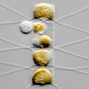
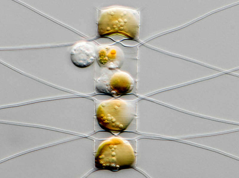
Sampling date 2/2022. Scale bars indicate 25 µm.Four images. One cell is being attacked by a parasite (Chytridiomycota).First couple:Synoptic representation of the valves. The second image is a zoom-in of the first.Second couple:Optical cross-section. The fourth image is a zoom-in of the third.Please click on < or > on the image edges or on the dots at the bottom edge of the images to browse through the slides!Place name: Baltic Sea, Kieler Förde, Kiel Fjord (Germany)Latitude: 54.3894126 Longitude: 10.1749055Microscope Zeiss Axioplan, camera Olympus OM-D E-M5 II. DOF image.© Wolfgang Bettighofer,images under Creative Commons License V 3.0 (CC BY-NC-SA).For permission to use of (high resolution) images please contact
postmaster@protisten.de.For further information about the image, please click here:
Link to protisten.de page