-
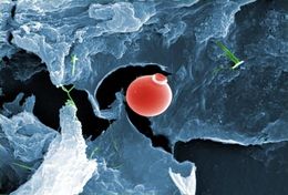
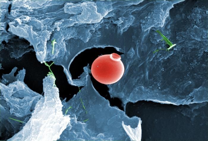
This digitally-colorized scanning electron micrograph (SEM) of an untreated water specimen extracted from a wild stream mainly used to control flooding during inclement weather, revealed the presence of unidentified organisms, which included bacteria, protozoa, and algae. Clearly visible in the center of this image, was a exquisitely-formed unidentified round vescicle-shaped microorganism, which may have been algal, or diatomic. Shaped like an ancient Grecian urn, the almost perfectly rounded smooth, flawless surface was made even more beautiful given its delicate structure.Created: 2009
-
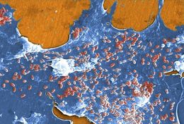
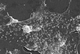
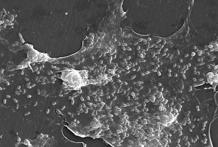
This digitally-colorized scanning electron micrograph (SEM) of an untreated water specimen extracted from a wild stream mainly used to control flooding during inclement weather, revealed the presence of unidentified organisms, which included bacteria, protozoa, and algae. In this particular view, a mass of gelatinous biofilm had enmeshed numbers of microorganisms, including amoebae and bacteria.Created: 2009
-
This digitally-colorized scanning electron micrograph (SEM) of an untreated water specimen extracted from a wild stream mainly used to control flooding during inclement weather, revealed the presence of unidentified organisms, which included bacteria, protozoa, and algae. In this particular view, a mass of gelatinous biofilm had enmeshed numbers of microorganisms, including amoebae and bacteria. Note at right, the stalk of what is suspected to be an algal specie.Created: 2009
-
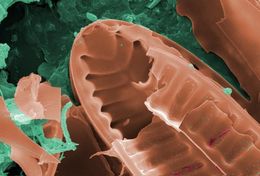
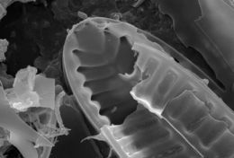
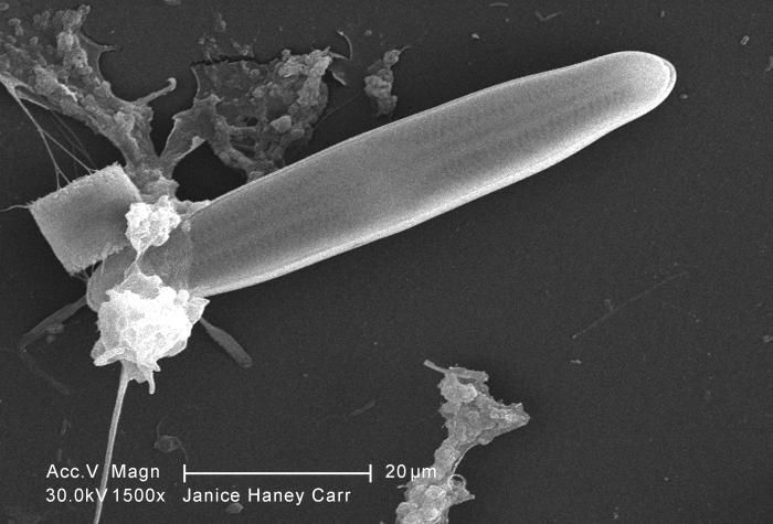
This digitally-colorized scanning electron micrograph (SEM) of an untreated water specimen extracted from a wild stream mainly used to control flooding during inclement weather, revealed the presence of unidentified organisms, which included bacteria, protozoa, and algae. In this particular image, a single unidentified diatomic microorganism was depicted revealing a wondrous symmetrical ultrastructural morphology.Created: 2009
-
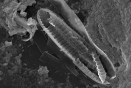
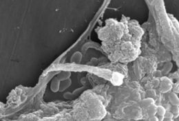
This digitally-colorized scanning electron micrograph (SEM) of an untreated water specimen extracted from a wild stream mainly used to control flooding during inclement weather, revealed the presence of unidentified organisms, which included bacteria, protozoa, and algae. In this particular view, it appears that a diatom had been fractured in the processing of this specimen. Surrounding this diatomic microorganism, was a large biofilm mass within, and around which were numerous protozoan amoeboid and bacterial microorganisms.Created: 2009
-
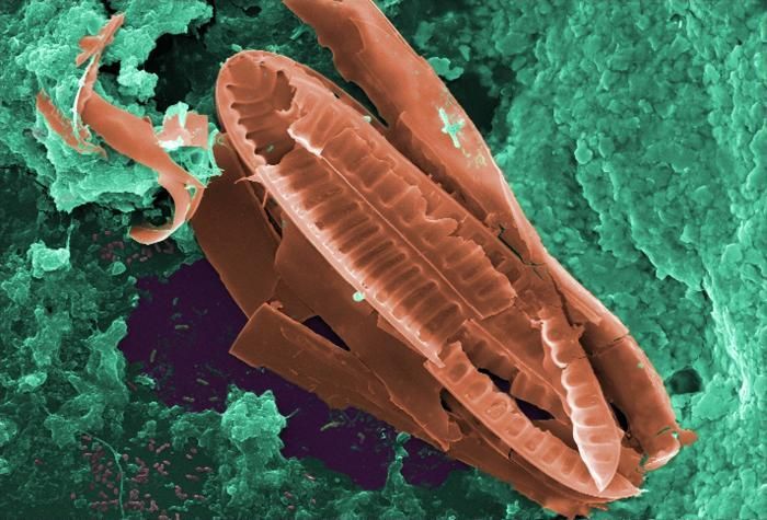
This digitally-colorized scanning electron micrograph (SEM) of an untreated water specimen extracted from a wild stream mainly used to control flooding during inclement weather, revealed the presence of unidentified organisms, which included bacteria, protozoa, and algae. In this particular view, it appears that a diatom had been fractured in the processing of this specimen. Surrounding this diatomic microorganism, was a large biofilm mass within, and around which were numerous protozoan amoeboid and bacterial microorganisms. See PHIL 11686 for a larger view of this diatom.Created: 2009
-
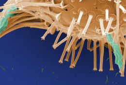
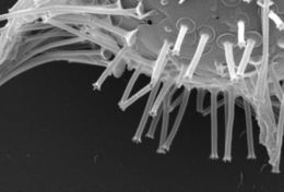
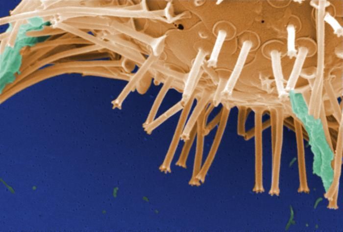
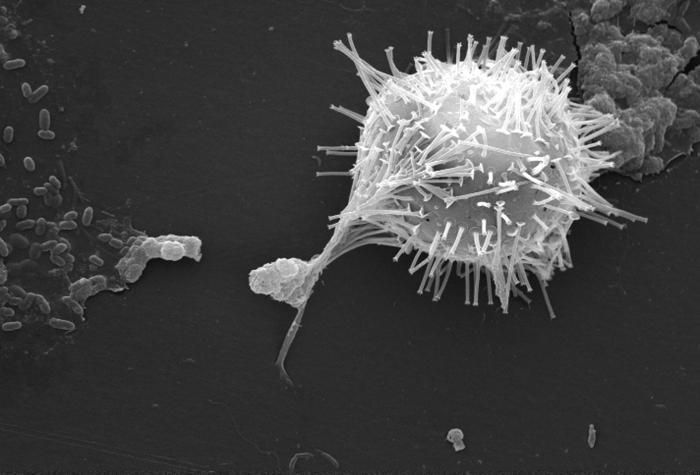
This digitally-colorized scanning electron micrograph (SEM) of an untreated water specimen extracted from a wild stream mainly used to control flooding during inclement weather, revealed the presence of unidentified organisms, which included bacteria, protozoa, and algae. In this particular view, a microorganism is featured, the exterior of which is covered by numerous projections imparting an appearance of a sea urchin. This microscopic pin cushion was teathered to its surroundings by a biofilm within which many bacteria, and amoeboid protozoa could be seen enmeshed as well. See PHIL 11682 and 11683, for additional views of this creature under successively greater magnifications.Created: 2009
-
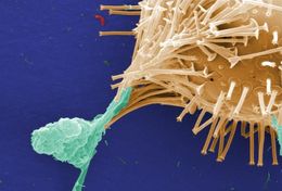
This digitally-colorized scanning electron micrograph (SEM) of an untreated water specimen extracted from a wild stream mainly used to control flooding during inclement weather, revealed the presence of unidentified organisms, which included bacteria, protozoa, and algae. In this particular view, a microorganism is featured, the exterior of which is covered by numerous projections imparting an appearance of a sea urchin. This microscopic pin cushion was teathered to its surroundings by a biofilm within which many bacteria, and amoeboid protozoa could be seen enmeshed as well. See PHIL 11682 and 11684, for additional views of this creature under successively greater magnifications.Created: 2009
-
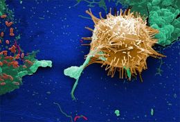
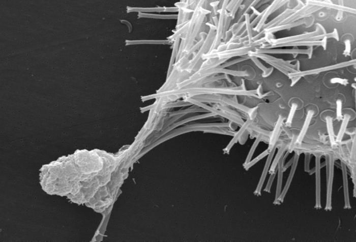
This digitally-colorized scanning electron micrograph (SEM) of an untreated water specimen extracted from a wild stream mainly used to control flooding during inclement weather, revealed the presence of unidentified organisms, which included bacteria, protozoa, and algae. In this particular view, a microorganism is featured, the exterior of which is covered by numerous projections imparting an appearance of a sea urchin. This microscopic pin cushion was teathered to its surroundings by a biofilm within which many bacteria, and amoeboid protozoa could be seen enmeshed as well. See PHIL 11683 and 11684, for subsequent views of this creature under successively greater magnifications.Created: 2009
-
This digitally-colorized scanning electron micrograph (SEM) of an untreated water specimen extracted from a wild stream mainly used to control flooding during inclement weather, revealed the presence of unidentified organisms, which included bacteria, protozoa, and algae.Created: 2009
-
This scanning electron micrograph (SEM) of an untreated water specimen extracted from a wild stream mainly used to control flooding during inclement weather, revealed the presence of unidentified organisms, which included bacteria, protozoa, and algae. In this particular view, it appears that a diatom had been fractured in the processing of this specimen. Surrounding this diatomic microorganism, was a large biofilm mass within, and around which were numerous protozoan amoeboid and bacterial microorganisms. For a colorized version of this image see PHIL 11703.Created: 2009
-
This scanning electron micrograph (SEM) of an untreated water specimen extracted from a wild stream mainly used to control flooding during inclement weather, revealed the presence of unidentified organisms, which included bacteria, protozoa, and algae. In this particular view, it appears that a diatom had been fractured in the processing of this specimen. Surrounding this diatomic microorganism, was a large biofilm mass within, and around which were numerous protozoan amoeboid and bacterial microorganisms. See PHIL 11686 for a larger view of this diatom. For a colorized version of this image see PHIL 11701.Created: 2009
-
This scanning electron micrograph (SEM) of an untreated water specimen extracted from a wild stream mainly used to control flooding during inclement weather, revealed the presence of unidentified organisms, which included bacteria, protozoa, and algae. In this particular view, a microorganism is featured, the exterior of which is covered by numerous projections imparting an appearance of a sea urchin. This microscopic pin cushion was teathered to its surroundings by a biofilm within which many bacteria, and amoeboid protozoa could be seen enmeshed as well. See PHIL 11682 and 11683, for additional views of this creature under successively greater magnifications. For a colorized version of this image see PHIL 11700.Created: 2009
-
This scanning electron micrograph (SEM) of an untreated water specimen extracted from a wild stream mainly used to control flooding during inclement weather, revealed the presence of unidentified organisms, which included bacteria, protozoa, and algae. In this particular view, a microorganism is featured, the exterior of which is covered by numerous projections imparting an appearance of a sea urchin. This microscopic pin cushion was teathered to its surroundings by a biofilm within which many bacteria, and amoeboid protozoa could be seen enmeshed as well. See PHIL 11682 and 11684, for additional views of this creature under successively greater magnifications. For a colorized version of this image see PHIL 11699.Created: 2009
-
This scanning electron micrograph (SEM) of an untreated water specimen extracted from a wild stream mainly used to control flooding during inclement weather, revealed the presence of unidentified organisms, which included bacteria, protozoa, and algae. In this particular view, a microorganism is featured, the exterior of which is covered by numerous projections imparting an appearance of a sea urchin. This microscopic pin cushion was teathered to its surroundings by a biofilm within which many bacteria, and amoeboid protozoa could be seen enmeshed as well. See PHIL 11683 and 11684, for subsequent views of this creature under successively greater magnifications. Also, see PHIL 11698 for a colorized version of this image.Created: 2009
-
This scanning electron micrograph (SEM) of an untreated water specimen extracted from a wild stream mainly used to control flooding during inclement weather, revealed the presence of unidentified organisms, which included bacteria, protozoa, and algae. In this particular view, a mass of gelatinous biofilm had enmeshed numbers of microorganisms, including amoebae and bacteria. Note at right, the stalk of what is suspected to be an algal specie. For a colorized view of this image see PHIL 11705.Created: 2009
-
This scanning electron micrograph (SEM) of an untreated water specimen extracted from a wild stream mainly used to control flooding during inclement weather, revealed the presence of unidentified organisms, which included bacteria, protozoa, and algae. In this particular view, a mass of gelatinous biofilm had enmeshed numbers of microorganisms, including amoebae and bacteria. See PHIL 11706 for a colorized version of this image.Created: 2009
-
This scanning electron micrograph (SEM) of an untreated water specimen extracted from a wild stream mainly used to control flooding during inclement weather, revealed the presence of unidentified organisms, which included bacteria, protozoa, and algae. In this particular view, a mass of gelatinous biofilm had enmeshed numbers of microorganisms, including amoebae and bacteria. For a colorized version of this image, see PHIL 11707.Created: 2009
-
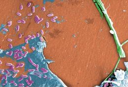
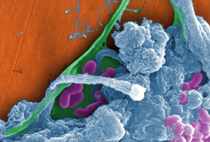
This scanning electron micrograph (SEM) of an untreated water specimen extracted from a wild stream mainly used to control flooding during inclement weather, revealed the presence of unidentified organisms, which included bacteria, protozoa, and algae. Here we see what appeared to be examples of diatomic, bacterial, and protozoan species, which were but a few of the inhabitants of this fresh water stream. Note also the amorphic gelatinous biofilm within which were embedded bacteria, and amoebic microorganisms. For a colorized view of this image, see PHIL 11710.Created: 2009
-
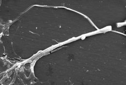
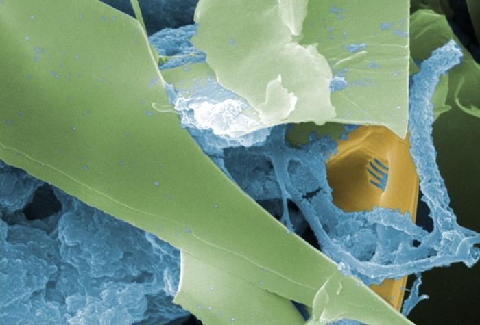
This scanning electron micrograph (SEM) of an untreated water specimen extracted from a wild stream mainly used to control flooding during inclement weather, revealed the presence of unidentified organisms, which included bacteria, protozoa, and algae. This view captured what may have been an algal specie, which had become enmeshed in a gelatinous biofilm at left.Created: 2009
-
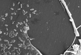
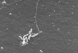
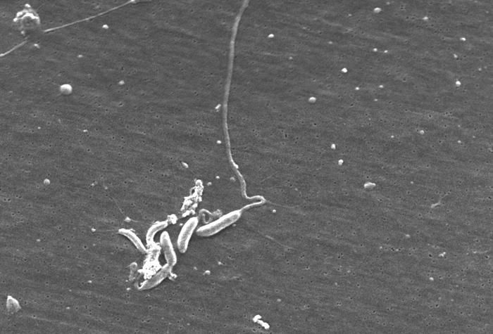
This scanning electron micrograph (SEM) of an untreated water specimen extracted from a wild stream mainly used to control flooding during inclement weather, revealed the presence of unidentified organisms, which included bacteria, protozoa, and algae. In this particular image, a number of flagellated microorganisms were grouped together with other steam water particulates.Created: 2009
-
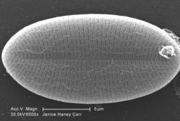
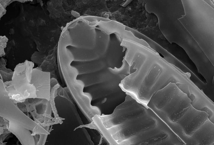
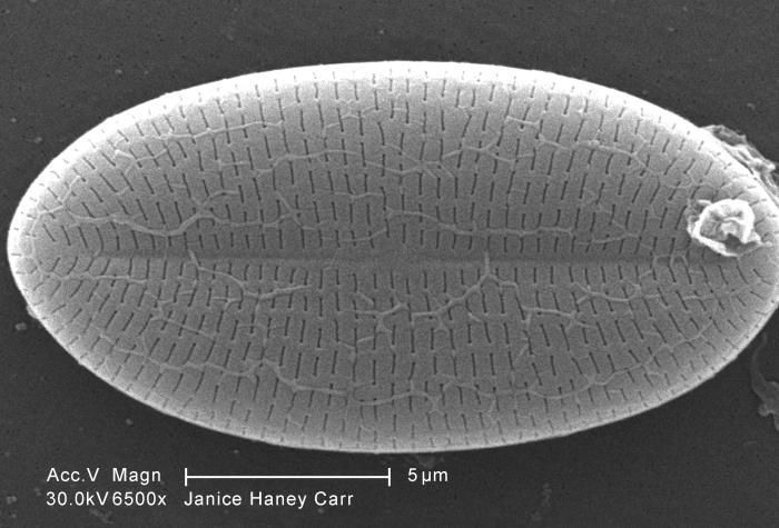
Magnified 6500X, this scanning electron micrograph (SEM) of an untreated water specimen extracted from a wild stream mainly used to control flooding during inclement weather, revealed the presence of unidentified organisms, which included bacteria, protozoa, and algae. In this particular image, a single unidentified diatomic microorganism was depicted revealing its wondrous symmetrical ultrastructural morphology. For a colorized version of this image see PHIL 11704.Created: 2009
-
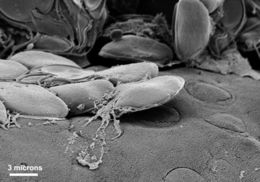
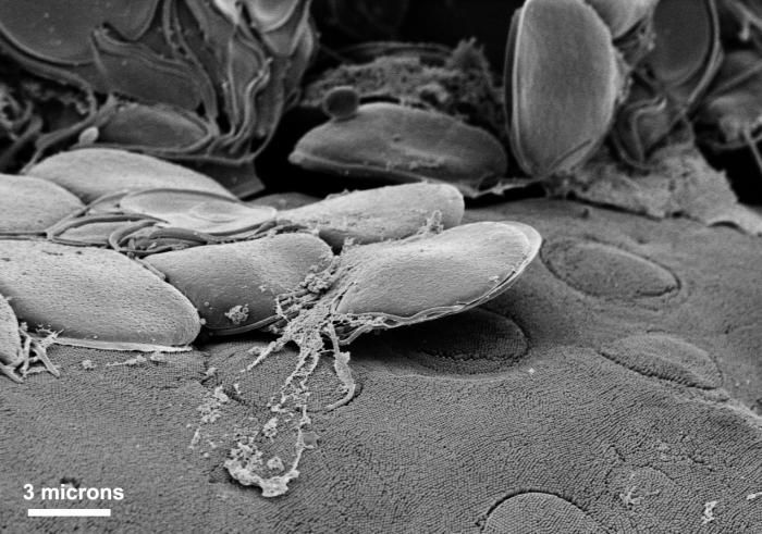
This scanning electron micrograph (SEM) depicted a group of Giardia trophozoites that were clustered on the intestinal mucosal surface. Immediately adjacent to these organisms were a number of the characteristic circulars lesions that can be left on surface as a result of the tight adhesion of the organisms ventral adhesive disk.Created: 1999