Description of Sagittaria hyalina
(
anglais
)
fourni par BioPedia
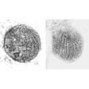
Cylindroid, swimming specimens pyriform, tapered anteriorly.Contractile vacuole in posterior end. Oral aperture ellipsoid. No elongated caudal cilium. 15-16 somatic kineties, C-shaped right paraoral membrane, 3-4 adoral organelles on left of cytostome. Lives in tubular mucus case which is readily deserted. Cell 30-40 x 12-16 microns, when contracted about 15-30 x 15-20 microns. Cylindrical, both ends broadly rounded, transverse section almost circular. While the cell is building the lorica it is obovoid and easily confused with contracted swarmers of peritrichs. Nuclear apparatus in cell centre. Macronucleus about 8 microns in diameter; nucleolus reticulate, sometimes only 1 large, central nucleolus or a few smaller (about 3 microns) nucleoli. Micronucleus 1.5-2 microns in diameter, presumably in perinuclear space of macronucleus. Cortex colourless, very flexible, usually distinctly furrowed by somatic kineties. Cytoplasm hyaline, contains several 2-4 microns sized, yellowish inclusions. Food vacuoles 5-6 microns in diameter, not very compact, contain bacteria and flat, reddish crystals. Rests quietly in case, front third protruding; disturbed cells move hastily back and forth in tube. Individuals having deserted their cases swim slowly to rapidly by rotation about longitudinal axis. Case tubular, brownish, consists of soft, flexible, mucous substance, 25-40 microns wide and up to 500 microns long, anterior end widened goblet-like; never ramified, straight to worm-like bent, attached to various substrates (water surface, microscope slide, soil particles etc.) by button-like posterior end. Cases under construction are funnel-shaped to cylindrical. Interior wall smooth, exterior rough due to adhering detritus, bacteria and zooflagellates. Case usually vacated when a cell is transferred from sample to slide! Specimens held under coverslip for longer periods (about 20 min) begin to build new cases by apparently secreting approximately 1 x 0.31 microns sized, cylindroid mucocysts on which defecated food residue adheres. Feeding commences already during case building. Some kineties slightly shortened anteriorly or posteriorly. Oral aperture slightly subpolar, obovoid with narrower end directed ventrally, somewhat depressed giving apex notched appearance. Paroral membrane semicircular, consists of ciliated dikinetids, on its left a row of argyrophilic granules marking nematodesmata originating from dikinetids. These, as well as the nematodesmata of the adoral organelles, are almost half body-length and form a sacculate structure when food vacuole is pinched off. On left slope of oral aperture 3-4 adoral organelles consisting of 2 rows of 3 basal bodies each. Resting cysts 15-17 microns in diameter, in basal bulge of case, cyst wall thin and smooth.