-
Joenia (joe-een-ee-a) is a lophomonad hypermastigid flagellate - this group being distinguished by having a pad of very long flagella inserting to one side of the anterior apex of the cell. The lophomonads are a subset of the trichomonad flagellates, and the trichomonad beat profile is evident in this image. There is a very substantial axostyle,which anteriorly expands to envelope the nucleus and the flagellar area. Posteriorly with many food vacuoles containing small particles of ingested wood. From the termite Kalotermes. Phase contrast.
-
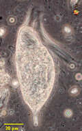
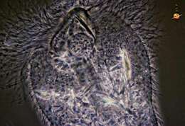
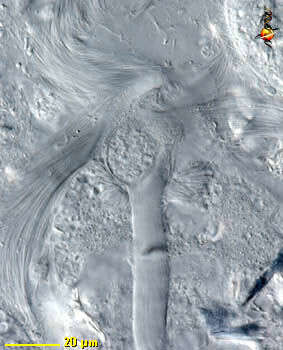
Joenia (joe-een-ee-a) is a lophomonad hypermastigid flagellate - this group being distinguished by having a pad of very long flagella inserting to one side of the anterior apex of the cell. Joenia has a very substantial axostyle, and this detailed image shows how it expands to envelope the nucleus and the flagellar area. Just below the nucleus, the mustachios of dictyosomes extend out to either side. The flagella insert into a curving pad to the upper left side of the cell, but they sweep around, below and to the side. From the termite Kalotermes. Differential interference contrast.
-
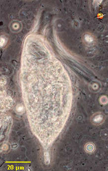
Joenia (joe-een-ee-a) is a lophomonad hypermastigid flagellate - this group being distinguished by having a pad of very long flagella inserting to one side of the anterior apex of the cell. The lophomonads are a subset of the trichomonad flagellates, and the trichomonad beat profile is evident in this image. There is a very substantial axostyle, forming a calyx or sheath that envelopes the nucleus and the flagellar area, and projecting from the back of the cell. From the termite Kalotermes. Phase contrast.
-
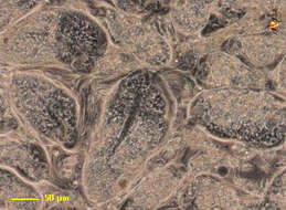
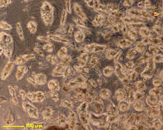
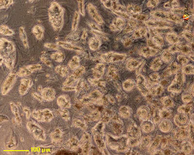
Joenia (joe-een-ee-a) is a lophomonad hypermastigid flagellate - this group being distinguished by having a pad of very long flagella inserting to one side of the anterior apex of the cell. This is an image of a small drop of fluid from the gut of the termite Kalotermes, and showing the very high concentrations of cells which may occur there. Phase contrast.
-
Joenia (joe-een-ee-a) is a lophomonad hypermastigid flagellate - this group being distinguished by having a pad of very long flagella inserting to one side of the anterior apex of the cell. This is an image of a small drop of fluid from the termite gut, and showing the very high concentrations of cells which may occur there. From the termite Kalotermes. Phase contrast.
-
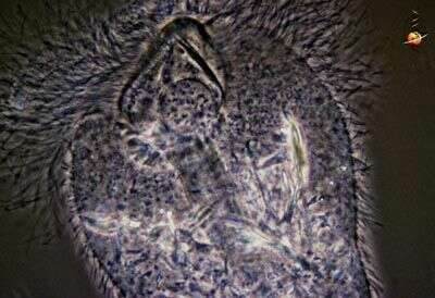
Joenia (joe-een-ee-a) is a lophomonad hypermastigid flagellate - this group being distinguished by having a pad of very long flagella inserting to one side of the anterior apex of the cell. The lophomonads are a subset of the trichomonad flagellates, and the trichomonad beat profile is evident in this image. There is a very substantial axostyle,which anteriorly expands to envelope the nucleus and the flagellar area. Posteriorly with many food vacuoles containing small particles of ingested wood. This image is of a fixed cell, and shows the anterior flagellum,. the axostyle and the region of the nucleus. Phase contrast.
-
-
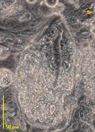
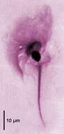
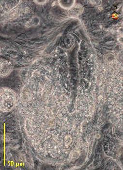
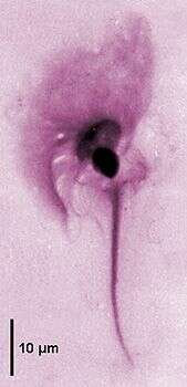
Joenia is a parabasalid cristamonad flagellate (50-500 µm) with a conical rostrum partly covered with the flagellar area. Axostyle forming a calyx enveloping the nucleus and the flagellar area; axostyle trunk stout protruding posteriorly. Parabasal body composed of several branches bearing Golgi dictyosomes around the nucleus. Joenia annectens from Kalotermes flavicollis: anterior flagellar area, nucleus and axostyle tapering to the posterior end (Giemsa staining).
-
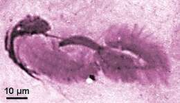
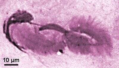
Joenia is a parabasalid cristamonad flagellate (50-500 µm) with a conical rostrum partly covered with the flagellar area. Axostyle forming a calyx enveloping the nucleus and the flagellar area; axostyle trunk stout protruding posteriorly. Parabasal body composed of several branches bearing Golgi dictyosomes around the nucleus. Joenia annectens dividing cell showing the stout paradesmose of the division spindle between the two flagellar areas (Giemsa staining)
-
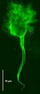
Joenia is a parabasalid cristamonad flagellate (50-500 µm) with a conical rostrum partly covered with the flagellar area. Axostyle forming a calyx enveloping the nucleus and the flagellar area; axostyle trunk stout protruding posteriorly. Parabasal body composed of several branches bearing Golgi dictyosomes around the nucleus. Joenia annectens from Kalotermes flavicollis: anterior flagellar area, nucleus and axostyle tapering to the posterior end (immunofluorescence).
-
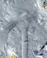
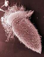
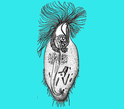
Scanning EM showing the anterior tuft of flagella and the cell body covered with rod-shaped bacteria.