-
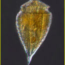
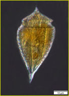
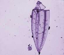
Oxytoxum nanum
-
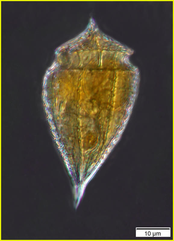
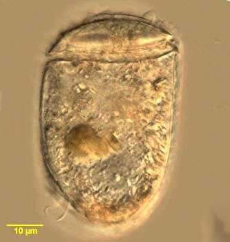
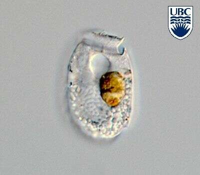
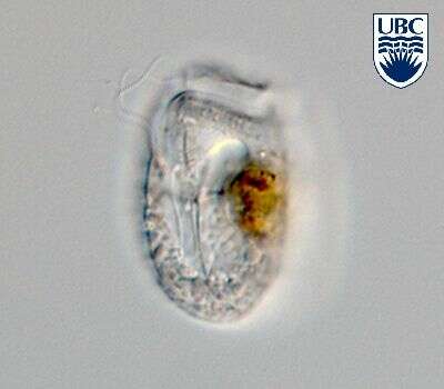
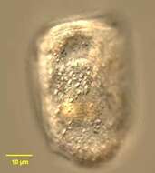
Planodinium (plane-o-din-ee-um) striatum Saunders & Dodge 1984. The image shows a cell in left lateral view. The cell contains no plastids, however a reddish food particle is present. the cell is thecate. The cell has no processes.
-
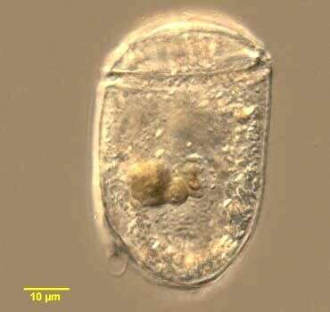
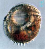
Planodinium striatum Saunders et Dodge 1984
-
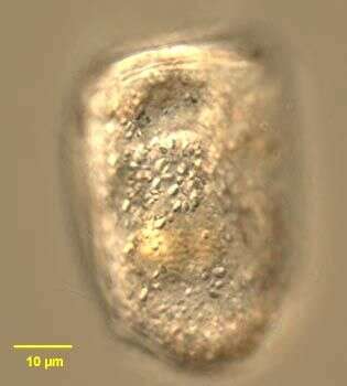
Planodinium striatum Saunders et Dodge 1984
-
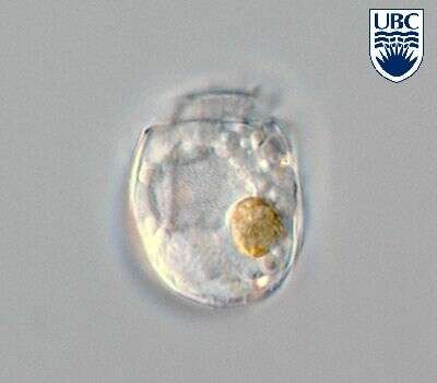
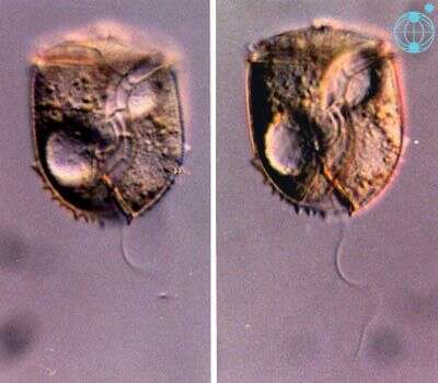
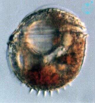
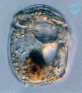
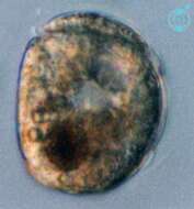
Amphidiniopsis (am-fee-din-ee-op-sis) hirsutum (Balech) Dodge 1982. The image on the left shows the ventral view oft a cell, with antapical spines visible. The cingulum is near the anterior end of the cell and it is slightly ascending. The image on the right shows a mid-focus plane through a cell, with two large pusules, one near the anterior and the second near the posterior end. The nucleus lies in the lower cell half, on the left cell side (= right image side).
-
Thecadinium, observed in marine muds and sandy sediments in the vicinity of Broome, Western Australia in September 2003. This image was taken using differential interference contrast optics. This work was supported by the Australian Biological Resources Study.
-
Thecadinium, showing apical lists, observed in marine muds and sandy sediments in the vicinity of Broome, Western Australia in September 2003. This image was taken using differential interference contrast optics. This work was supported by the Australian Biological Resources Study.
-
Thecadinium, observed in marine muds and sandy sediments in the vicinity of Broome, Western Australia in September 2003. This image was taken using differential interference contrast optics. This work was supported by the Australian Biological Resources Study.
-
Thecadinium, showing cingular spine, observed in marine muds and sandy sediments in the vicinity of Broome, Western Australia in September 2003. This image was taken using differential interference contrast optics. This work was supported by the Australian Biological Resources Study.
-
Thecadinium, observed in marine muds and sandy sediments in the vicinity of Broome, Western Australia in September 2003. This image was taken using differential interference contrast optics. This work was supported by the Australian Biological Resources Study.
-
Thecadinium (theek-a-din-ee-um) acanthium Hoppenrath 2000. The image shows a cell in right lateral view. No plastids are present, however a food particle is visible near the posterior of the cell. The cell is laterally compressed. A theca is present, from which several posterior spines protrude. There is a very small apical list present.
-
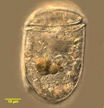
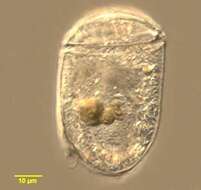
Thecadinium (theek-a-din-ee-um) neopetasatum Saunders & Dodge 1984. The image shows a cell in right lateral view. The cell is laterally compressed. The cell contains no plastids. The cell is thecate, but has no processes.
-
Thecadinium (theek-a-din-ee-um) ornatum Hoppenrath 2000. The image shows a cell in left lateral view. The cell is laterally compressed. The cell contains no plastids, but several food particles are present. The cell is thecate, and several spines are present posteriorly. There is a very small apical list.
-
This image was made from samples taken during a scientific cruise in the Pacific. Water was filtered to concentrate the organisms that were present, then dried onto a thin sheet of plastic and then shadowed with a fine layer of metal to provide contrast. The preparation was then observed with an electron-microscope. This technique has been used to document the diversity of marine microbes, especially, protists in the oceans.
-
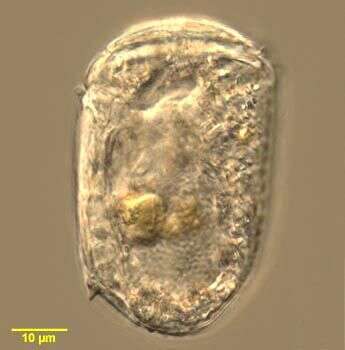
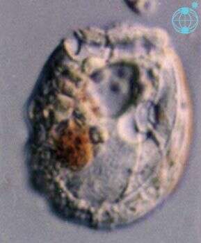
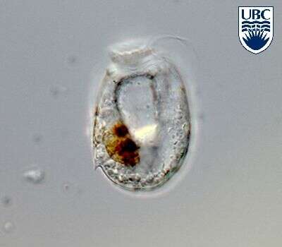
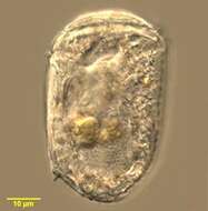
Sabulodinium (sab-you-low-din-ee-um) undulatum Saunders & Dodge 1984. The image shows a cell in left lateral view. The cell is laterally compressed. The cingulum is near the anterior end of the cell. There are no plastids visible, however a reddish food particle is visible. The cell is thecate. The nucleus is in the posterior of the cell.
-
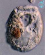
Sabulodinium undulatum Saunders et Dodge 1984
-
Sabulodinium undulatum Saunders et Dodge 1984
-
Sabulodinium undulatum Saunders et Dodge 1984