-
Diana M. P. Galassi, Paola De Laurentiis, Barbara Fiasca
Zookeys
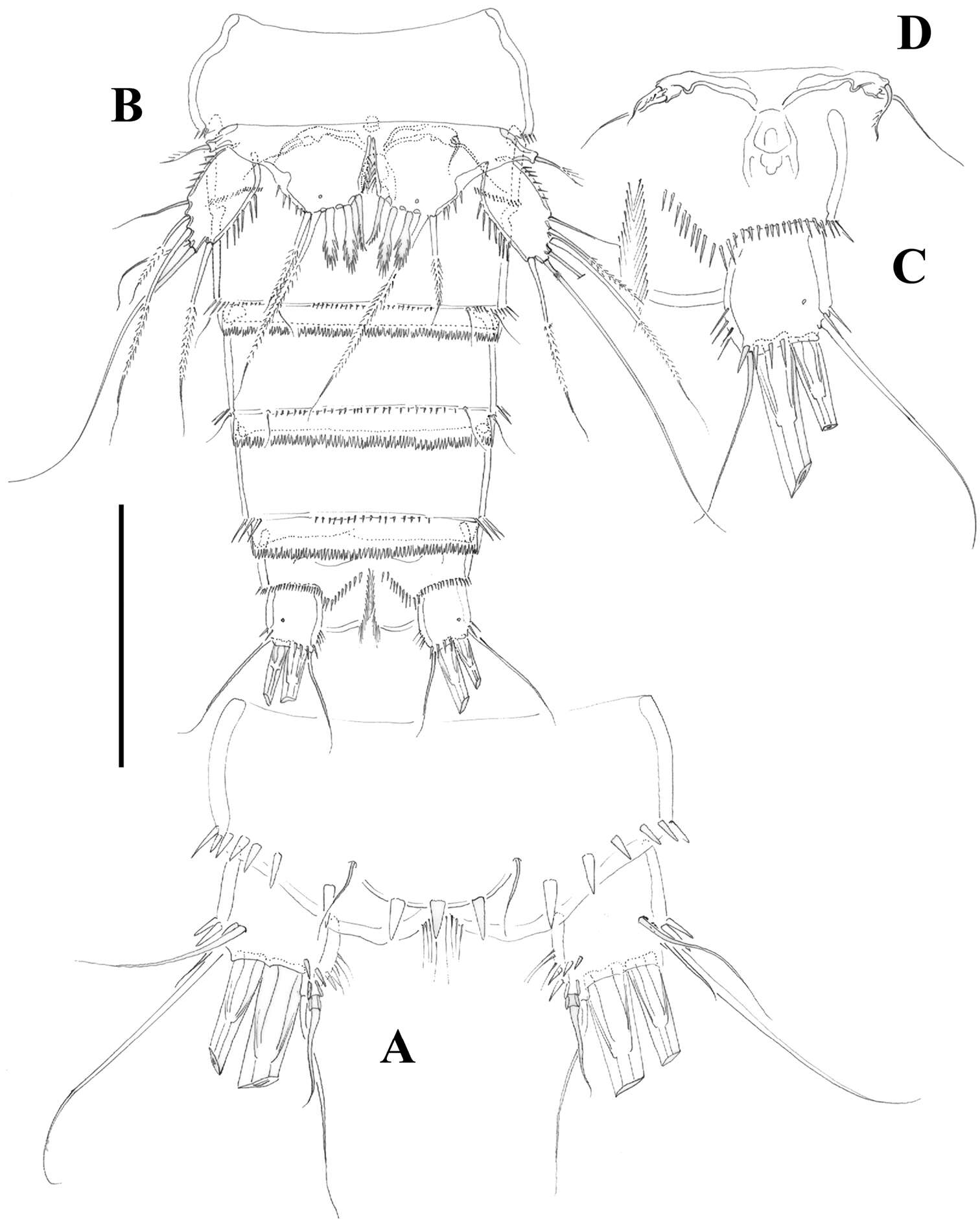
Figure 10.Phyllognathopus inexspectatus sp. n. (♀). A habitus, dorsal view B abdomen, ventral view C caudal ramus, ventral view (scale bars in μm).
-
Samuel Gómez, Nicola K. Carrasco, Francisco Neptalí Morales-Serna
Zookeys
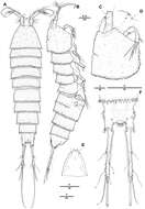
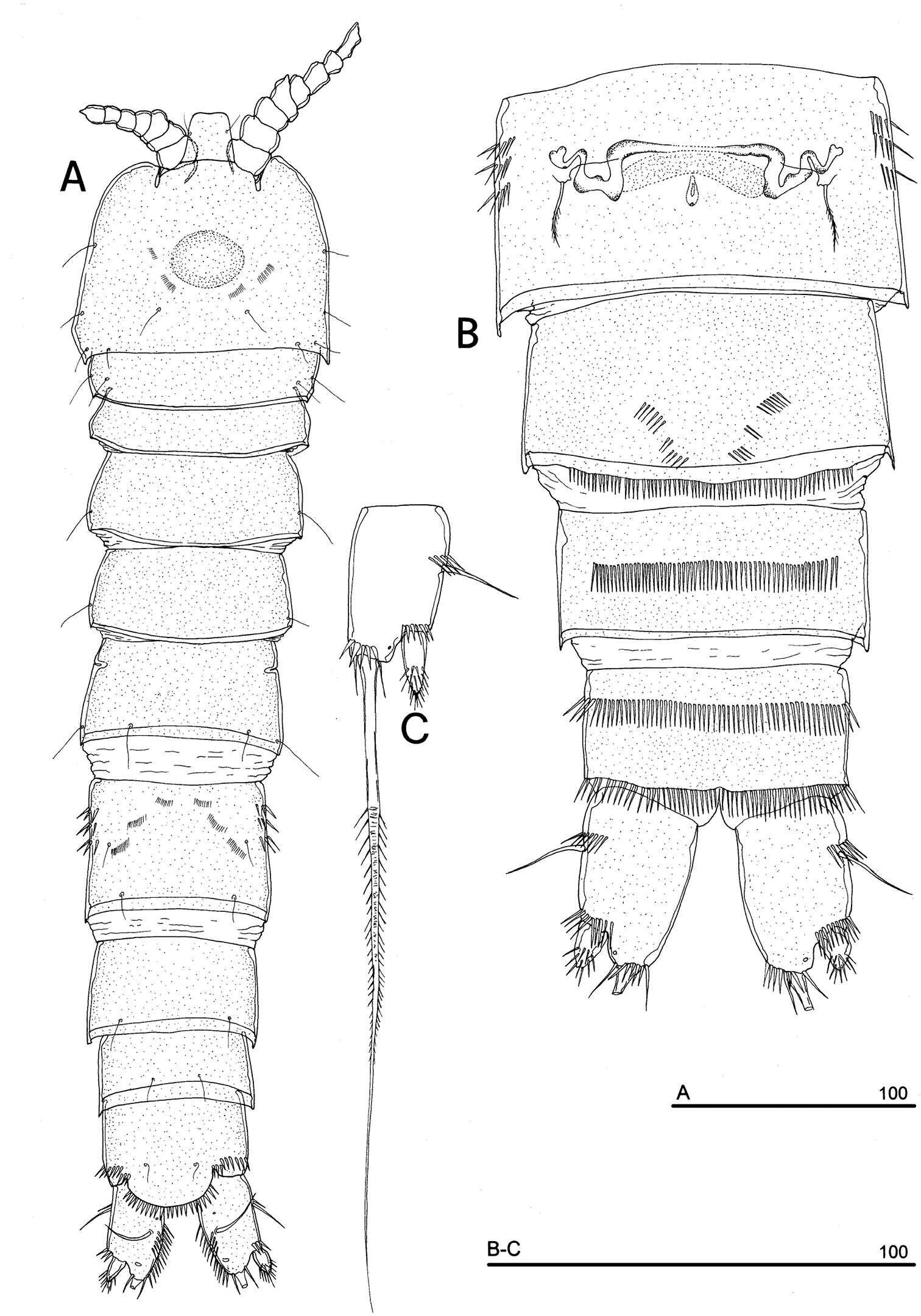
Figure 2.Nitocra taylori sp. n. Female. A habitus B rostrum, dorsal C urosome, dorsal. Scale bar: A=300 µm; B=75 µm; C=150 µm.
-
Eduardo Suárez-Morales, Jani Jarquín-González
Zookeys
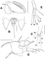
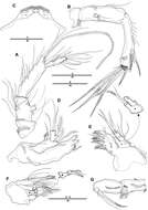
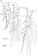
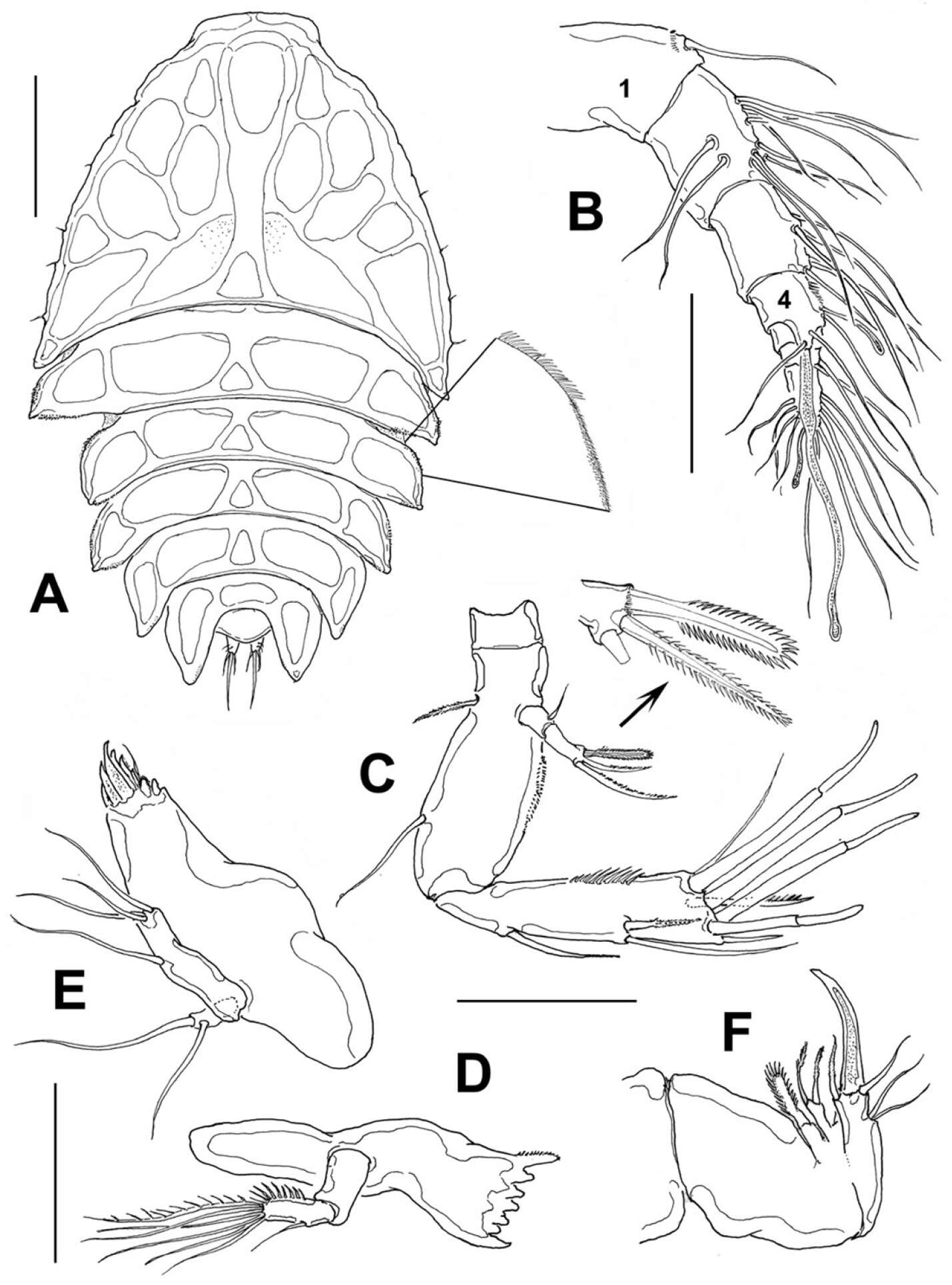
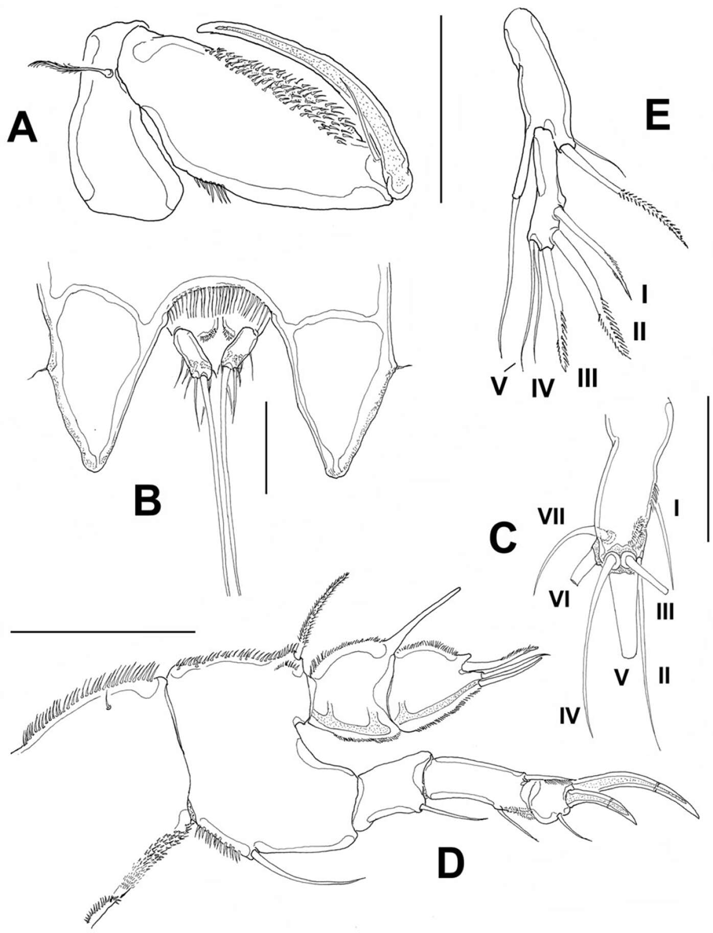
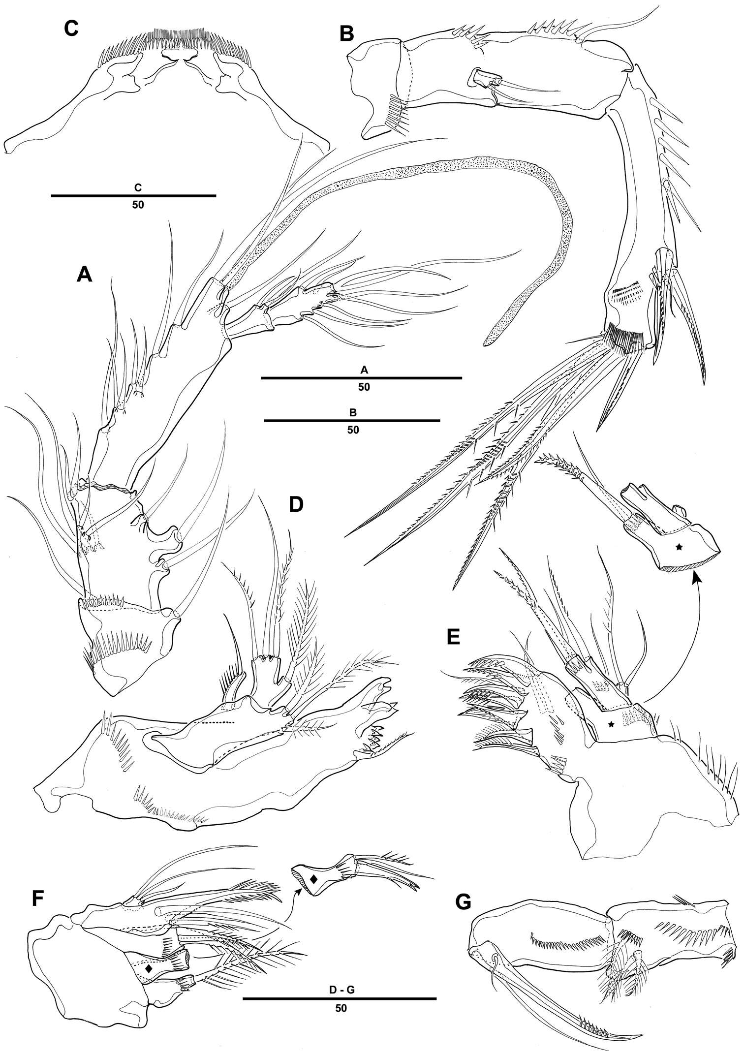
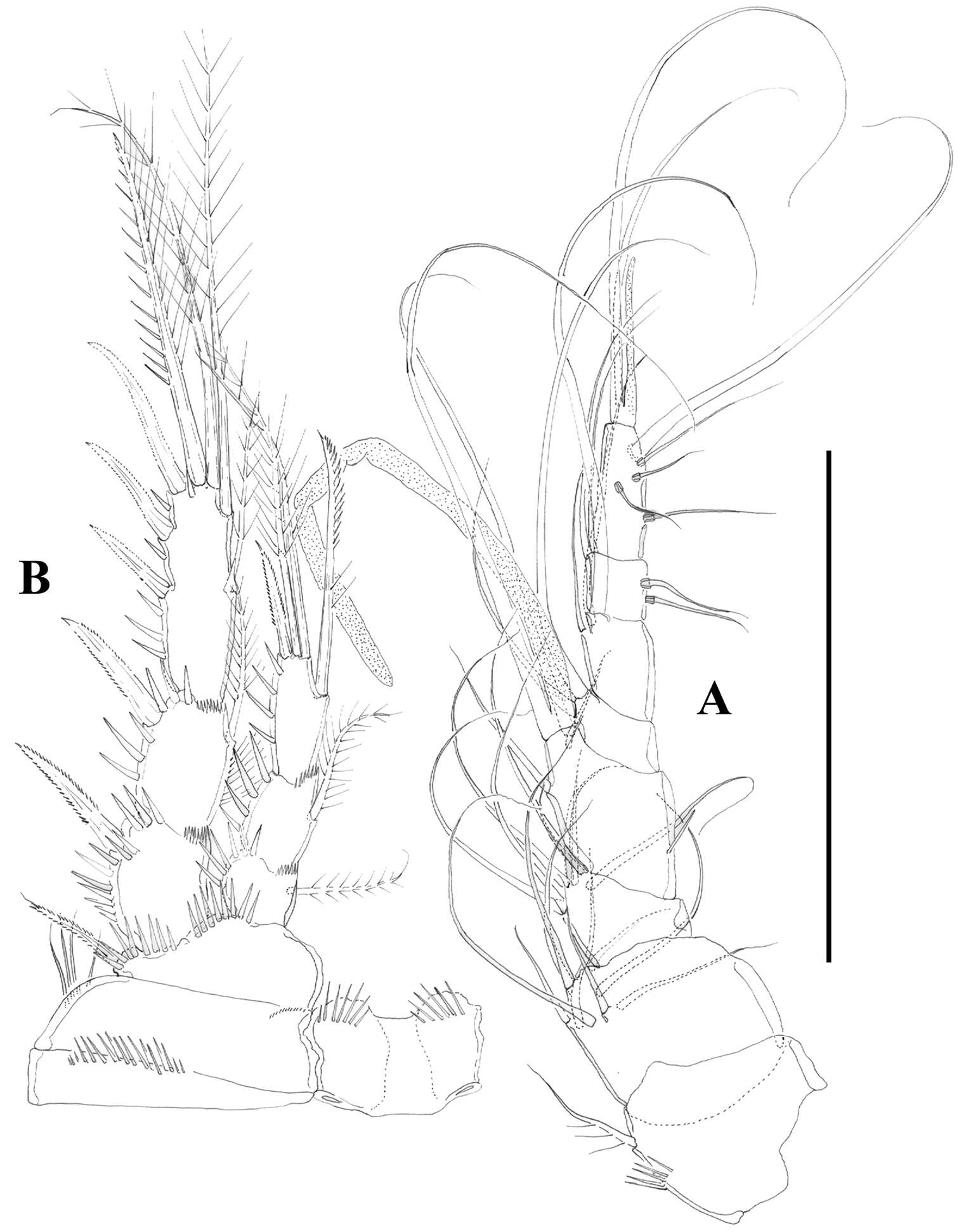
Figure 2.Peltidium nayarit sp. n., from Playa Careyeros, Nayarit, Mexican Pacific. A adult female, habitus, dorsal view, showing detail of ornamentation of epimeral processes of cephalothorax B antennule C antenna D mandible E maxillule F maxilla. Scales bars: A= 250 μm, B–F=100 μm.
-
Juan M. Fuentes-Reinés, Eduardo Suárez-Morales
Zookeys
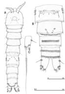
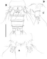
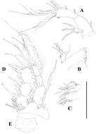
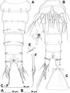
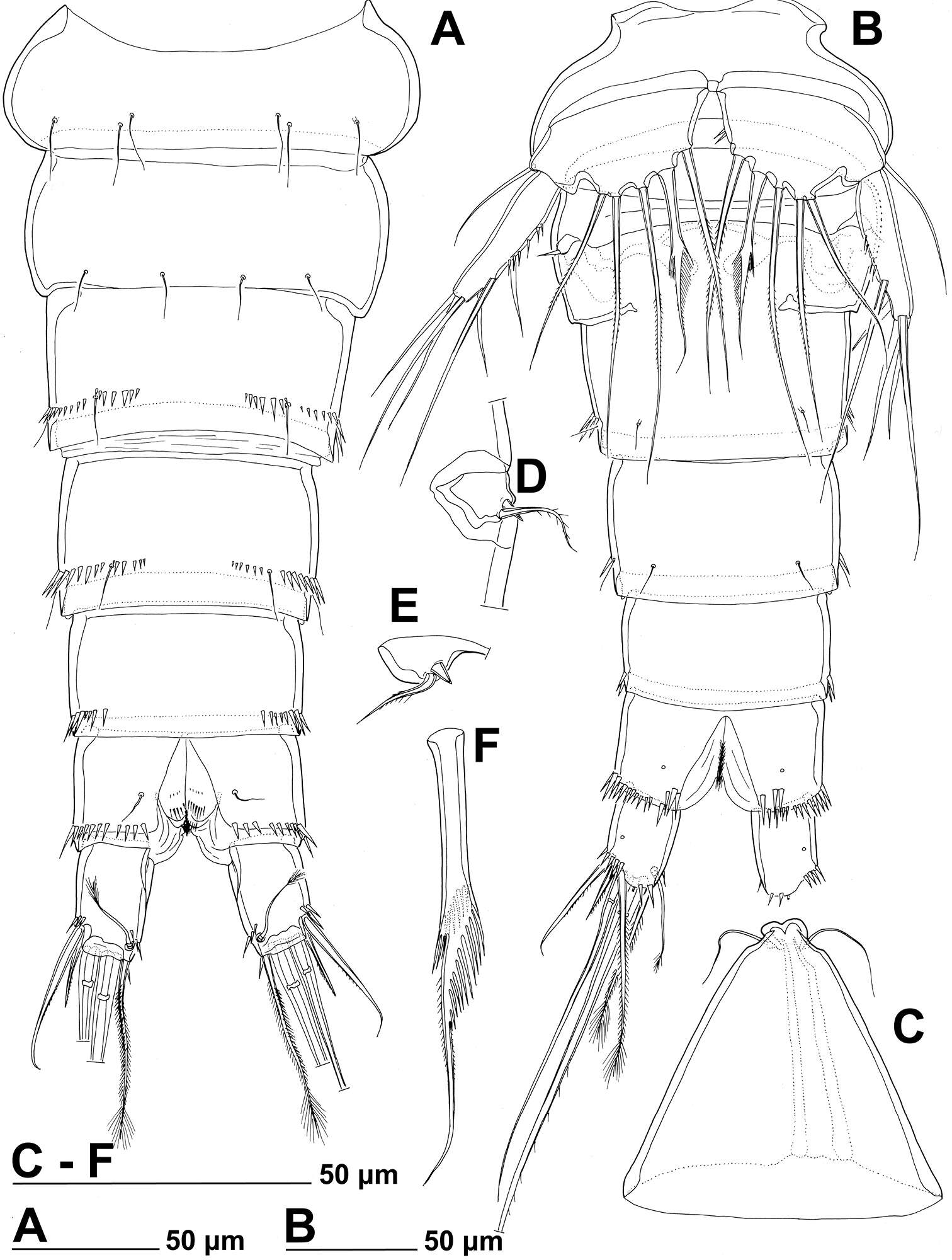
Figure 1.Nitokra affinis colombiensis ssp. n., from northern Colombia. A adult female, habitus, lateral view B adult male, habitus, lateral view C female, anal somite showing ornamentation of anal operculum D male, urosome, ventral view E male, third urosomite, ventral view F male, caudal rami, ventral view. Scale bars: A, B = 100 μm, C, E, F = 10 μm, D = 50 μm.
-
Hyun Woo Bang, Jeffrey G. Baguley, Heejin Moon
Zookeys
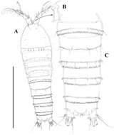
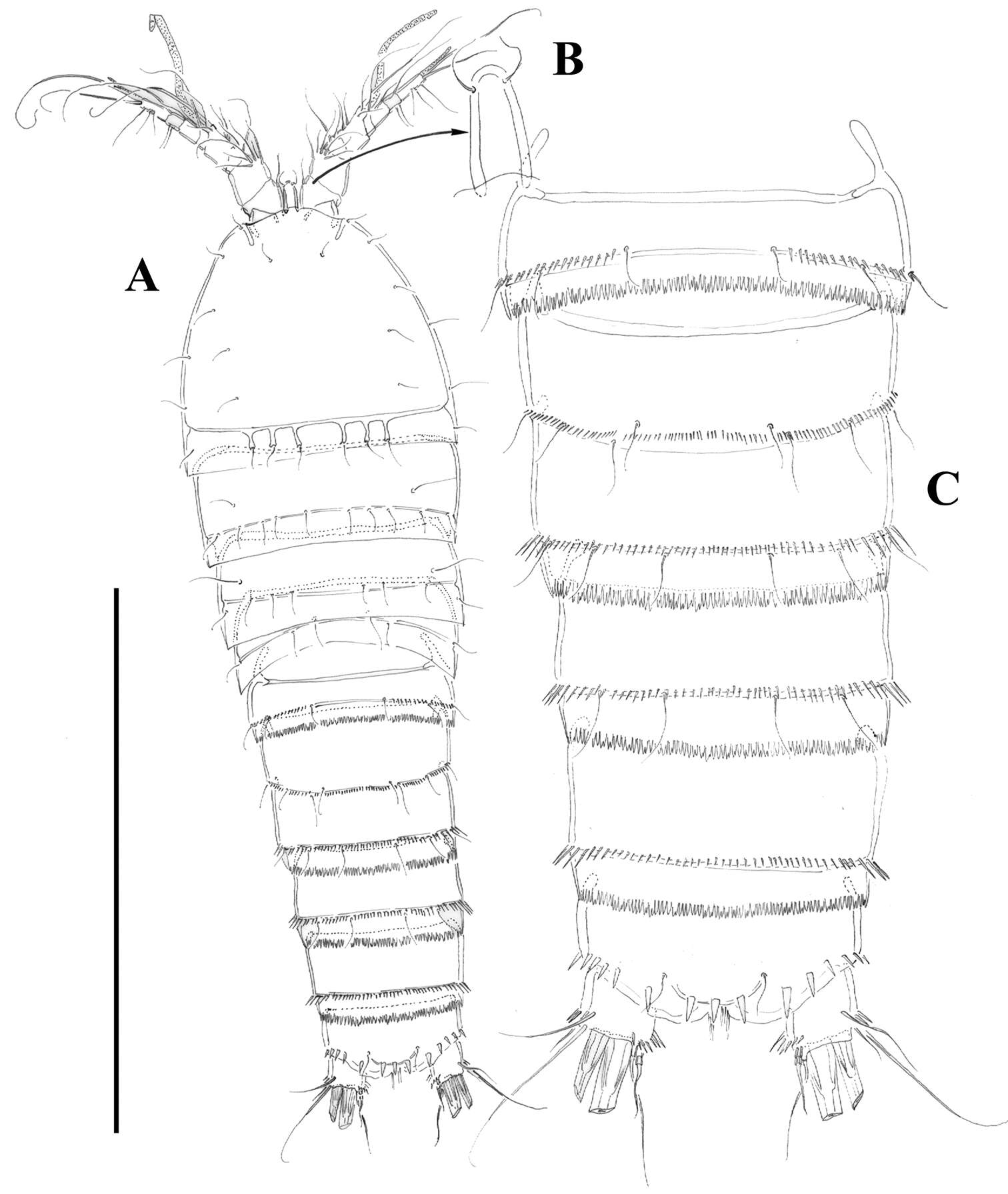
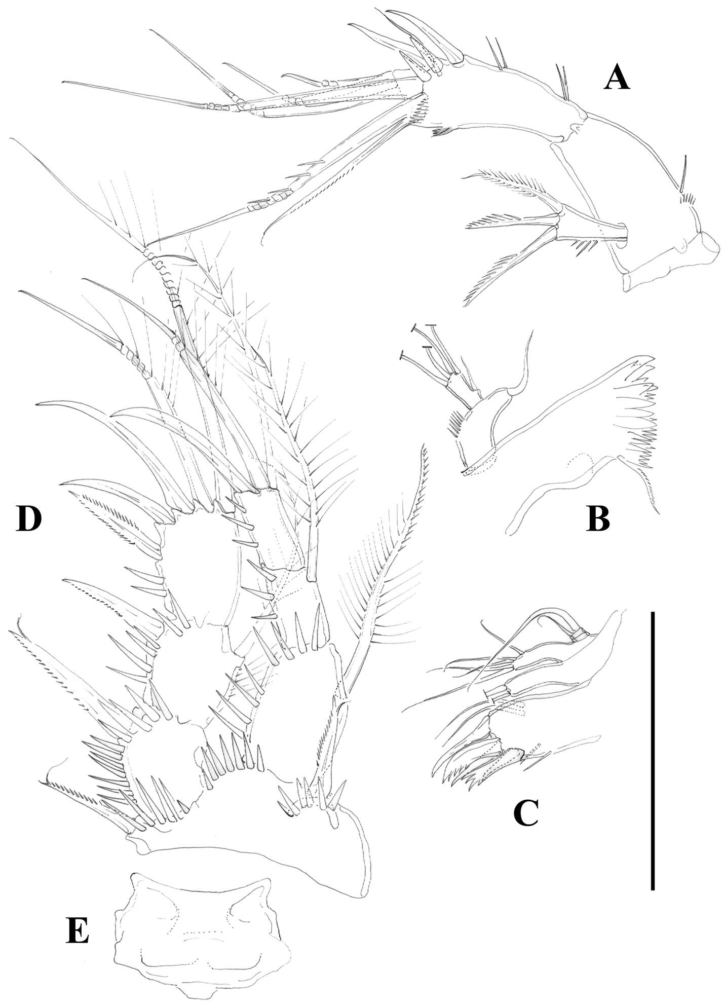
Figure 1.Pentacletopsyllus montagni gen. et sp. n. female: A habitus, dorsal B habitus, lateral C cephalothorax, lateral D tooth-like process of cephalothorax lateral anterior margin E rostrum, dorsal F caudal ramus, dorsal.
-
Tomislav Karanovic, Kichoon Kim, Wonchoel Lee
Zookeys
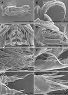
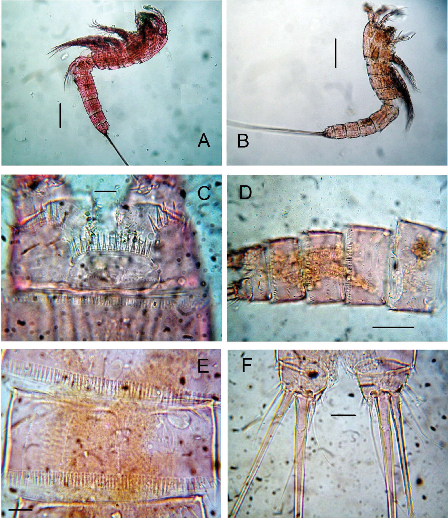
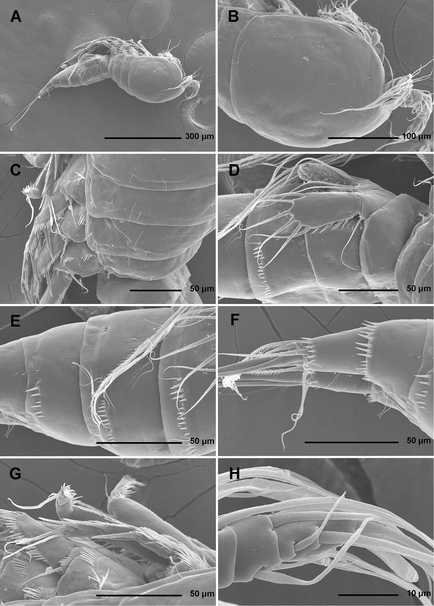
Figure 1.Stenhelia pubescens Chislenko, 1978, scanning electron micrographs, female 1: A habitus, lateral B cephalothorax, lateral C free thoracic somites, lateral D fifth pedigerous somite and genital double-somite, lateral, with one spermatophore attached on ventral side E fourth and fifth urosomites, lateral F anal somite and caudal rami, lateral G first legs and proximal part of second and third legs, lateral H distal part of right antennula, dorsal.
-
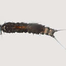
Figure 2.Mesocletodes elmari sp. n., adult female, paratype 2. CLSM photograph of a Congo-red stained specimen, lateral view. Scale bar: 100 µm
-
Diana M. P. Galassi, Paola De Laurentiis, Barbara Fiasca
Zookeys
Figure 11.Phyllognathopus inexspectatus sp. n. (♀) A urosome, dorsal view B antennule C antenna (scale bars in μm).
-
Samuel Gómez, Nicola K. Carrasco, Francisco Neptalí Morales-Serna
Zookeys
Figure 3.Nitocra taylori sp. n. Female. A anal somite and caudal rami, dorsal B urosome, ventral, showing P5 C left caudal ramus, ventral D P6 and genital complex. Scale bar: A=44 µm; B=100 µm; C=50 µm; D=71 µm.
-
Eduardo Suárez-Morales, Jani Jarquín-González
Zookeys
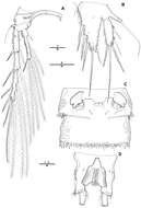
Figure 3.Peltidium nayarit sp. n., adult female from Playa Careyeros, Nayarit, Mexican Pacific. A maxilliped B urosome showing anal somite and caudal rami, ventral view C right caudal ramus, ventral view showing setation following nomenclature by Huys et al. (1996) D leg 1 E leg 5 showing setal nomenclature of exopodal setae following Wells (2007). Scales bars: A, D, E=100 μm, B= 50 μm, C=20 μm.
-
Juan M. Fuentes-Reinés, Eduardo Suárez-Morales
Zookeys
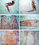
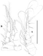
Figure 3.Nitokra affinis colombiensis ssp. n., adult female from northern Colombia. A antennule B antenna C mandible blade D mandibular palp E maxillule F maxilla G maxilliped H rostrum with rostral process. Scale bars: A–G = 50 μm, H = 10 μm.
-
Hyun Woo Bang, Jeffrey G. Baguley, Heejin Moon
Zookeys
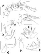
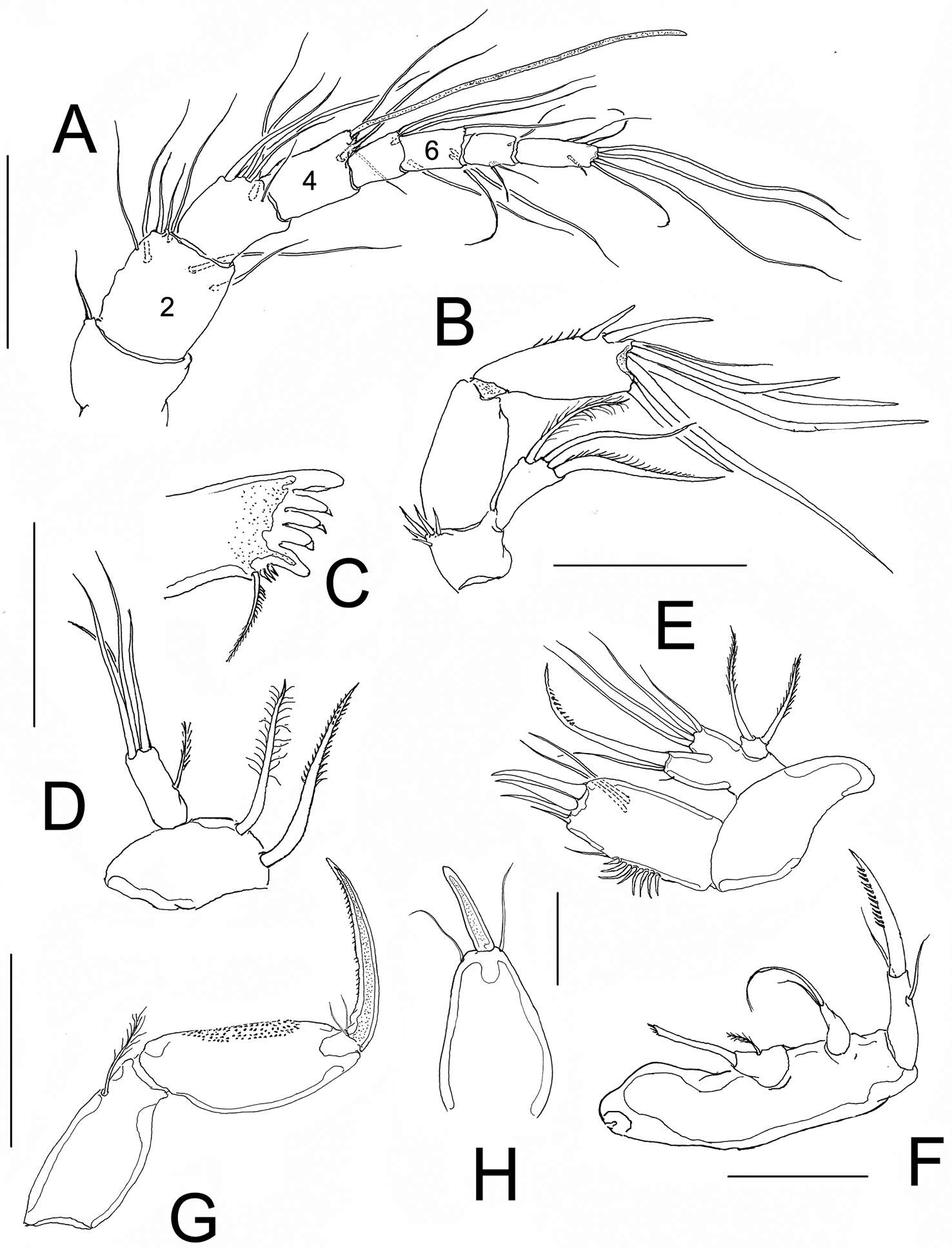
Figure 2.Pentacletopsyllus montagni gen. et sp. n. female: A antennule, dorsal B antenna, dorsal C labrum, posterior D mandible E maxillule (inset showing armature on coxa) F maxilla (inset showing armature on middle endite) G maxilliped.
-
Tomislav Karanovic, Kichoon Kim, Wonchoel Lee
Zookeys
Figure 2.Stenhelia pubescens Chislenko, 1978, scanning electron micrographs, ovigerous female 2: A habitus, ventral B rostrum and left antennula, ventral C mouth appendages, ventral D first leg, anterior E second, third, and fourth legs, anterior F exopod of fifth leg and sixth leg, ventral G anal somite and caudal rami, ventral H posterior part of left caudal ramus, ventral.
-
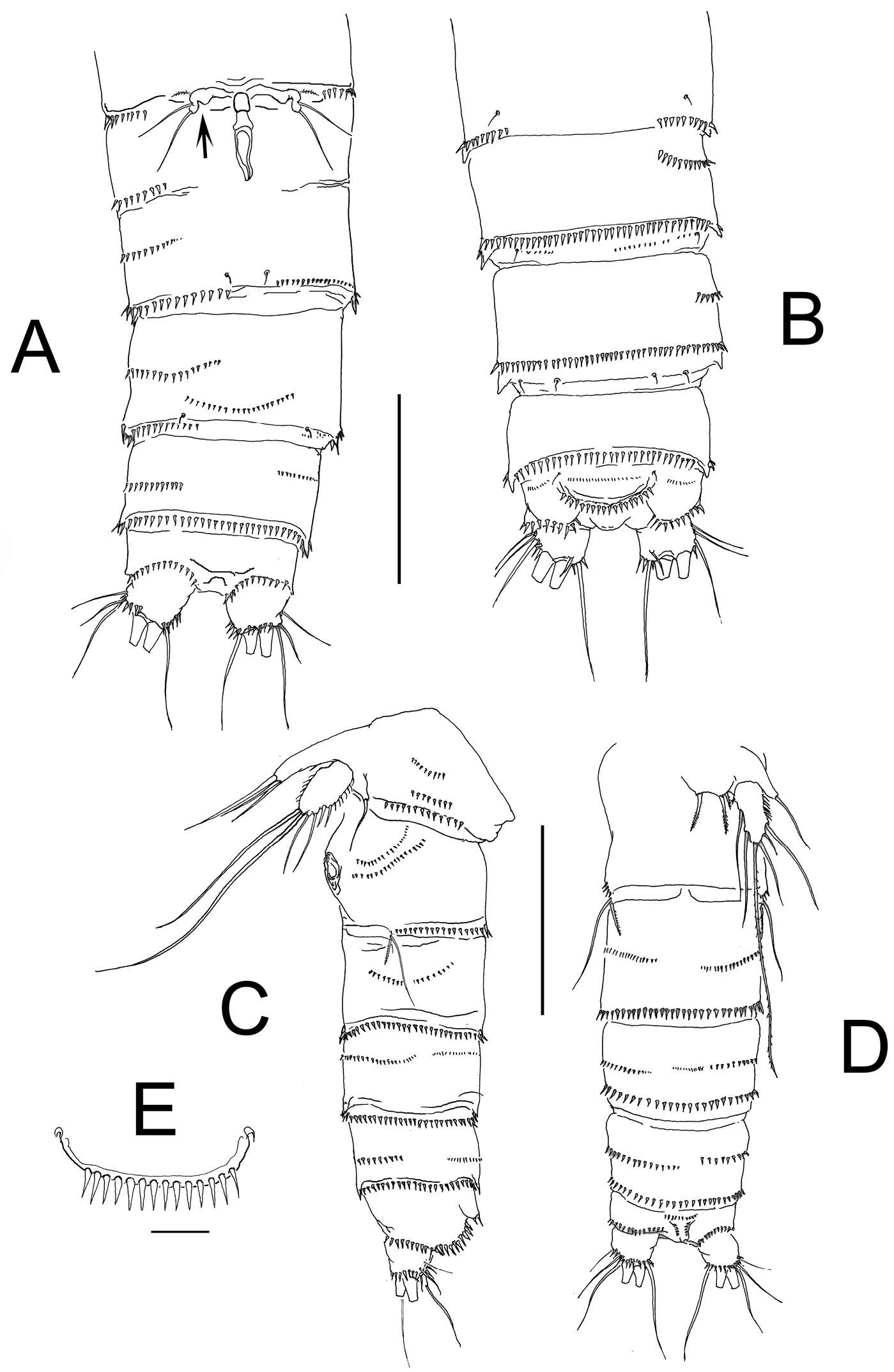
Figure 14.Mesocletodes elmari sp. n. A CIV male paratype 5, A1 dorsal view B CIII paratype 6, P1 C CIII paratype 6, P2, outer basal seta supplemented according to counterpart D CIII paratype 6, P3 E CIII paratype 6, P4 F CIII paratype 6, A1. Missing setae indicated by arrows. Scale bars: 50 µm.
-
Diana M. P. Galassi, Paola De Laurentiis, Barbara Fiasca
Zookeys
Figure 12.Phyllognathopus inexspectatus sp. n. (♀). A mandible B maxillule C maxilla D maxilliped E P1 (scale bars in μm).
-
Samuel Gómez, Nicola K. Carrasco, Francisco Neptalí Morales-Serna
Zookeys
Figure 4.Nitocra taylori sp. n. Female. A antenna B mandible C maxillule D P1, anterior E intercoxal sclerite of P1, anterior. Scale bar: A–E=50 µm.
-
Eduardo Suárez-Morales, Jani Jarquín-González
Zookeys
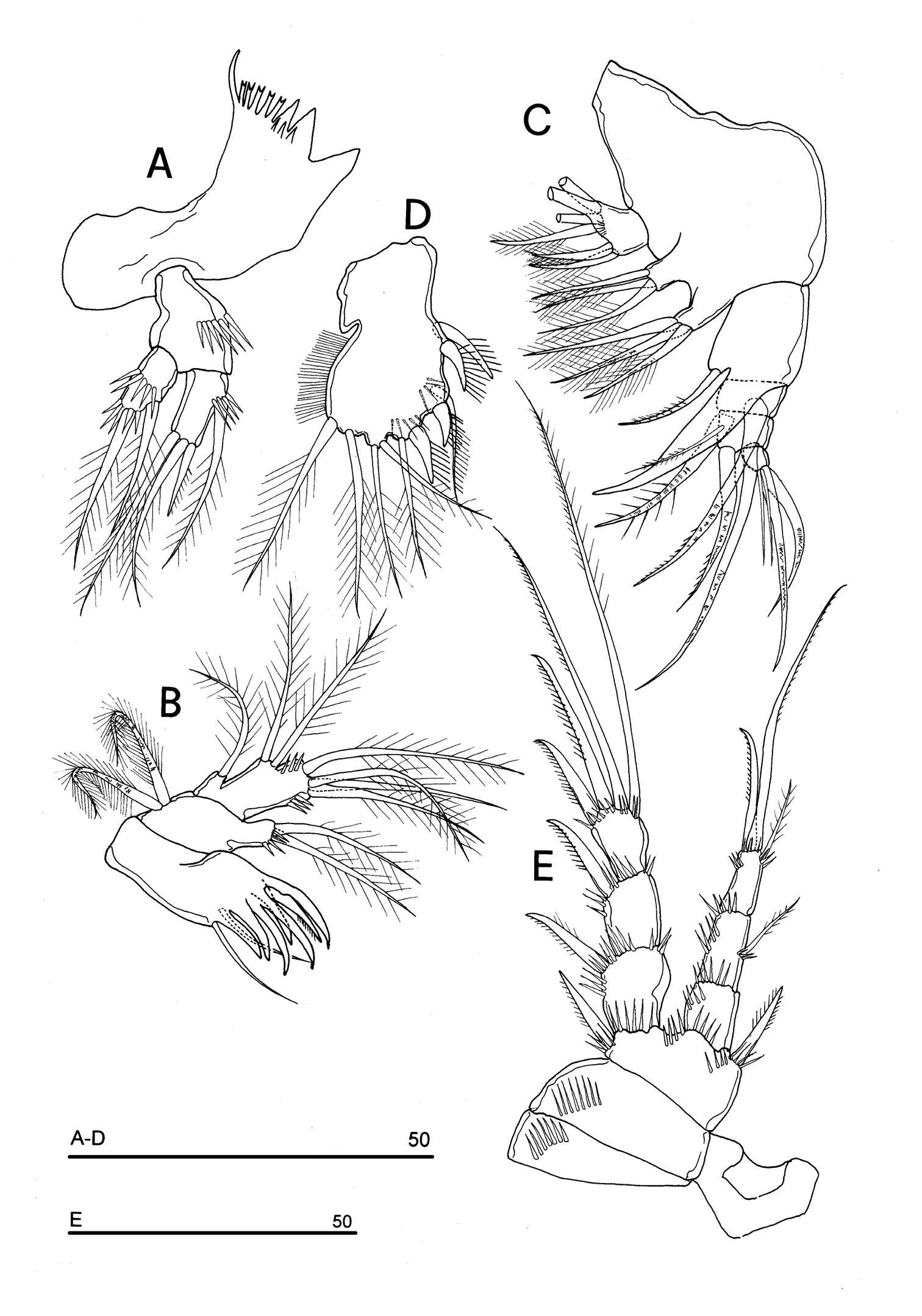
Figure 4.Peltidium nayarit sp. n., adult female from Playa Careyeros, Nayarit, Mexican Pacific. A leg 2 B leg 3 C leg 4. Scales bars: A–C=100 μm.
-
Juan M. Fuentes-Reinés, Eduardo Suárez-Morales
Zookeys
Figure 2.Nitokra affinis colombiensis ssp. n., from northern Colombia. A female, urosome, ventral view showing genital field and P6 B same, dorsal view, showing genital field and sixth leg plate, arrowed C male, urosome, lateral view showing P5 and P6 plate D same, ventral view E male, detail of ornamentation of anal operculum. Scale bars: A–D = 100 μm, E = 10 μm.
-
Hyun Woo Bang, Jeffrey G. Baguley, Heejin Moon
Zookeys
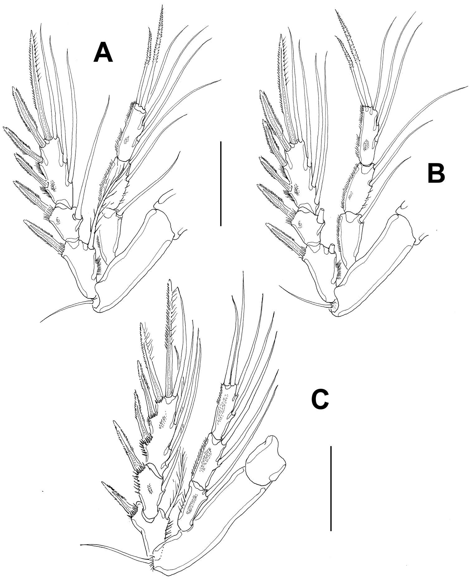
Figure 3.Pentacletopsyllus montagni gen. et sp. n. female: A P1, anterior B P2, anterior C P3, anterior.
-
Tomislav Karanovic, Kichoon Kim, Wonchoel Lee
Zookeys
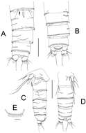
Figure 3.Stenhelia pubescens Chislenko, 1978, line drawings, female 3: A urosome, dorsal B urosome, ventral (armature on left caudal ramus omitted) C rostrum, dissected and compressed, dorsal D sixth leg, dorso-lateral E sixth leg, ventro-lateral F fifth leg second endopodal seta from inner side, anterior.
-
Diana M. P. Galassi, Paola De Laurentiis, Barbara Fiasca
Zookeys
Figure 13.Phyllognathopus inexspectatus sp. n. (♀). A P2 B P3 C P4 D P5 E P6 (scale bars in μm).
-
Samuel Gómez, Nicola K. Carrasco, Francisco Neptalí Morales-Serna
Zookeys
Figure 5.Nitocra taylori sp. n. Female. A antennule B P2, anterior. Scale bar: A=70 µm; B=100 µm.
-
Juan M. Fuentes-Reinés, Eduardo Suárez-Morales
Zookeys
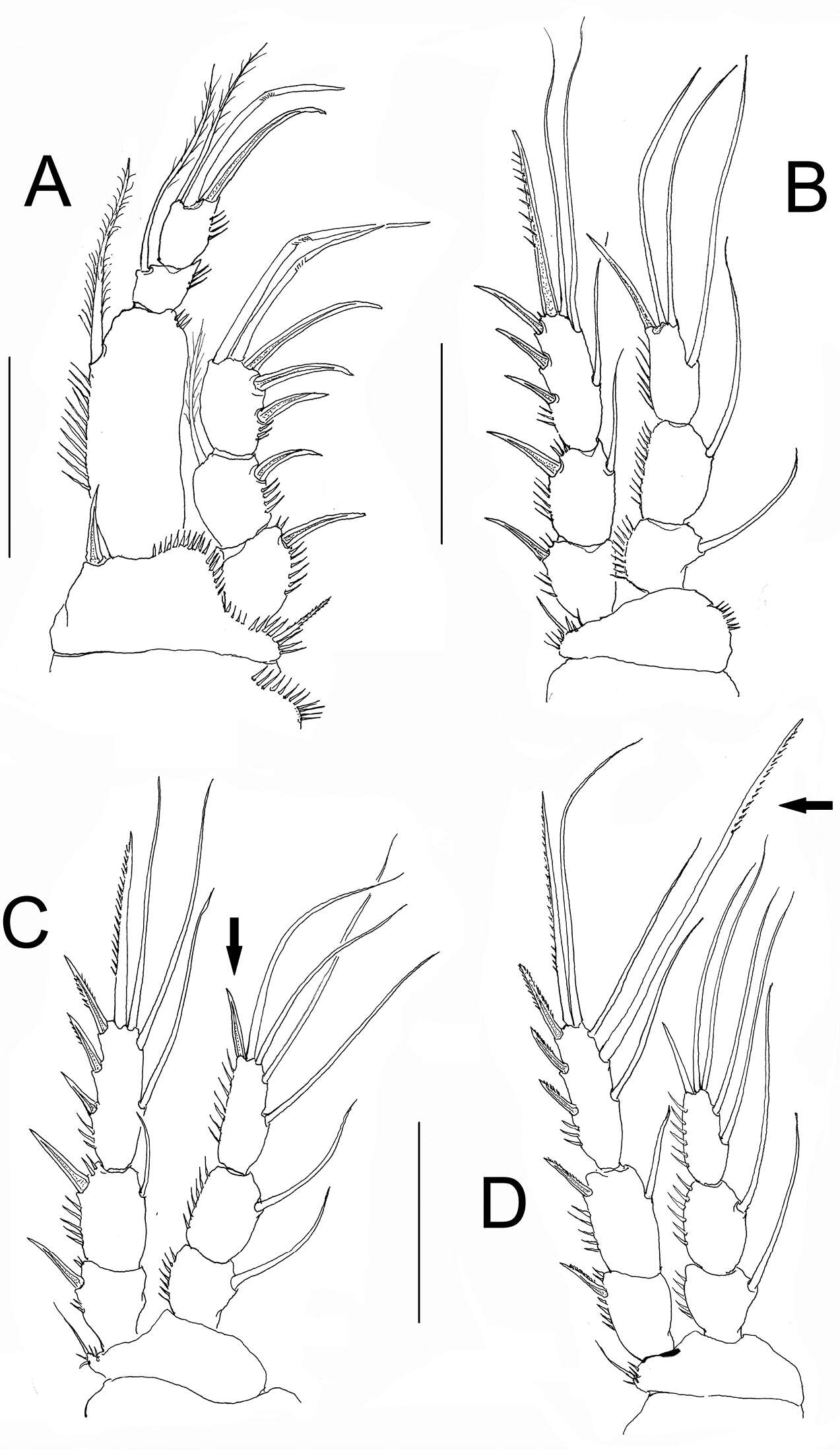
Figure 4.Nitokra affinis colombiensis ssp. n., adult female from northern Colombia. A first swimming leg (P1) B second swimming leg (P2) C third swimming leg (P3) D fourth swimming leg (P4) showing modified pectinate element (arrowed). Scale bars: A–D= 50 μm.
-
Hyun Woo Bang, Jeffrey G. Baguley, Heejin Moon
Zookeys
Figure 4.Pentacletopsyllus montagni gen. et sp. n. female: A P4, anterior B P5, anterior C genital field, ventral D anal somite, ventral.