Description
(
anglais
)
fourni par Zookeys
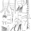
Female. Total length, 1.29–1.41 mm (mean±SD = 1.37±0.04 mm, N=5; holotype, 1.29 mm); prosome length, 0.75–0.82 mm (0.79±0.02 mm; holotype, 0.75 mm); prosome width, 0.31–0.34 mm (0.32±0.01 mm; holotype, 0.32 mm). Habitus (Figs 2A, B) with anterior margin of cephalosome rounded in dorsal view. Rostrum with paired filaments (Fig. 2C). Cephalosome and first pedigerous somite completely fused; fourth and fifth pedigerous somites totally fused. Prosomal ends rounded; dorso-lateral spines on fifth pedigerous somites. Urosome 4-segmented. Genital double-somite asymmetrical in dorsal view, elongate, ca. 2.54 times as long as wide; postero-dorsal and lateral margins with somewhat irregular row of spinules; in ventral view, genital area furnished with blunt, linguiform process midway, transverse rows of spinules anteriorly and paired flaps originating from genital opercula (see Fig. 5); each of paired egg-sacs consisting of 9–14 eggs, attached to lateral of genital opening (Fig. 2A). Proportional lengths of urosomites and caudal ramus 43:15: 15: 7: 20 (=100); length to width ratios 2.5, 1.3, 1.3, 0.4, and 3.6, respectively. Second and third urosomites with row of minute spinules along postero-dorsal and lateral margins. Caudal rami with hair on inner margin and symmetrical with 6 setae: seta I absent, seta II with fine setules only along inner margin; setae III-VI plumose; seta VII located dorsally.
Antennule (Fig. 2D) reaching beyond posterior end of genital double-somite, symmetrical, 22-segmented; segments 6-7 incompletely fused; segments 6, 15, 16, 18-21 each without aesthetasc (ae). Fusion pattern and setal elements as follow: 1 - 1 + ae, 2 - 3 + ae, 3 - 2 + ae, 4 - 3 + ae, 5 - 3 + ae, 6 - (1 spiniform element), 7 - 2 + ae, 8 - 2 + ae, 9 - 2 + ae, 10 - (1 spiniform element) + ae, 11-14 - 2 + ae, 15-16 - 2, 17 - 2 + ae, 18-19 - 1, 20-21 - 2, 22 - 6 + ae.
Antenna (Fig. 2E) coxa with single seta; basis with 2 setae at inner corner; endopod 2- segmented, first segment with 2 setae, second segment with 7 and 8 setae on terminal and subterminal lobes, respectively, and lateral row of fine setules; exopod 4-segmented, first segment with 1 seta, second segment with 1 proximal, 2 medial and 1 terminal setae; third segment with 3 setae; fourth segment with 1 medial and 3 terminal setae.
Mandible (Fig. 2F) with basis bearing 4 setae along inner margin; endopod 2- segmented, first segment with 4 setae, second with 9 setae; exopod 5-segmented, first to fifth segments with 1, 1, 1, 1, 2 setae, respectively. Gnathobase (coxa) with serrate dorsal seta and 3 cuspidate and 4 blunt teeth.
Maxillule (Fig. 3A) with preacoxal arthrite bearing 9 strong and 6 fine setae and small spinules; coxa with 4 setae on endite and 9 setae on epipodite; basis with 4 and 5 setae on proximal and distal endites, respectively; basal exite with 1 seta; endopod 3-segmented, with 4, 4 and 6 setae from first to third segments, respectively; exopod foliaceous with 10 setae along outer margin.
Maxilla (Fig. 2G) with first and second praecoxal endites having 4 and 3 setae, respectively; first coxal endite with 3 long setae, second endite with 1 short strong and 2 long setae; basis with 1 short and 2 long setae; endopod with 9 setae.
Maxilliped (Fig. 3B) with praecoxa and coxa completely fused; endites with 0, 2, 3, 4 setae, respectively; basis with 3 setae; endopodal segment having 6 segments, first segment with 2 setae, second segment with 2 bifurcated setae and 1 seta, third and fourth segments with 1 bifurcated seta and 1 seta, fifth and sixth segments with 3 and 4 setae, respectively.
Legs 1–4 (Figs 3C–F) biramous with 3-segmented rami; coxa and basis of both rami with spinules on distal corner. Seta and spine formula as follows:
Leg 5 (Fig. 3G) uniramous and almost symmetrical; in posterior view, basis with short medial seta and spinular rows; exopod 3-segmented, first segment produced into small pointed process at inner subterminal corner, with distolateral spine and one or two rows of spinules; second segment having short and thickned disto-lateral process and medial serrate spine; third segment spiniform, tapering distally with inner spinules and proximo-medial spine.
Male. Total length 0.94-1.02 mm (mean±SD = 0.97±0.03, N= 4; allotype, 1.02 mm). Prosome length 0.62-0.66 mm (mean±SD = 0.64±0.01, allotype, 0.66 mm), width 0.26-0.27 mm (mean±SD = 0.26±0.005, allotype, 0.26 mm).
Habitus (Figs 4A, B) similar to that of female, except for urosome. Urosome 5- segmented; proportional lengths of urosomites and caudal ramus 13: 25: 21: 17: 11:13 (=100); length to width ratios 0.5, 1.1, 1.2, 1, 0.6 and 1.7. Genital somite nearly symmetrical with one or two rows of spinules ventrally. Urosomites 2–4 with spinular row along posterior margin. Caudal rami symmetrical, with six setae as in female.
Right antennule (Fig. 4C) geniculate and indistinctly 20-segmented; setal formula as follows: 1 -1 + ae, 2 - 2 + ae, 3 - 2 + ae, 4 - 1, 5 -1 + ae, 6 - (1 spiniform element), 7 - 1 + ae, 8 - (1 spiniform element), 9 - 2 + ae, 10 - (1 spiniform element), 11 - 1 + ae, 12 - (1 spiniform element) + ae, 13 -1 + ae, 14-16 - 2 + ae, 17-18 - 1 + (1 process), 19 - 2 + (1 process), 20 - 9 + ae.
Leg 5 (Figs 4D, E, F, G) highly asymmetrical and biramous; intercoxal sclerite and both coxae fused; coxa with fine spinular rows on anterior surface. Right leg (Figs 4D, E) with basis having outer spinular row; endopod rudimentary, represented by knob-like process with fine setule at tip; exopod (Fig. 3F) 3-segmented, first segment protruded into outer process reaching middle of third segment, proximal process with 1 spine and spinular row; second segment expanded midway, each side with spine; third segment curved inward with 3 rows of spinules on anterior surface and middle swelling, distal to which tapering distally. Left leg (Figs 4D, E) with elongated basis having triangular process at midlength; endopod (Fig. 4G) highly developed, bifurcated, inner medial process smoothly curved outward reaching distal tip of second exopod, outer process thickened, foliaceous with 1 subterminal and 4 thin terminal protrusions; exopod 2-segmented, first segment as long as basis, irregularly sinuated along inner margin; second segment triangular with hirsute process proximally and stout serrated protrusion at medio-lateral margin, with 3 processes of unequal length terminally.
- licence
- cc-by-3.0
- droit d’auteur
- Khwanruan Srinui, Shuhei Nishida, Susumu Ohtsuka
- citation bibliographique
- Srinui K, Nishida S, Ohtsuka S (2013) A new species of Pseudodiaptomus (Crustacea, Copepoda, Calanoida, Pseudodiaptomidae) from the Prasae River Estuary, Gulf of Thailand ZooKeys 338: 39–54
- auteur
- Khwanruan Srinui
- auteur
- Shuhei Nishida
- auteur
- Susumu Ohtsuka